The formation of a purulent sac near the root system can lead to serious consequences, including loss. Therefore, today we will talk about granulomas on the root of the tooth, tell you what they are, and determine what treatment is necessary.
The main feature of dental disease is its frequent asymptomatic course. Typically, the occurrence of compaction is not noticed by the patient, while complete ignorance of the situation can lead to the growth of a source of infection.
What is granuloma
Granuloma is a small nodular inflammation of soft tissue. Its size usually does not exceed 5–7 mm in diameter, but if left untreated for a long time, it begins to increase and can lead to destruction of bone tissue. Pus accumulates inside it. The granuloma is localized at the root of the tooth, more often at its apex. It is the most common apical lesion1 (that is, located at the apex of the tooth root).
The photo shows the process of pathology formation
Did you know that a dental microscope is a powerful aid in the treatment of cysts and granulomas? With the help of multiple magnification achieved by the device, the doctor treats not by touch, but with certainty. And what is very important is that the presence of the device and an experienced endodontist in the clinic where you go means a high chance that a tooth with a similar problem will be saved, after which it will serve for many more years.
Possible complications
Complications of granuloma can be both local and systemic.
These include:
- tooth loss;
- maxillary osteomyelitis;
- cyst development;
- formation of migratory granuloma;
- abscesses and fistulas of soft tissues of the face;
- meningitis;
- encephalitis;
- inflammation of peripheral nerves;
- sinusitis;
- sepsis;
- pyelonephritis;
- bacterial endocarditis;
- phlegmon of the soft tissues of the face.
Signs of the disease
People who have this pathology do not notice it for a long time because they have no painful sensations. If acute pain appears, this indicates that the disease is advanced or has worsened. But then it is accompanied by other symptoms: swelling and redness of the gums in the area where the pathology is located, the release of pus from the periodontal pocket, a general deterioration in well-being, and darkening of the tooth.
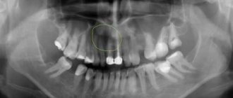
How to detect pathology at an early stage of development if it does not manifest itself with any symptoms for a long time? A dental granuloma is clearly visible on an x-ray. Accordingly, it can only be detected at a dentist’s appointment. If you undergo preventive examinations twice a year and take a panoramic photo of the jaw, the problem will be detected in time. It is also important to treat teeth promptly and competently - caries, pulpitis, periodontitis precede the development of a tumor.
Dental granuloma is clearly visible on x-ray
Stages of development
Tooth granuloma goes through several stages in its development. As the disease progresses, a gradual proliferation of connective tissue is observed due to the penetration of infection into the soft tissues. The walls of the purulent sac protect the body from the spread of pathogenic flora to healthy areas.
The following stages of development of pathological growth should be noted:
- Primary. Infectious pathogens penetrate the gums due to the presence of dental diseases. Pathogenic particles provoke the gradual death of nerve fibers and blood vessels.
- Secondary. The granuloma capsule breaks through, which causes the element to move and expose its root.
- Spread of infection to the bone structures of the jaw.
- Final. The bone is separated from the inflamed area. A dense capsule consisting of connective tissues gradually forms. The capsule is filled with dead immune cells.
In acute pathology, granulation cells quickly replace healthy periodontal tissue.
Why does granuloma need to be treated?
Many patients think that granuloma does not pose a serious danger, because the formation is very small and does not cause much discomfort. There are also people who are sure that pus on the root of a tooth with such a pathology can resolve on its own. But all these judgments are wrong.
In fact, if left untreated, granuloma can lead to complete destruction of the tooth root and the need for its removal, but this is only the mildest of complications.
Granuloma on the root of a tooth most often develops if a person has not undergone treatment or it was of poor quality. For example, when caries is complicated by pulpitis, and pulpitis by periodontitis. The risk of a tumor increases significantly if the patient has concomitant factors: hypothermia, injury, decreased immunity, hormonal changes or other diseases of the body.
A small purulent sac begins to enlarge and can gradually degenerate into a cyst, which in advanced cases reaches 5–6 cm in diameter. Pus can seek a way out, and then a fistula tract is formed. It can open not only into the oral cavity, but also inside the body, which can lead to infection of internal organs and the development of sepsis.
The following purulent complications are also possible: perimandibular abscess, or phlegmon, osteomyelitis. If bacteria penetrate other organs (and with a granuloma this is not difficult, because it consists of tissue with a large number of capillaries and blood vessels), then sinusitis, meningitis, arthritis of the jaw joint, and otitis may develop.
Tooth root granuloma in an advanced stage often causes complications on the cardiovascular system and kidneys, becoming a provoking factor for the development of pyelonephritis and myocarditis.
Symptoms
In most cases, for a long time a person does not suspect that he is developing a granuloma, since there are no symptoms. The tooth does not hurt, except that sometimes there is a slight discomfort when clenching the teeth tightly or when chewing hard food. Sometimes this can happen when eating hot and cold.
At those moments when the body's defenses weaken, the pathology worsens. This can occur after infections, surgeries, or during menstruation in women. The following symptoms appear:
- sharp pain when biting;
- swelling of the gums;
- gum tissue pain.
Often periods of exacerbations and chronic manifestations follow each other for a long time. But sooner or later the granuloma will make itself known in such a way that the symptoms can no longer be ignored. The signs will be as follows:
- sharp pain;
- swelling of the gums;
- prostration;
- high body temperature;
- swelling in the neck or cheek (depending on which tooth hurts);
- the appearance of pus – periostitis or flux.
In later stages, resorption of bone tissue in the jaw is observed. The consequences can be different, ranging from tooth loss to sepsis. This is a very dangerous condition that poses a risk not only to the health, but also to the life of the patient.
Conservative and drug treatment
Timely detection of a granuloma on an x-ray allows the dentist to apply the most loyal approach to the treatment of this pathology. Here are the actions that will be taken then:
- cleaning and antiseptic treatment of tooth root canals,
- placing a drug based on calcium hydroxide inside the channels,
- applying a temporary filling,
- installation of a permanent filling.
The second and third points can be repeated repeatedly until the formation finally resolves. Additionally, the doctor prescribes home therapy for the patient, which consists of taking antibiotics and painkillers, and performing antiseptic mouth rinses, for example, with Chlorhexidine.
The photo shows the process of conservative treatment
Many doctors believe that this measure is not always effective; often, after conservative therapy, the disease can recur. But patients do not always like that treatment can last for 4–6 months. However, if you don't want to resort to more radical solutions, then it's worth a try.
“A year ago, I began to notice that when I chew on one tooth, it hurts, but after eating, the pain goes away immediately. I didn’t think that I needed to go to the dentist with such a problem: the tooth was treated and filled. But one fine day he began to get so sick, even regardless of food intake, that he ran to the doctor. A diagnosis of granulomatous periodontitis was made. They treated him for a very long time, put medicine inside him for almost a year, but the tumor at the root did not resolve, and it had to be removed surgically. I don’t know, maybe I should have deleted it right away instead of suffering like that...”
Helga32, review from the dental portal gidpozubam.ru
Physiotherapeutic methods
In some clinical cases, when the granuloma is small and the inflammatory process is not acute, treatment is possible using the following methods.
Electrophoresis
This procedure involves introducing iodine-based medications into the infected canals using current pulses, designed to kill bacteria and also promote the resorption of the formation. True, physiotherapy requires patience and a lot of free time, because it will take about 10–20 procedures performed daily or every other day to get rid of the pathology.
Depophoresis
Another physiotherapeutic method is depophoresis. The principle of introducing medication into the root canals here is similar to that of electrophoresis. Copper/calcium hydroxide is used only as the main drug. Depophoresis can only be performed on teeth that lack pulp. The number of procedures is usually three, with a break of a week between them.
This method does not require complex surgical procedures.
Destruction of granuloma with laser
The method is more expensive, but fast and effective. Suitable even for curved root canals. Before this, the specialist cleans the canals and sterilizes them. Penetrating through the root, the doctor treats the granuloma with a laser, and it resolves. The laser has a powerful disinfecting effect, does not injure surrounding tissues, on the contrary, promotes their rapid healing, stops bleeding, because cauterizes blood vessels. Doctors who practice this type of innovative equipment claim that relapses of the disease are excluded.
The laser has a powerful disinfecting effect
Contraindications to the use of laser: oncology, cracks and fractures at the root.
Surgical methods
If a patient has a granuloma on the root of a tooth, and conservative treatment and physiotherapy have not yielded results, there is only one way out - surgery. This also applies to severely advanced cases.
If the pus on the root of the tooth has led to an acute inflammatory process, then it is first removed using drainage and only after that the removal of the granuloma begins. Most often, the granulomatous nodule is curetted using cystectomy; the procedure is combined with resection of the apex of the tooth root, i.e. part of the root is excised along with the neoplasm. Such measures allow you to save the tooth itself.
The photo shows the procedure for removing pus
Another less gentle type of surgical intervention, which nevertheless allows you to save the tooth, is hemisection. This operation can only be performed on multi-root elements. Indicated when it is not possible to save one of the roots, then the root is removed along with part of the crown adjacent to it. After the procedure, the patient is required to undergo prosthetics with an artificial crown as soon as possible to restore shape, aesthetics and functionality.
There is another surgical method that is least desirable for any patient - removal of the granuloma along with the affected tooth. This is an extreme measure, but with extensive destruction of hard tissue and severe inflammation, it is not always possible to avoid it.
Surgical treatment is always accompanied by taking antibacterial and painkillers, and rinsing the mouth with antiseptic solutions.
Is it possible to cure granuloma using traditional methods?
Treatment of pathology with folk remedies is ineffective. Decoctions of various herbs can only bring temporary relief when there are signs such as inflammation and swelling of the tissues surrounding the tooth, and pain. It must be remembered that rinsing with herbs or soda acts locally, and the granuloma is located at the root of the tooth, and beneficial substances from decoctions or solutions do not penetrate to it.
It is unacceptable to warm up a diseased tooth, on the root of which pus has accumulated, because then this is fraught with the spread of infection deep into the body, the breakthrough of purulent contents into the circulatory and lymphatic systems, into the surrounding tissues.
Notice
: Undefined variable: post_id in
/home/c/ch75405/public_html/wp-content/themes/UltraSmile/single-item.php
on line
45 Notice
: Undefined variable: full in
/home/c/ch75405/public_html/wp-content /themes/UltraSmile/single-item.php
on line
46
Rate this article:
( 1 ratings, average: 5.00 out of 5)
flux
- Mammadzade R.E. Non-surgical treatment of teeth with large periapical destruction // Modern dentistry. – 2017.
Expert “Granuloma can remain dormant for a long time. But a viral infection, hypothermia, and even a severe stressful situation can cause tumor growth. The best measure to prevent disease is timely examinations and treatment of oral diseases.”
Dentist-therapist Belyaeva Olga Aleksandrovna
Consulting specialist
Dzhutova Aida Vladimirovna
Doctor rating: 8.8 out of 10 (5) Specialization: Implant surgeon, periodontist Experience: 8 years
Causes
Dental granuloma occurs due to various reasons, infectious and non-infectious:
- Caries, as when it occurs, the canal and root of the tooth become infected.
- Periodontitis is an inflammatory process of tissues located in the dental socket of the jaw.
- Complications of pulpitis are inflammation of the dental nerve.
- Tooth fracture and the possibility of contamination.
- Injuries to the oral cavity and gums, in which microbes penetrate the dental tissues.
- Infection through unsterile instruments when filling teeth.
- Failure to comply with oral hygiene rules.
- Abnormal development of teeth from birth.
Factors that increase the risk of formations and associated complications:
- Hypothermia.
- Frequent stressful situations.
- Sudden climate change.
- Past flu, infections.
- Excessive physical exertion.
- Factors that weaken the immune system.
Comments
Once a year I definitely go for an examination with a dentist, and this time he discovered a granuloma... and recommended removing the tooth right along with it!!! How is it possible, why deletion if it was discovered in a short time????
Yulia V. (03/12/2020 at 06:22 pm) Reply to comment
- Yulia, it can be assumed that conservative treatment, as well as surgery with preservation of the tooth, is impossible due to the presence of a root crack, or the inflammation has progressed greatly this year under the influence of other factors (general diseases of the body, stress, hypothermia). Another reason is severe destruction of tooth tissue, in which its preservation is not possible. Try contacting another doctor to compare the results of the study and the proposed methods for solving the problem.
Editorial staff of the portal UltraSmile.ru (03/20/2020 at 09:09) Reply to comment
If the dentist advises surgical removal of the granuloma, how long does the healing process take and what complications may occur? I’m worried that I’ll have to take time off from work; after all, this is an operation, albeit of a local nature.
Tatyana (03/20/2020 at 07:53) Reply to comment
Recently at an appointment I was diagnosed with a granuloma. But the dentist suggested only one way - tooth extraction. He said that during treatment there are no guarantees that in a year the tooth may have to be removed anyway. How justified is this? Maybe you should contact another specialist?
Lydia (03/20/2020 at 08:37) Reply to comment
Is it really necessary to take a panoramic photo twice a year? I think that a competent dentist, during an annual examination if there are complaints, should recommend this procedure. And then do it.
Peter (03/20/2020 at 10:00) Reply to comment
Good afternoon! I don’t quite understand whether granulomas can appear due to caries? I have small caries on several teeth, as the dentist told me that there is no need to rush into treating them. Is it so?
Dmitry Ch. (03/20/2020 at 10:50) Reply to comment
Please tell me, is there any way to prevent the appearance of this granuloma? Are there any methods of prevention? Is surgery performed under local anesthesia? How immediately after detecting a source of inflammation?
Ksenia (03/20/2020 at 11:42 am) Reply to comment
I went to the doctor for dental treatment, a granuloma was discovered, and the tooth had to be removed. Although literally six months ago there was a scheduled examination. Couldn't granuloma have been detected then?
Elizaveta (03/20/2020 at 11:46 am) Reply to comment
Hello. So what causes a granuloma to appear initially? And is it necessary to check with a dentist twice a year if you have no caries, no plaque, etc.? For example, I didn’t just go to the dentist at all.
Baht (04/23/2020 at 07:39) Reply to comment
I had granuloma; it could only be cured surgically and the process was very long and painful. Now I'm worried that the disease may return. Is there such a chance if I lead a healthy lifestyle and do not have chronic diseases?
Olga (04/23/2020 at 08:13) Reply to comment
That is, it is also visible only in the picture? Horror. I undergo preventive maintenance at the dentist every six months, but I don’t take a picture, just a superficial examination. Looks like I'll have to take some photos too.
Valentin (04/23/2020 at 09:05) Reply to comment
Write your comment Cancel reply
Prevention
It is recommended to carry out preventive actions in a comprehensive manner. The measures are aimed at preventing the development of dental granuloma. Preventive measures include the following:
- Maintaining cleanliness in the oral cavity. It is important to brush your teeth and rinse your gums thoroughly every day.
- Treat bleeding gums.
- Visit your dentist regularly at least twice a year.
- Regularly replace toothbrushes to avoid spreading bacteria throughout your mouth.
- Mild pain in the teeth indicates the need to visit a doctor. It is important to get checked in time so that if there is a pathology, it is detected in a timely manner.
- Closely monitor the formation of caries, pulpitis and periodontitis. They provoke the development of granuloma.
- Use exclusively medicated toothpastes for preventive measures.
- Constantly rinse your mouth with herbal infusions.
- It is recommended to add foods with the maximum amount of calcium to your diet. It is important to fill food with useful microelements and vitamins.
