It is important to know what the lungs are, where they are located in a person, and what functions they perform. The respiratory organ is located in the chest in humans. The chest is one of the most interesting anatomical systems. The bronchi, heart, some other organs and large vessels are also located here. This system is formed by the ribs, spine, sternum and muscles. It reliably protects all important internal organs and, thanks to the pectoral muscles, ensures the uninterrupted functioning of the respiratory organ, which almost completely occupies the chest cavity. The respiratory organ expands and contracts several thousand times a day.
Where are the human lungs located?
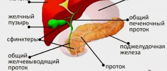
The localization of the structure in question is the chest cavity. The organ occupies the right and left halves, limiting the mediastinal complex (esophagus, heart, trachea and other formations) on the sides. Where the human lungs are is clearly shown in the figure below. Each part of the paired organ has the shape of a semi-cone. The base is located on the diaphragm, and the apex reaches the shoulder girdle, protruding upward 1-3 cm from the collarbone.
The structure of the human lungs
The described paired organ has a complex structure that allows it to perform several functions. To understand how the human lungs are structured and how their contraction and gas exchange are ensured, it is necessary to study the external and internal parts of these physiological formations. An interesting feature of the organ is its porosity, due to which it occupies a small volume in the body with an impressive surface area comparable to the size of a tennis court.
Structure of the lungs - lobes and segments
The formation presented is paired, but not symmetrical. The human lungs are divided into lobes separated from each other by slits. There are two of them on the left side of the organ - upper and lower. The right lung contains an additional, middle (medial) lobe. Each of them consists of smaller structural units, segments. Such areas are separated from neighboring similar areas by connective tissue layers.
The number of segments is also different for the left and right lungs (8 and 10 pieces). In the center of each of them there is its own bronchus and artery, providing ventilation and blood supply to the structure. The segments are “composed” of lobules (lobules) - small pyramid-shaped formations. They contain bronchial branches, from which up to 2 dozen bronchioles are formed. The diameter of these air-carrying “tubes” does not exceed 1 mm. The end of each of them represents a structural unit that forms the human lungs, the acinus.
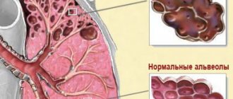
The respiratory bronchioles continue to branch into thin passages ending in special sacs. The smallest element of the acini are the alveoli of the lungs - protrusions or hemispherical vesicles. It is in them that the exchange of gases occurs between the inhaled and expelled air and the circulatory system, thanks to the interweaving of each section with a miniature capillary network.
External structure of the lungs
The organ in question is not muscular, so its contraction is ensured by external structures. The serous membrane is the pleura of the lungs. It consists of 2 layers - parietal and visceral. The first, outer layer is connected to the chest wall. The visceral or internal pleura covers the outer surface of the human lungs. Between the leaves there is a small space (cavity) filled with a serous viscous substance. This is a pleural fluid that is necessary to hold the layers together during inhalation and exhalation, preventing friction.
Internal structure of the lungs
The presented organ in cross-section resembles an inverted tree, crown down. The structure of the lungs inside begins with the “trunk” - the trachea or windpipe. The large “branches” are the bronchi. They are successively divided into smaller and thinner tubes (bronchioles). The “leaves” are alveoli, tiny air bubbles. They are arranged in clusters, forming bags.
What determines the quality of an x-ray image?
Chest X-ray is one of the most informative methods for diagnosing the respiratory system, if it is done correctly and interpreted correctly by a doctor. In this case, it is necessary to follow the rules of installation and procedure.
Factors influencing the result of X-ray diagnostics:
- Symmetrical body position. If the patient does not stand straight during the procedure, the sternoclavicular joints will be located asymmetrically, which can be considered rotation of the thoracic vertebrae.
- Image hardness . Medium hardness is preferable, since with a soft image some formations may not be visible, but with a hard image, on the contrary, unnecessary shadows (artifacts) will appear, which a specialist may mistake for pathology.
- Concomitant diseases that may affect the chest.
- Completeness of coverage (a good image contains the apexes of the lungs above and the costophrenic sinuses below).
- The shoulder blades should be positioned on the outside of the chest to avoid distortion of the image.
- Image clarity . The patient is asked not to breathe during the shooting to immobilize the musculoskeletal system of the chest.
- Contrast . Adjusted by a doctor on the device; The radiation power is set depending on the patient’s muscle and fat mass.
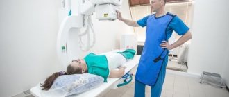
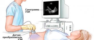
Photo 2. To obtain a good result, the patient is required to stand straight and hold his breath for a short time.
Lung functions
The main task of the presented paired organ is to provide all living cells with oxygen. The human lungs constantly carry out gas exchange. Oxygen enters the blood during inhalation, through the capillary network that encircles the alveoli. Similarly, when you exhale, carbon dioxide is removed. In addition to the main function, the lungs in the human body are responsible for a number of secondary, but very important tasks:
- They protect the heart from mechanical shocks from the outside and provide shock absorption.
- They regulate the level of partial pressure of carbon dioxide by adjusting the pH of the blood.
- Participate in maintaining hormonal balance.
- Prevents the penetration of airborne pathogens. On the mucous membrane of the respiratory tract there are cilia (ciliated epithelium), which capture dust and bacteria from the air and move them back upward, enveloping them in a viscous secretion. It contains antimicrobial components (glycoproteins) and immunoglobulins type A.
- They slightly contribute to thermoregulation in the human body.
- They provide air flow for extracting sounds, forming a voice, and singing.
- Serve as a reserve blood storage facility. The lungs of an adult contain about 450 ml of biological fluid, but this figure can double. If large-scale blood loss occurs in a large circle, the organs will release it to replenish its volume. This can be life-saving if the body is seriously injured.
Human lung capacity
The absolute or total capacity is 5-6 liters in an adult male. The lungs of a healthy person have a larger volume than the organs of a smoker or a patient with some kind of respiratory pathology. This indicator is additionally influenced by other factors:
- height;
- floor;
- body type;
- age;
- geographical location (relative to sea level height).
The maximum capacity of a person's lungs is never fully utilized. The deepest inhalation and exhalation is about 2 liters. In a calm state and measured breathing, this indicator is significantly lower. After exhalation, about 3 liters of air are present in the human lungs. This is the functional residual capacity necessary to maintain a stable ratio of oxygen and carbon dioxide in the alveoli. Accurate volume measurement for diagnostic purposes is called spirometry.
Preventive measures
An important component of the period of recovery and cleansing of the lungs is a complete and balanced diet containing the necessary microelements and substances. It is necessary to add garlic to the daily diet of a former smoker, as it is a powerful stimulator of restorative and cleansing processes. Along with garlic, it is necessary to include in the diet fruits containing significant amounts of ascorbic acid, which ensures the restoration process of connective tissues of organs.
Experts recommend drinking at least 1.5-2 liters of water during the day. Water helps remove all toxins and cell breakdown products from the body. However, on the recommendation of a doctor, medications may be prescribed. However, you should not prescribe the drug yourself. This is fraught with serious consequences.
Thus, the smoker's lungs, of course, evoke deep sympathy. But if you make the right decision in time and quit smoking and follow the recommendations given, then the internal organs and systems, including the lungs, can be returned to their previous state within a certain time.
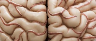
Everyone saw this terrifying photo. Smoker's lungs! Recently, people have been writing online that this is part of anti-tobacco propaganda. Is it so? Let's try to figure it out.
How do the lungs work?
The breathing mechanism is carried out not only with the help of the described paired organ. The functioning of the human lungs depends on the coordinated work of several structures:
- As you inhale, the diaphragm straightens and “goes” down. In parallel, due to several groups of nearby muscles, the ribs diverge.
- Air enters the bronchi through the trachea. The lungs expand, increase in size, and the distance between them increases.
- The air spreads through the bronchial branches, travels through the bronchioles and reaches the alveoli.
- Due to the difference in partial pressures in the bubbles, exchange occurs. The alveoli “give” oxygen into the blood and “take” carbon dioxide.
- As you exhale, the diaphragm relaxes, other muscles contract, and the ribs come together. The lungs push the “waste” air with carbon dioxide out and take their original position.
- The process is repeated.
Blood supply
Two types of blood circulate in the lungs: venous and arterial. This respiratory organ is very closely surrounded by blood vessels of different sizes. The most basic is the pulmonary artery, which then gradually divides into small vessels. At the end of the branching, capillaries are formed that entwine the alveoli. Very close contact and allows for gas exchange in the lungs. Arterial blood nourishes not only the lungs, but also the bronchi.
This main respiratory organ contains not only blood vessels, but also lymphatic vessels. In addition to the various branches, nerve cells also branch in this organ. They are very closely interconnected with blood vessels and bronchi. Nerves can create vascular-bronchial bundles in the bronchi and lungs. Because of this close relationship, doctors sometimes diagnose bronchospasm or pneumonia due to stress or another malfunction of the nervous system.
Lung diseases in humans
Like other organs, the presented physiological structure is susceptible to many congenital and acquired pathologies. There are very rare and almost unstudied lung diseases, for which no treatment has even been developed. The most common ailments:
- developmental defects;
- tuberculosis;
- cysts;
- respiratory distress syndrome;
- cancer;
- syphilis;
- pneumonia;
- asthma;
- parasitic infestations;
- cystic fibrosis;
- chronic obstructive disease (the lungs of a smoker are more affected);
- bronchiolitis;
- alveolar hemorrhagic syndromes;
- bronchiectasis;
- mesothelioma and other pathologies.
Diagnosis of pulmonary diseases
If you have symptoms indicating damage to the lower respiratory tract, you should consult a pulmonologist. Differential diagnosis of pulmonary diseases is performed using a variety of laboratory, instrumental and hardware techniques. First, the doctor performs a functional examination - visually assesses the condition of the respiratory tract, palpates nearby areas, uses percussion (tapping), and auscultation. If necessary, different research methods are prescribed:
- magnetic resonance, computed tomography;
- spirometry;
- fluorography;
- radiography;
- Ultrasound;
- bronchography;
- analysis of gases and sputum;
- fluoroscopy;
- angiography;
- gas mediastinography;
- pleurography;
- X-ray electrokymography.
Treatment and prevention of pulmonary diseases
Therapy for pulmonary pathologies is developed based on the diagnosis, the severity of symptoms, and is aimed at eliminating the causes that provoked them. In the treatment of diseases, conservative, surgical and physiological methods, and complex schemes can be used. Any ailment requires an individual approach, which is drawn up by the doctor. Prevention of pulmonary diseases includes:
- balanced diet;
- to give up smoking;
- scheduled fluorographic examination;
- regular medical examinations;
- playing sports;
- ensuring the purity of inhaled air.
Is close friendship with tobacco a cause of cancer?
Recently, there has been an increase in cancer diseases. One of the reasons is the increase in the number of smokers, including in younger age groups.
Tobacco smoke contains more than 3,500 chemicals. Many have a carcinogenic effect: they can change the genetic information of a cell and lead to tumor transformation.
This is why lung cancer and smoking go hand in hand. Clinical studies have confirmed that 90% of patients with malignant lung tumors are tobacco lovers.
What does tobacco smoke consist of?
- hydrogen sulfide;
- carbon monoxide;
- butane;
- methanol;
- hydrocyanic acid (used by the Nazis in gas chambers);
- acetone;
- ammonia.
Tobacco smoke also contains other substances that pose a danger to human health.
Cigarette smoke contains particulate matter that settles in the lung parenchyma, causing the death of alveolar epithelial cells.
These include:
Attention! Manufacturers never indicate the full composition of cigarettes on the packaging. The sale of tobacco products brings huge profits every year. Remember that this is the price of your health.
Signs of lung cancer
Early detection of the disease gives a chance for cure and long life. Be attentive to yourself and your loved ones. Don't miss the first symptoms.
Seek help immediately if:
- worried about constant weakness;
- noticed blood in the sputum;
- the nature of the cough has changed;
- there is significant weight loss;
- shortness of breath appeared.
The photos and videos in this article will show all the dangers that await a smoker.
Remember! Annual fluorography will help detect the disease at an early stage.