Outline of the article: 1. How do metastases make themselves known? 2. Methods for detecting lung cancer and metastases 3. What do lung metastases look like? Oncological diseases of the lungs are very often complicated by metastases, which are secondary foci of malignant neoplasms. They arise as a result of migration of atypical cells from the lesion by hematogenous and lymphogenous routes.
Tumors become secondary as a result of their accelerated growth from malignant cells of the affected organs: the second lung (if the primary focus of malignant localization is in the right or left bronchus), bladder, liver, breast, stomach, etc.
Through the blood through the bronchial arteries, tumor cells infect the pulmonary parenchyma and reach small arterioles.
Leading clinics in Israel
Assuta
Israel, Tel Aviv
Ikhilov
Israel, Tel Aviv
Hadassah
Israel, Jerusalem
Lung metastases can spread from almost any cancer.
Most often they occur in such primary oncological diseases as:
- Skin melanoma;
- Breast tumor;
- Bowel cancer;
- Stomach cancer;
- Liver cancer;
- Kidney cancer;
- Bladder tumor.
The abbreviated name for metastases is MTC (MTS - from the Latin “metastasis”).
What types of metastases can there be in the lungs?
Secondary lesions can occur in both the left and right lungs. Pulmonary metastases are divided according to characteristics into groups such as:
- Single-sided and double-sided;
- Large and small;
- Solitary (single) and multiple;
- Focal and infiltrative;
- Nodal metastases;
- In the form of tissue strands.
If SUSP is suspected of secondary oncology, you should undergo examination.
Classification
There are several classification criteria by which metastases are divided into different groups. By type they can be focal or infiltrative, and by diameter they can be large or small. Other classifications:
- by localization
- one- or two-sided; - by quantity
- single (solitary), single (if not more than three) and multiple (if more than 3); - according to the characteristics of distribution
- disseminated and mediastinal.
Symptoms of lung cancer due to metastases
The tumor can also suppress the lymphatics.
For example, a pancoat tumor often closes off the arm's lymphatic system, causing swelling. Metastasis is the removal of cells from a tumor from its original site of formation to another organ or tissue. Metastases also grow and displace healthy tissue at the site. Depending on where the patient has metastases, different symptoms occur. Lung cancer often spreads to the brain, as well as the bones and adrenal glands. Because large metastases also make the bones unstable, patients sometimes experience fractures. Nausea and epileptic seizures. Thus, metastases in the adrenal glands can disrupt hormonal balance.
- Distribution of bile pigment in the blood during liver infection.
- The result is yellowing of the eyes and eyes, light stools, and dark urine.
- In addition, loss of appetite and possible symptoms.
- Hormonal imbalance.
These are just a few common examples of possible symptoms. Lung cancer can also spread to other organs and tissues and cause complaints.
Symptoms and signs of pulmonary metastases
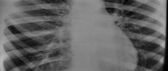
Metastases in the lungs (Dynamics) X-ray
In the early stages, metastases in the lungs do not manifest themselves in any way, the disease is asymptomatic . As cancer cells decay, they release toxic substances that poison the body. The patient seeks medical help more often at the last, terminal stage of cancer.
The presence of secondary foci of oncology in the lungs is accompanied by the following symptoms:
- Frequent shortness of breath, which appears not only during physical activity, but also at rest;
- Regular dry cough, turning into a wet one, which can be confused with another disease;
- Sputum mixed with blood;
- Chest pain that does not go away even with painkillers. Only narcotic drugs can reduce pain;
- Swelling of the face and upper extremities with localization of a secondary lesion in the right lung, headaches.
What do lung metastases look like?
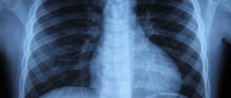
Metastases in the lung can be determined using radiography.
Secondary foci of oncology on X-ray images appear in nodular, mixed and diffuse forms. Nodal metastases appear in single or multiple forms. Single or solitary formations look like rounded nodules, reminiscent of the primary focus of oncology. Most often they form in the basal tissue.
If the secondary genesis is pseudopneumatic, then on x-ray it appears as thin linear formations.
With metastasis to the pleura, large tuberous formations are visible on x-rays, as a result of the progression of which the condition of the cancer patient worsens and pulmonary failure develops.
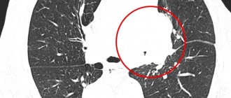
Foci in the lungs on CT scan for a tumor, types of cancer and its incidence
This pathology can be caused by a number of respiratory diseases. The lesions discovered during the examination are local areas characterized by reduced transparency of the lung tissue. These include darkening or compaction, the size of which may vary. They are detected during diagnostics when X-rays penetrate the body.
Adenocarcinoma
Adenocarcinoma is a subtype of non-small cell disease (NSCLC). This cancerous nodule develops in small airways such as bronchioles and is located more along the outer edges of the organ.
Adenocarcinoma accounts for 40% of all respiratory cancers, is more common in women, and tends to grow more slowly than other similar pathologies. Most cases of lung tumors in people who have never smoked are adenocarcinomas.
Squamous cell carcinoma
Squamous cell carcinoma is a type of NSCLC. It is also called epidermoid carcinoma. This type begins in squamous cells—thin, flat cells that look like fish scales when viewed under a microscope.
Squamous cell tumors - by localization, these are central tumors that occur in one of the main respiratory tracts (left or right bronchus). If the tumor grows to a large size, a CT scan may reveal a cavity in the respiratory system. A cavity is a gas- or fluid-filled space within a tumor mass or nodule and is a classic feature of squamous cell carcinoma. Squamous cell adenocarcinoma can spread to several sites, including the brain, spine and other bones, adrenal glands, and liver.
About 30% of all cancers are classified as squamous cell carcinoma. It is more associated with smoking than other types of non-small cell cancer. Other risk factors for developing a malignant nodule include age, family history, and exposure to secondhand smoke, mineral and metal dust, asbestos, or radon.
Large cell lung cancer
When imaging, this lung cancer does not appear on CT as adenocarcinoma or squamous cell carcinoma. Large cell cancers differ from small cell cancers in that they have larger cell sizes.
In the past, about 10% of all lung cancers were classified as large cell cancer. However, as more accurate diagnostic methods were used, this figure decreased. This type of lung cancer can be found in any part of the lungs, although it is more common in the periphery. Most cases of large cell cancer are found in men.
Small cell lung cancer
This type is one of the main types of cancer. Small lung cancer cells appear flat and smaller on CT scans, unlike normal, healthy structures.
Small cell carcinoma begins in the bronchi in the center of the chest, although in about 5% of cases it occurs in the periphery of the lungs and is a type of neuroendocrine tumor.
This disease accounts for about 15% of all malignant tumors and is most often found in smokers.
Metastases in the lungs on CT
Metastases are nodular formations in the lung tissue. When interpreting CT scans, they are determined by the following criteria:
- Hematogenous metastases are characterized by smooth edges, clear contours and a homogeneous structure. With swelling of the pulmonary parenchyma, the edges of the node become blurred. Located on the periphery.
- Lymphogenic metastases are multiple small foci that are located in the interlobular septa, in the pleural layers, and are associated with damage to the lymph nodes.
- Contact metastases are soft tissue space-occupying formations, single formations growing from an adjacent organ: esophagus, trachea, larynx.
How long do people live with pulmonary metastases?
Life expectancy for lung metastasis depends on how quickly the secondary cancer is detected.
If you experience at least one of the above symptoms, you should immediately consult a doctor and undergo an examination. In medical practice, there have been cases of detection of pulmonary metastases long before the detection of the primary tumor site.
The progression of a secondary tumor causes intoxication of the body as a whole. To identify the presence of metastases, you should know how the symptoms of the disease manifest themselves. The first signs of progression of secondary lung cancer are:
- Decreased appetite and, as a consequence, body weight;
- General malaise, fatigue and decreased performance;
- Increased body temperature, becoming chronic;
- Dry cough with metastases becomes constant.
The above signs may also indicate primary lung cancer. This rather dangerous disease is more often observed in smokers. Metastases in small cell lung cancer spread rapidly, grow quickly, and if they are not detected in a timely manner, the prognosis for the patient will be dismal. Primary lung cancer is treated with chemotherapy. If the procedure is carried out in a timely manner, there is a chance to cure oncology completely . But this form of the disease is usually detected in the final stages, when it is no longer possible to cure it. Taking strong analgesics, you can live from four months to a year.
Would you like to receive an estimate for treatment?
*Only upon receipt of data on the patient’s disease, a representative of the clinic will be able to calculate an accurate estimate for treatment.
There are forms of primary lung cancer that do not progress as rapidly as small cell cancer. These are squamous cell carcinoma, large cell carcinoma and adenocarcinoma. These forms of cancer are treated with surgery. If the operation is performed in a timely manner, the prognosis for recovery will be good. If metastases occur in other organs, the patient will face death.
Treatment
The course of the treatment algorithm is determined by the type of initial tumor formation and the tumor’s response to therapy. The course of action depends on the patient’s condition and whether metastases are detected in other organs.
Chemotherapy
Oncology treatment comes down to destroying cancer cells and preventing their spread. The main method is chemotherapy - the introduction of antineoplastic substances through the blood and stomach. They are treated with chemotherapy alone and in combination with other methods.
According to the tasks to be solved they are classified:
- Adjuvant (remaining fragments of tumors and relapses of metastases are excluded);
- Neoadjuvant (the size of the tumor is reduced and the degree of cell acceptance to drugs is determined);
- Therapeutic (reduction in the size of the secondary tumor, general improvement in the patient’s well-being).
Radiation therapy
The purpose of ionized radiation is to apply high doses to the tumor locally, minimally affecting other tissues. To do this, it is necessary to identify exactly where the malignant formation is located in order to correctly determine the direction and depth of irradiation.
Hormone therapy
If the lung is damaged due to the dissemination of hormone-dependent neoplasms (breast cancer, prostate cancer), treatment with medications is prescribed. Such therapy is not carried out separately, but in combination with other types of treatment.
Radiosurgery
The method is indicated for people if open surgery is accompanied by risk. The patient is exposed to high doses of radiation. When the tumor is located in places difficult to reach with a scalpel or near vital organs.
Surgery
Statistics show that patients with metastases in the lungs are being operated on much more often, taking into account the fact that surgery is prescribed extremely rarely. This indicates the prevalence of this type of disease.
Surgery is indicated if:
- there is no initial focus of cancer or relapse;
- no metastases to other parts of the body;
- no more than three foci of metastases in the lung;
- after initial treatment, tumors do not appear within a year;
- the time interval between the first and second metastases is at least six months;
- the patient will be able to tolerate prolonged exposure to anesthesia.
The size of the tumor determines the extent of the operation. Surgical intervention can be aimed at excision of a fragment of the lung - lobectomy, or complete removal of the organ - pneumonectomy. The radical method increases the survival rate for lung cancer.
Brachytherapy
This type of contact radiotherapy involves placing a radioactive implant directly in the bronchi.
Diagnosis of metastases in the lungs
To identify the presence of secondary genesis in the lung, the following diagnostic methods are used:
- X-ray – examines the structure of lung tissue, reveals dark spots, location of metastasis and its size. To do this, two photographs are taken - from the front and from the side. In the images, multiple metastases appear as round nodules;
- Computed tomography is a complement to radiography. A CT scan shows the areas where metastatic tumors are located, their size and shape. Using CT, momentary changes in the lungs are detected;
- Magnetic resonance imaging – is prescribed for people who have previously been exposed to radiation, as well as for children. This study allows us to identify secondary tumors, the size of which barely reaches 0.3 mm.
Diagnostics
Radiography. Treatment of a primary malignant tumor is regularly accompanied by x-rays of the thoracic organs. But this is not the most informative type of examination - the description reveals only lesions larger than 1 cm.
CT scan. If darkening is detected in the lungs, it is more correct to prescribe a computed tomography scan using a contrast agent. This diagnostic method detects lesions less than 5 mm. In the future, to monitor the dynamics of the process after treatment, it will be necessary to periodically repeat CT scans so as not to miss a relapse in the form of small formations. X-ray will not notice them or will determine them as benign.
Cytological examination of sputum. The result of the analysis differentiates malignant formations. A portion of morning sputum is examined using microscopy. After six to seven days, the result will show the presence or absence of atypical cells.
Needle biopsy of the lung. Targeted puncture to obtain biomaterial – cell biopsy. It is performed under X-ray or ultrasound guidance with anesthesia. A needle is inserted through the chest wall into the tumor and captures a piece of affected tissue.
Pneumonia, tuberculosis, lung cysts and benign lung tumors have similar symptoms.
Methods of treatment of secondary foci of oncology in the lungs
How to treat secondary lung cancer?
In modern medicine, the following methods are used to treat metastases in the lungs:
- Surgical intervention - the affected area is removed. This method of treatment is effective only if there is a single focal lesion, so it is used quite rarely;
- Chemotherapy is an addition to other treatment methods. The duration of the chemotherapy course depends on the main method of treatment and the patient’s well-being. In medical practice, chemotherapy is used in conjunction with radiation therapy. To raise the level of leukocytes in the blood after the procedure, dexamethasone is prescribed;
- Radiation therapy helps slow down the active growth of cancer cells and reduces pain. Irradiation is carried out in stationary conditions using a remote method;
- Hormonal therapy - used in the presence of a hormone-sensitive primary lesion in the prostate or mammary glands. Serves as an addition to primary therapy;
- Radiosurgery - a procedure that allows you to remove hard-to-reach tumors using a cyber-knife (beam of rays).
Disability for lung cancer is issued in the case of removal of one lobe.
Causes
Metastases are divided into primary in cancer of the lungs themselves and secondary, which can be detected when other organs are affected, such as:
- uterus;
- ovaries;
- kidneys;
- stomach;
- esophagus;
- thyroid;
- colon or rectum;
- mammary gland;
- prostate.
Causes may include peripheral lung cancer, skin melanoma and various sarcomas. Doctors believe that the source of this type of malignant tumors is almost all oncological diseases, it’s just that in some types they are diagnosed much more often. Cancer cells spread through transport in lymph fluid or blood. Since lung tissue has an extensive capillary network and a loose structure, its metastases are one of the first to strike.
- Proper nutrition for weight loss: menu for every day
- Pumpkin pancakes: delicious recipes with photos
- Causes and treatment of chest pain
Are metastases treated with folk remedies?
Treatment of secondary oncology in the lung can also be done using traditional methods.
The most common folk remedy is celandine. It is necessary to pour boiling water over a tablespoon of dried herb and leave in a thermos for about an hour and a half. Then strain the infusion and take it twice a day, two tablespoons before meals. In conclusion, we can say that there are various forms of lung cancer. This includes both primary cancer and metastases that have spread from other foci. The disease can be asymptomatic, which means that the patient can seek help when treatment no longer gives the desired result.
The survival prognosis depends on the stage of the disease, the type, shape and location of the tumors.
Types of metastases and their distribution
Metastases can reach the lungs in the following ways:
- lymphogenous (with lymph flow through lymphatic vessels);
- hematogenous;
- contact
Metastases are classified according to their form:
- solitary;
- ribbed;
- lytic.
- spherical.
Globular ones are more common than others and are easier to identify in an image. These are shadows of varying intensity, having a spherical shape. This may be one metastasis, or there may be several spherical shadows. Metastases grow over time. But this happens evenly, the shape of the ball is preserved.