April 12, 2020 Admin Home page » Ultrasound during pregnancy and childbirth Views: 959
Many methods are used to establish pregnancy. The tests, which can be purchased at any pharmacy, are accurate enough to show the presence of pregnancy. The same can be said about visiting an obstetrician-gynecologist, who can establish the fact of its presence quite early. But this can only be accurately determined using ultrasound.
However, it also happens that a woman has all the signs of pregnancy: a delay in the menstrual cycle, a positive test and an examination by an obstetrician-gynecologist, who confirms the presence of a pregnant uterus, but an ultrasound does not determine pregnancy. Next, we’ll try to figure out whether it could be that an ultrasound does not show pregnancy if there is a delay in menstruation and a positive test.
Why do you need an ultrasound during pregnancy?
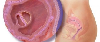
One of the most accurate ways to determine pregnancy is ultrasound. With it, the doctor can see the fertilized egg in the uterine cavity and determine the approximate period.
Ultrasound is divided into types:
- Transrectal – carried out through the rectum. This procedure does not apply to pregnant women, since it is most often performed on girls who are not sexually active.
- Transvaginal - done through the vagina, suitable only for women in early pregnancy. It is believed that examination by this method is the most reliable.
- Transabdominal is aimed at examining the pelvic organs and is carried out through the anterior abdominal wall.
- Combined - combines the last two ultrasound methods, carried out for a complete diagnosis.
- 3D, 4D - allow you to get a realistic image of the baby. And thanks to the 4D ultrasound method, you can see the child’s movements and even facial expressions in real time. Usually carried out to clarify the condition of the fetus.
Advice from doctors if pregnancy is not confirmed by ultrasound
You cannot trust the 1st ultrasound examination one hundred percent. To determine a successful pregnancy in the early period, highly qualified uzist and extensive experience are required. When the fetal sac is already large, it is difficult to confuse it with a tumor. An inexperienced specialist may well mistake a small embryo for a neoplasm.
In addition, the cause of the error may be inflammation in the uterus. At the same time, it swells greatly, and it becomes difficult to see the still small fetal sac. Even if the first test showed two stripes, gynecologists advise waiting a couple more weeks and then doing an ultrasound (even if it was performed earlier and did not confirm pregnancy).
During this time, you can do several more tests. Moreover, it is better to take them from different manufacturers and from an expensive line. If even at 4-6 weeks the ultrasound does not confirm pregnancy, then it is worth checking for oncology.
Also, equipment errors cannot be ruled out. Any technique can make mistakes, especially old ones. If the tests show pregnancy and at the same time all the signs inherent in it are observed, then you can do a repeat ultrasound in another clinic using more modern equipment. Early diagnosis (before the first screening - 10th week) is recommended for women at risk:
- the parents of the unborn child are close relatives;
- women who became pregnant after 35 years of age;
- if there were relatives in the family with severe hereditary diseases or deformities;
- women with diabetes mellitus and cardiovascular pathologies;
- the parents of the unborn baby are alcoholics, drug addicts;
- complicated gynecological and obstetric history (stillborn children, miscarriages, ectopic pregnancy, etc.);
- women who live in environmentally polluted areas with high background radiation, working in hazardous industries.
If not in the third week of pregnancy hCG is more than 1800 mU/ml, then this indicator may indicate the presence of an ectopic location of the fertilized egg if it is not visible in the uterine cavity. When the embryo is already noticeable and an ultrasound examination has confirmed the fact of a successful conception, then it is imperative to register with a gynecologist. The doctor will prescribe mandatory screenings, which are done to monitor the proper development of the fetus and exclude a frozen pregnancy.
At what week is pregnancy visible on an ultrasound?
Transvaginal ultrasound is usually used to confirm early pregnancy. Many mothers are tormented by the question: “At what week is pregnancy visible on an ultrasound?” Let's figure it out. This method allows you to determine the fertilized egg at 3-4 weeks of pregnancy. At this time, the level of hCG (human chorionic gonadotropin) should be at least 1800 units. This indicator is one of the important ones, as it allows you to more accurately determine the presence of pregnancy and its course.
Why can't you see an embryo on an ultrasound?
If a pregnancy test shows two lines, but the embryo is not visualized on an ultrasound, the reason for this may be:
- Incorrect calculation of gestational age from the moment of conception. The embryo may not be visible because the woman conducts the examination too early.
- Ultrasound diagnostics were carried out on an old device or the specialist did not have the proper level of qualifications.
- The examination was done through the abdomen and not transvaginally.
- A pregnant woman had a miscarriage, but she did not pay attention to it (she confused it with the beginning of her period), while the level of hCG in the blood had not yet decreased to its previous value.
If an ultrasound does not show an embryo in the ovum, do not immediately panic. For a number of reasons, the diagnosis of anembryonia can be made incorrectly, so it is necessary to monitor the level of hCG in the blood and undergo diagnostics again.
October 10, 2020 by anna
Can a doctor not detect pregnancy?
But let's return to the question of whether an ultrasound may not show pregnancy. Such cases occur quite often. If the hCG is positive and the ultrasound does not show pregnancy, then in most cases this may indicate an ectopic pregnancy. It is also worth noting that even if there is a fertilized egg, you cannot be completely sure of pregnancy, since it may be empty. The ultrasound specialist will be able to confidently confirm the “interesting position” only if he finds the embryo itself. At 3 weeks from conception, the size of the fertilized egg is 4 mm. At this time, only this is displayed on the ultrasound, and closer to the fifth week the embryo itself can be detected.
conclusions
- When a woman has such signs of pregnancy as: changes in the emotional background, appetite, taste preferences, nausea, fatigue, weakness and, of course, delay, and an ultrasound does not show pregnancy, this does not mean that there is no pregnancy. Here you should turn to other methods. First of all, the test, if it is positive more than twice, then this is a very weighty argument, which in the early stages has much more weight than an ultrasound. It is equally important to visit an obstetrician-gynecologist, who will conduct an examination and confirm pregnancy or suspect any pathology.
- Another important study is an analysis for the presence of human chorionic gonadotropin. Regular analysis will indicate the development of pregnancy or its fading.
- Ultrasound can also detect hormone-producing tumors, the only sign of which is a false-positive hCG test result and signs of pregnancy.
- Another pathology that requires urgent intervention is ectopic pregnancy. If, for example, it is localized in the fallopian tube, then failure to detect it can lead to rupture of the tube, which is a very life-threatening condition. In the future, a woman may have problems trying to get pregnant.
- It is worth understanding that one ultrasound, which detected pathology, is not enough. And after a few weeks, if the condition does not require urgent surgical intervention, it will be necessary to repeat the examination.
- There is no need to worry about the safety of ultrasound methods. They are all absolutely safe and do not cause any harm to the body of the mother or child, which is confirmed by the experience of using ultrasound over more than half a century.
Ultrasound diagnostics to determine the presence of pregnancy is a very important diagnostic measure, which is highly not recommended to be ignored, because only this method in many cases not only detects pregnancy, but also saves lives, without endangering either the expectant mother or her baby .
Early ultrasound
Ultrasound of pregnancy in the first weeks is considered uninformative. Moreover, doctors do not recommend performing it in the absence of medical indicators. These indicators include: threat of miscarriage, problems with fertilization, bleeding, suspicion of ectopic pregnancy. Experts explain this by saying that when the fertilized egg is attached, the uterus should be at rest, since unnecessary intervention can provoke termination of pregnancy. That is why it is advisable to carry out the first ultrasound in the early stages of pregnancy no earlier than the fifth week.
Is ultrasound wrong with the timing?
A gynecologist prescribes a routine ultrasound examination for a pregnant woman at 11-12 weeks. It is this examination of the first trimester that the doctor needs to confirm the presence of a fertilized egg. Even at this stage, the embryo is so small that ultrasound can make an error when diagnosing the woman’s position. There are cases when doctors confused a hematoma or polyps with an embryo.
Until the 8th week, experts determine the gestational age by measuring the length of the embryo. Until this stage, the development of all fertilized eggs occurs in the same way. With this period of development, an ultrasound examination can show the exact period, plus or minus 1-2 days.
There are cases when diagnosis shows a period longer than obstetric. There is an explanation - perhaps the last discharge of blood, which the woman mistook for her next menstruation, was minor bleeding. Thus, the woman does not even realize her situation for some time.
When determining the period, experts allow a difference between the terms (obstetric and ultrasound diagnosis) of about 2 weeks.
There are cases when an ultrasound shows a period less than obstetric. Perhaps this is the norm, or perhaps a deviation in the development of the embryo. In such cases, the specialist prescribes either Doppler sonography or repeated ultrasound diagnosis. A Doppler examination will help eliminate oxygen starvation of the fetus.
If there is such a difference in timing, your obstetrician-gynecologist may offer you:
- Be examined again after some time;
- Consult another specialist;
- Recommend hospitalization.
It is better not to ignore the recommendations of a gynecologist. The life of your unborn baby will depend on their implementation.
In addition to the fact that an ultrasound examination may not detect pregnancy at the initial stage, it may make an error in determining the sex of the child and the term. The timing error is explained by the individual size of each woman’s uterus. If an error is made, the deviation becomes about two weeks.
Why ultrasound does not show pregnancy: main reasons
Can an ultrasound fail to show pregnancy not only in the early stages, but also in later stages? This also happens in practice. For example, at 10-11 weeks of pregnancy, a doctor may send a woman for an abortion without identifying an “interesting situation”, but suspecting an ectopic pregnancy. Why is this happening?
We propose to identify a number of reasons:
- Lack of proper experience and qualifications of a doctor.
- Due to the anatomical features of the structure of the uterus (for example, an unusual shape), the device may not detect the fertilized egg.
- The gestation period is too short. The doctor will be able to examine the yolk sac, which is part of the fertilized egg, no earlier than the fifth week. That is why there is no point in conducting an ultrasound scan in a antenatal clinic before this date.
Anyone can make a mistake, so if such a situation arises, you should not panic, but it is better to double-check again: take a second hCG test, contact another specialist.
The entire period of pregnancy is very important not only for the woman, but also for the baby himself, since every day he undergoes changes and develops at tremendous speed. As a rule, the ultrasound specialist determines the gestational age based on the size of the fetus. Let's consider the size of the fetus by week of pregnancy using ultrasound
How are expectant mothers examined?
If the pregnancy test is positive, this can be confirmed by ultrasound examination - diagnosis is carried out in a commercial center or in a antenatal clinic. It is important to know that equipment with a high level of resolution and functionality, as well as the qualifications of a specialist, plays an important role in obtaining reliable examination results.
Up to 9 obstetric weeks, two methods are used to examine pregnant women:
- Transabdominal – through the area of the anterior abdominal wall.
- Transvaginal - using a transducer that is inserted into the vagina.
Up to 5 weeks, the formed fertilized egg is very small - its size is only about two millimeters. The transvaginal method is considered an effective method for diagnosing the embryonic period - its high-frequency sensor makes it possible to get as close as possible to the uterine cavity and transmit the smallest dimensions of the examined organs to the monitor screen.
The technique of examining the expectant mother using high-frequency waves is non-invasive and absolutely harmless - it allows the doctor to safely monitor the development of the fetus
During the entire gestation period, a woman undergoes at least three ultrasound scans. The examination session is short-term; the doctor tries not to hold the sensor in one place for a long time, especially during the formation of the most important organs and systems of the unborn baby.
First trimester
1. 1 week is counted not from the moment of conception, but from the date of the beginning of the last critical days. It is from this moment that the doctor announces the preliminary date of birth. This calculation is made because at the beginning of the next menstruation an egg is formed, the fertilization of which, under a successful combination of circumstances, occurs in about two weeks. Therefore, it is too early to talk about the size of the fetus.
2. Around the end of the second week, conception occurs. At 2.5 weeks of pregnancy according to ultrasound, the size of the unborn baby is no more than 0.1-0.2 mm. At this time, the child has not yet acquired human traits, but is more comparable to a poppy seed. It is likely that even at 2.5 weeks of pregnancy the size of the fetus will not be determined, so conducting an examination at this time can be considered a pointless exercise.
3. The embryo is 5 weeks old, but according to obstetric calculations we are talking about 3 weeks. We mentioned earlier that gynecologists begin counting from the first day of critical days. During this period, the process of movement of the zygote through the fallopian tube to the uterus continues. The fruit is 0.15 mm.
4. At the beginning of the 4th week, the zygote reaches the uterus and implants into its mucous membrane, which may cause slight bleeding. The size of the fertilized egg is 1 mm, comparable to a sesame seed.
5. Miscarriages often occur during this period, so at this time the expectant mother needs to carefully monitor her health. Meanwhile, the fertilized egg is growing rapidly; at the 5th week of pregnancy, the size of the fetus reaches 1.25 mm.
6. At week 6, the embryo can already be seen using ultrasound. At this time, he begins to move, but his movements are not yet noticeable by the mother, since the embryo is very small. Its size is 2-4 mm.
7. At this time, the embryo’s heart beats, the tail gradually begins to disappear, which will finally disappear by the end of the tenth week. The weight of the fetus at this stage is 0.8 g, and the size is 4-5 mm, comparable in size to a pea.
8. This week the fetus reaches 1.6 cm in length, and its weight is already 3 g. At this time, changes occur not only with the unborn baby, but also with his mother. A woman may notice breast enlargement, darkening of the nipple halos, and abdominal pain associated with rapid enlargement of the uterus. The pain should go away soon. A baby at 8 weeks of pregnancy is similar in size to a bean fetus.
9. A nine-week embryo develops a swallowing reflex and can already clench and unclench its fists. The established systems and organs continue to rapidly form. At this stage, the size of the baby reaches 2.3 cm, and its weight can vary from 5 to 15 g.
10. From this period, the mother’s belly begins to grow, especially if this is not the first pregnancy. The placenta is formed in the mother's body, thanks to which the baby will receive nutrition. The length of the fetus reaches 3.1 cm, and the weight may remain unchanged. You can compare the size of a baby with an average plum.
11. This week, the fetus’s intestines are forming. Now he already knows how to yawn, turn around, move his legs and arms. But his mother cannot feel his movement yet, since he is still very small. The size of the baby is about 4.1 cm in length, and its weight is 7 g.
12. By this time, the baby has already formed the urinary and circulatory systems. Now the coccygeal-parietal size (CPS) reaches 5.4 cm.
13. This week is the final week of the first trimester. As a rule, the first ultrasound is prescribed at this time. The size of the fruit is about 7.4 cm and the weight is 20 grams.
What methods are there to confirm pregnancy?
Several methods are used to confirm conception. This is a home test that can be purchased at a regular pharmacy, a blood test from a vein to determine the level of human chorionic gonadotropin, as well as an ultrasound examination.
These methods have varying degrees of reliability and have their own characteristics. The expectant mother often knows exactly the date of conception, determining it by the date of ovulation.
The main sign of pregnancy, amenorrhea, or absence of menstruation, does not always indicate conception. Menstruation may not begin for other reasons not related to pregnancy. If you don’t have your period and there is no reason to suspect pregnancy, you should not put off visiting the gynecologist’s office.
Home test
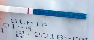
A pharmacy (aka home) test is the most popular method that every woman resorts to when she thinks she is pregnant.
The essence of the test is to immerse a strip soaked in a special chemical composition in morning urine that reacts to human chorionic gonadotropin (hCG), which appears in the urine. This hormone is produced in the body only with the onset of pregnancy or the development of serious pathologies (oncological diseases).
The production of the hormone does not begin at the moment of fertilization, but when the fertilized egg attaches to the wall of the uterus. For hCG to show up as two lines on the test, it needs to accumulate in the body. After conception, its amount grows exponentially every day.
The tests have different sensitivities and also differ in how they are used. They are divided into classic strips, impregnated with substances sensitive to hCG, tablet strips (the indicator is placed in a plastic housing) and inkjet strips. Inkjet is considered the most reliable. It requires no preparation and can be used anywhere.
Laboratory blood test
This method is more accurate than a home test. Human chorionic gonadotropin is detected in the blood before its amount reaches the required concentration in the urine, to which the indicator strip reacts.
You can donate blood from a vein to the laboratory 3-5 days from the start of a missed period or 12-14 days from the expected date of conception. Each week of pregnancy corresponds to a certain level of the hormone. In the laboratory, the results of the study are presented in the form of a table of values corresponding to each week of pregnancy.
Gynecological examination
During the examination, the gynecologist determines the presence of a developing fetus based on the following signs:
- Congestion of the pelvic organs. After conception, blood flow in the uterus increases, causing the labia, vagina and cervix to change color. They darken and become swollen.
- Changes in the condition of the uterus. The non-pregnant uterus is dense and pear-shaped. After conception, it becomes round, soft, and its size increases.
- Mild irritability of the uterus. During a two-handed medical examination, it easily contracts, reacting to the actions of a specialist, then returns to its normal state.
- Asymmetry of the uterus. One side of the organ is larger than the other, depending on which side the embryo is attached to.
- Soft neck. The neck (isthmus) softens, the doctor practically does not feel its tissue during examination.
Signs of pregnancy, which the gynecologist sees during examination, appear at 4-7 weeks. If the supposed “interesting situation” lasts less than 3 weeks, it is better to postpone the visit to the doctor.
Ultrasonography
A high-resolution ultrasound machine is able to detect pregnancy on the 5-6th day of a missed period (this corresponds to 3 weeks of gestation). An ultrasound examination at this stage detects a tiny tubercle measuring 2-4 millimeters in the uterine cavity. This is the growing embryo.
Only a highly qualified specialist can determine whether it is endometrial folds or a growing embryo.
Confirmation of the beginning of the pregnancy period by ultrasound examination is carried out at the request of the woman or according to indications.
Indications for early ultrasound:
- a woman’s history of ectopic fetal development;
- examination by a gynecologist, which did not confirm changes in the uterus characteristic of pregnancy in the absence of menstruation.
Second trimester
14. This week begins the second trimester. From this time on, significant external changes appear: the fetus’s hair begins to grow, its eyes come closer together and facial expressions appear. Now the baby is approximately 10 cm tall and weighs 30 grams.
15. At this time, the fetus represents a miniature copy of the newborn. The future baby is growing rapidly, reaching 11 cm in length and weighing 50 grams. At this stage, the size of the fruit is comparable to that of an avocado.
16. At this time, the mother can feel the first movements of her baby. The fetus's face continues to develop, the bladder is actively working, and the nails are completing their formation. The baby reaches 15 cm in length and weighs about 100 grams. A baby at 16 weeks can be compared to a small carrot.
17. At this stage, the baby hears sounds and distinguishes between the voices of mom and dad. Now the baby reaches up to 18 centimeters in length and 150 grams in weight. At week 17, the baby is the size of a medium potato.
18. At this time, a qualified doctor can use an ultrasound to reveal the gender of the unborn child. During its entire stay in the tummy, the baby has grown significantly stronger; it reaches 20 cm in length and 200 grams in weight. And in size the fruit is comparable to a medium-sized tomato.
19. Every day the baby’s movements become more and more distinct. Now not only the mother, but also other relatives can feel the movements if they put their hand on the stomach. Within a week, the fetus gained 30 g in weight and 2 cm in height. Now the fruit is comparable in size to a banana.
20. As a rule, at this time the gynecologist prescribes a second planned ultrasound examination. At week 20, the baby already has hair on his head, tiny nails and an expressive face. Its dimensions are about 25 cm, and its weight is approximately 300 g. The baby is similar in size to a small bunny.
21. Now the fruit reaches 26 cm in length and 350 g in weight. The baby weighs about the same as a large mango.
22. This week internal organs continue to develop. The growth rate of the fetus slows down a little as there is an emphasis on weight gain. So, the growth of the unborn baby remains unchanged, but his weight is already 475 grams. The size of the fetus at week 22 resembles an emperor penguin egg.
23. Now the fetus is growing by leaps and bounds. At this stage, its parameters are 500 grams of weight and 30 centimeters in length. An analogue for comparison would be an ear of corn.
24. The fetus is growing rapidly, so it becomes more crowded in the uterus every day. The baby's size at this stage is about 30 cm, and his weight has increased by as much as one hundred grams. Now the unborn child is similar in size to a green coconut.
25. At this time, the development and improvement of the baby continues. Now mom feels active kicks and regular tremors in her tummy. CTE at week 25 is 32 cm, and weight is 700 g.
26. At this stage, the rounded shape of the tummy is clearly visible to others. Now the baby’s weight reaches 800 g, and his height is 33 cm. The size of the fruit is comparable to a head of broccoli.
How to choose an ultrasound to determine pregnancy
The timing at which pregnancy can be diagnosed largely depends on the type of ultrasound. When carrying a baby, two types of scanning can be used - internal (transvaginal) and external (transabdominal).
Transvaginal is usually used in the first trimester, 4-9 weeks. At this time, internal ultrasound is the most informative, since during the examination the sensor is as close as possible to the uterus. A transvaginal examination can see the baby already on the 5-10th day of the delay, starting from the 4th week. The diameter of the fertilized egg at this time is only 2-4 mm.
Transabdominal ultrasound - through the outer wall of the abdomen - is preferably done at a later date. In the first trimester, it will show the embryo in the patient’s uterus only on the 21-22nd day after the delay, from the 6-7th week. The diameter of the fertilized egg with the embryo inside at this stage is 5-7 mm.
Third trimester
27. The final trimester of pregnancy has begun. After six months of being in the tummy, the baby already knows a lot: sucking fingers, recognizing the voices of his parents, recognizing salty and sweet foods. The baby is growing rapidly. At week 27, his weight reaches 900 g and his height is 34 cm.
28. At this time, a pregnant woman may experience training contractions. The most important thing for an expectant mother is to be able to distinguish between training contractions and real ones. As for the baby, now he smells, distinguishes tastes, can see and hear. The baby's CTE is 35 cm, and his weight has reached the one kilogram mark.
29. By this time, the baby in the tummy should be in cephalic presentation. The baby is getting even more crowded in his mother’s tummy, so he is no longer able to tumble as before. The CTE is 37 cm, and its weight has reached 1.2 kg. The weight of the fruit is comparable to an ostrich egg.
30. This week, the baby’s vision is actively developing and the nervous system is developing, and some character traits are beginning to develop. The baby's height can be approximately 37-38 cm, and its weight reaches 1.4 kg.
31. Around this time, the expectant mother needs to undergo a third scheduled ultrasound. Within a week, the baby has grown well and is about 40 cm tall and weighs 1.6 kg. A newborn lion cub weighs about the same.
32. At this time, the baby’s cheeks have become rounded, his skin has acquired a light pink tint, and very soon he will have folds on his arms and legs. The CTE of the fetus is 42 cm, and the weight is 1.8 kg.
33. The fetal indicators at 33 weeks are as follows: weight – 2 kg, height – 43 cm. The baby’s weight is comparable to a large pineapple.
34. The child’s heart is almost already formed. The baby is actively gaining weight, because of this, the mother may notice a significant weight gain, and also experience heartburn and digestive problems. Now the baby’s height varies from 42 to 43 cm, and her weight is 2.1 kg. The fruit is comparable in size to a melon.
35. There is very little time left to wait until the expected date of birth, but even if the baby is born earlier, there is nothing to worry about, since the baby is physically ready for this. Its activity is significantly reduced due to lack of space in the uterus, but still the mother needs to monitor and count the baby’s kicks. The baby's dimensions at 35 weeks are 46 cm and 2.5 kg, resembling the weight of an average pumpkin.
36. It is worth noting that the size of the fetus directly depends on genetic characteristics. The average parameters of the fetus this week are: height – 48 cm, weight – 2.7 kg.
37. This week the fetus is considered full-term. The average size of a baby can be about 3 kilograms and over 50 centimeters in height. The size of the baby resembles a medium-sized watermelon.
38. Abdominal prolapse is a harbinger of childbirth, and every mother should be ready for this at this time. There are no longer any changes in the baby’s development, and his weight and height at this time may remain unchanged.
39. The child can no longer be called a fetus, since in the tummy there is a full-fledged newborn who can be born at any minute. This week the baby's size can reach over 50 cm in height and 3.5 kg.
40. At this point, the journey of pregnancy comes to an end. As a rule, most children are born between 38 and 42 weeks, but 40 weeks is still considered to be the gestational age.
What should I do?
So, the answer to the question “Can an ultrasound not show pregnancy” has been received. Now we know that this situation is very real. But what should a woman do in this case? First of all, it is important not to be nervous and concentrate. To finally understand this issue, it is recommended to go to another clinic and undergo an ultrasound again. It is better to carry out the examination using expert-class equipment, since such equipment has high resolution. In addition, the examination should be accompanied by a blood test for hCG levels.