Fetal development in the first trimester is a complex process during which its main organs are formed. Carrying out an ultrasound scan at the 5th week of obstetric pregnancy is rarely practiced. They resort to it mainly to establish the fact of an accomplished conception. The use of ultrasound at the beginning of the second month is justified when a woman wants to be sure of the birth of a new life (for example, if the baby is long-awaited), but the decision to prescribe is made in agreement with the attending physician.
By the way, the obstetric period differs from the gestational period by two weeks. In the first case, the countdown is from the moment of the last menstruation, in the second - from the fact of conception (that is, during ovulation - approximately on the 14th day of the cycle). Therefore, at least 7 weeks must pass from the start date of the last menstruation. It is then possible to obtain some information about how the fetus develops.
Why and to whom is ultrasound diagnostics recommended?
In the fifth week of pregnancy, an ultrasound may not provide much information. During this period, research is needed to confirm that the delay in menstruation and changes in well-being are not at all accidental.
Many doctors believe that the expectant mother can do the first ultrasound examination later than in the fifth or sixth week of pregnancy (and it will already be more informative).
The only exceptions are those mothers who are expected to have problems with their pregnancy.
Also, ultrasound at such a time is necessary for those who have previously had an ectopic pregnancy, in order to prevent a frozen pregnancy and as one of the parts of prenatal genetic screening.
Possible problems
The most common problem of pregnancy during the first 3 months is miscarriage. As a rule, vaginal bleeding occurs, accompanied by or without abdominal pain.
The chances of miscarriage are higher for women who abuse alcohol, drugs, and caffeine. Other causes may include a genetic abnormality, excessive exercise in the first trimester, or abnormal hormones.
Anembryony
Often, pregnancy does not end in childbirth, but a reductive loss of the fetus occurs, this is the death of the embryo and anembryony. Anembryony is the absence of an embryo in the developing egg.
Possible causes of the pathology:
- Genetic anomalies.
- Diseases caused by viruses and infections.
- Endocrine disorders.
This process occurs as a result of the cessation of reproduction of the embryoplast, a group of cells that lay the foundation of the fetus. The result is an egg that continues to grow, but lacks an embryo. To make a diagnosis, an ultrasound examination is performed, but the symptoms are reliably determined only after the 8th week of pregnancy.
A woman develops warning signs:
- pain in the lower abdomen;
- bloody issues;
- deterioration of health.
Termination of pregnancy should take place in a hospital, so that after the removal of the fertilized egg, the woman is under the supervision of a doctor.
Biochemical pregnancy
Chemical pregnancy, or biochemical, is an early miscarriage that usually goes unnoticed and women often confuse it with the menstrual period. For fertilization, the sperm must meet the egg in the fallopian tube. If this happens, the embryo begins to develop until it reaches day 6, when it is time for it to join the woman's uterus.
After implantation, the female body begins to secrete the hormone hCG, which is measured by tests. In the case of a biochemical pregnancy, positive tests appear, but the pregnancy itself does not occur, because the embryo produces a sufficient amount of the hormone, but does not turn into a clinical pregnancy. Early miscarriage occurs along with the menstrual flow.
Ultrasound during this period does not show signs of gestation. Chemical pregnancy is not a definitive death sentence for infertility. Many women, after undergoing treatment, carried and gave birth to normal children.
Frozen pregnancy
A frozen pregnancy is the cessation of fetal development and growth. This can happen at any time, but is more often detected in the 1st trimester.
The reasons may be:
- inflammatory diseases in women;
- Rh conflicts;
- previous abortions;
- genetic predisposition;
- uterine pathology.
Symptoms that indicate the development of pathology:
- severe pain, pulling or cramping;
- bloody, clear or milky discharge;
- decreased sensitivity of the mammary glands;
- decrease in basal temperature.
Early toxicosis
Pregnancy of 5 obstetric weeks can lead to early toxicosis, which usually occurs in the first trimester. Toxic substances may be formed in the mother's body during this period as a result of fetal development. Another cause of early toxicosis is dysregulation of the neuroendocrine system.
Some hormones produced by the placenta and glands (pituitary gland, thyroid, gonads) are new to the female body and she cannot immediately adapt to them. For these reasons, a woman develops toxemia. That is why gynecologists call it a “disease of adaptation.” Some doctors believe that early toxicosis is the protection of the embryo from possible bad habits of the mother.
Ectopic pregnancy
This type of pregnancy occurs when a fertilized egg implants outside the uterus instead of inside it. Almost all ectopic pregnancies occur in the fallopian tube.
Sometimes the fertilized egg is pushed out by the tube and implants on the peritoneum or in the ovary. According to statistics, this type of pregnancy occurs in 1 in 50 women.
Pregnancy is always a big responsibility, and the beginning of its development is especially important. Any minor disturbance in the mother’s body can affect the embryo within the 5th obstetric week. This is one of the crucial periods, because it is at this time that the foundations for the development of the central nervous system are laid.
Author: Belyaeva Anna
The condition of the mother and the development of the fetus during this period
At the 5th week of pregnancy, the baby’s neural tube has already formed and the heart begins to beat; in addition, at this time the circulatory system, including the heart, is actively forming, and it will start working very soon.
All internal organs, bones and nervous system continue to form. The thyroid, pancreas and adrenal glands begin to develop. Moreover, they already secrete the first hormones, for example, thyroglobulin. From five weeks, the embryo can be called an embryo, as it enters the embryonic phase of development.
The size of the fetus is now 2-3 millimeters.
As for the mother’s condition, there are also serious changes in it. During this period, all signs of pregnancy may make themselves felt:
- may pull in the stomach;
- lethargy, constant fatigue;
- constant changes in mood;
- nausea;
- pain in the mammary glands;
- frequent urge to pee;
- changes in appetite;
- constant drowsiness;
- there may be menstruation, but it is quite scanty (their presence is not yet a bad symptom);
- sensitivity to odors.
But the absence of any signs and changes in well-being in the fifth week of pregnancy is the norm.
Important! During this period, the risk of miscarriage is high, in addition, the fetal nervous system is being formed, which means mothers should not be nervous.
Fetal development
Of course, the state of the fetus does not closely resemble the human form.
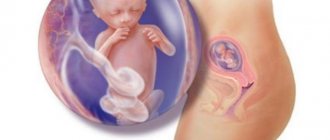
The size of the fetus can be seen in the ultrasound photo, and it is only 1 centimeter long.
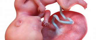
Its weight is usually no more than 2 grams. Therefore, you shouldn’t be surprised that your belly hasn’t grown at all. He just doesn't have anything to grow from yet. The fifth obstetric week of pregnancy creates a sickle-shaped fetus. The legs and arms are still in their infancy, but in the ultrasound photo you can already see where they are going to be. The face begins to develop. There are no characteristic signs yet, but outlines of the eyes, nose and mouth can already be seen on an ultrasound. During this period, the trachea and food tube separate, and the lungs form. The baby begins to grow a second layer of skin.
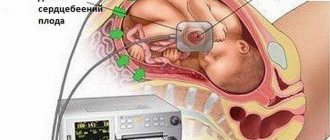
During an ultrasound, the doctor can already tell the mother how the fetal heart is beating. For some time, its rhythm will be disrupted every now and then, but when eight weeks of pregnancy arrives, its rhythm will be approximately 150 beats per minute. As an ultrasound shows, the fetal intestine in the fifth obstetric week looks like a tube, but soon it will begin to become covered with convolutions. At this stage, the thyroid gland and reproductive cells are formed. The sex of the fetus cannot yet be known, only time will tell who will be born.
What will the ultrasound show?
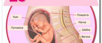
At the fifth week, the ultrasonologist can clearly see that the uterus has enlarged. But this increase may not yet be entirely uniform: for example, only the wall on which the vial with the embryo is attached can increase and become convex.
The amniotic sac is already clearly visible; its size is about a centimeter. If you enlarge the image of the amniotic sac, you will be able to see both the fetus itself and the yolk sac. A good specialist is able to recognize even the beating of a developing fetal heart on an ultrasound.
But in the ovary from which the egg came out and was fertilized, you can see the corpus luteum. This is another confirmation that pregnancy has occurred.
At this stage, the configuration of the fetus already allows us to make assumptions about where its head will be located and where its lower limbs will be located. By the way, their rudiments have also already appeared, but they have the form of bubbles. Typically, a five-week-old fetus is actively moving, which allows the specialist to draw a conclusion about its health and proper development.
But ultrasound examination does not yet allow us to examine anomalies in the structure of the fetus. But it can examine amniotic sac abruption, ectopic pregnancy, or uterine hypertonicity.
During an ultrasound, the specialist must measure and record the coccygeal-parietal dimensions of the fetus, as well as its position in the uterine cavity, the condition of the corpus luteum, ovaries and myometrium. All this data will allow you to calculate the size of the baby and the approximate date of his birth.
Important! If, at five weeks, the embryo is not visible in the amniotic sac and the doctor suspects a frozen pregnancy, but you feel fine (no pain or bleeding), do not rush to do a curettage. Check for an ultrasound in another ten days (at least a week).
Also, at a period of 5 weeks you can already see whether the expectant mother is expecting one baby or will have twins. The difference is that there will be two fertilized eggs, not one.
Preparing for an ultrasound
An ultrasound scan at 5 weeks of pregnancy can be performed either transvaginally or transabdominally. Usually the choice is made on the first method due to the small impact of vibration and heating on the organs of the woman and the unborn child.
- For an ultrasound scan at 5 weeks through the abdominal wall, it is recommended to follow a three-day diet that limits the intake of gas-forming drugs. The last meal should be at least 10-11 hours before the procedure, and the bladder should be filled 2 hours before the examination.
- If an ultrasound scan at 5 gestational weeks is prescribed through the vaginal cavity, then transvaginal ultrasonography only involves the prevention of flatulence and cleanliness of the genitals. The bladder should be empty.
After the woman lies down on the couch, the ultrasound doctor applies the gel to the abdominal area and examines the pelvic organs using a transducer sensor. He determines the presence of a fertilized egg, its location and the echostructure of the internal genitalia, after which he enters all data and indicators into a special protocol.
What is visible in the photo at 5 (7 obstetric) weeks?
As mentioned above, sometimes an ultrasound may not show anything. If the hCG level is normal, and rapid tests consistently show pregnancy, there is no need to worry: an ectopic pregnancy at this time can already be noticed. During this period, it is also often difficult to see anything in the photo.
The ultrasound photo at this stage may be blurry, and the embryo itself may resemble a shrimp or a small cylinder in shape. If the image and the equipment are of high quality, then you will be able to see the tail of the embryo and its head, as well as the rudiments of arms and legs, and sometimes even the oral and nasal slits, eyes, fingers and ears. As for the uterus, its shape should resemble an egg.
Twins at 5 weeks of pregnancy: photo
Indications and contraindications for ultrasound at 5 weeks
An ultrasound is performed at 5 weeks of pregnancy to confirm the occurrence of pregnancy and identify the location of the fertilized egg. By this time, the delay is at least 14 days, so it will no longer be possible to write it off as a normal menstrual cycle failure. As a rule, a “special condition” is indicated by the onset of toxicosis, but examination on a chair does not always help to understand whether the fetus is developing correctly.
Ultrasound in some cases is necessary to exclude a pathological course of pregnancy, for example, ectopic. It also happens that analyzes and “home” tests for hCG do not give an exact answer: conception has occurred, or a delay in menstruation is caused by other reasons. Then ultrasound diagnostics comes in handy.
Sometimes the most common marker of pregnancy - the absence of periods - does not work, and they come at the right time. In obstetrics, there are even special terms to describe this condition - “washing of the fetus” or “color pregnancy”. However, this is not the norm, since from the moment of fertilization the level of the hormone progesterone should increase, and therefore menstruation does not occur.
So, a study can be done at the beginning of the first trimester of pregnancy if:
- the woman herself wants to undergo such an examination;
- planning to terminate the pregnancy;
- it is not possible to detect the fact of conception using conventional methods (examination, pocket test, blood test);
- it is necessary to exclude ectopic pregnancy or fetal pathology;
- Bleeding began or pain appeared in the lower abdomen. Then emergency diagnosis can help save the child’s life;
- the expectant mother has previously encountered diseases of the genitourinary system or hormonal imbalances.
In other cases, ultrasound is not recommended for such a short period of time, since it is not very informative. In addition, ultrasound can harm the development of the baby, since in the 4th...5th weeks of intrauterine growth, differentiation and launch of most internal organs and life support systems begin. Organogenesis should ideally take place without outside interference and harmful external influences.
Preparation
Ultrasound diagnostics during pregnancy is performed transabdominally or transvaginally. The latter type, due to greater focus, is a priority in the early stages. In addition, it does not require filling the bladder, which is typical for examination through the abdominal wall. In this case, no special preparation is required, but it is advisable to exclude foods that cause increased gas formation from the diet a few days before visiting a specialist. These include fruits, carbonated drinks, legumes and bread.
What tests are needed?
When registering, a pregnant woman undergoes a number of mandatory tests: urine (general and bacterial culture), blood (general, determination of group and Rh factor, for the presence of antibodies for Rh negative factor, tests for syphilis, HIV infection, hepatitis B).
It is possible to establish the fact of conception with the simplest test for the gonadotropin hormone using blood sampling or at home (by urine). If the level of hCG in the blood corresponds to the deadline, then there is no need to worry. However, if the readings deviate significantly from the norm, then the obstetrician-gynecologist must figure out what was the reason. It is possible that there is a frozen or ectopic pregnancy. Sometimes a discrepancy with the norm may indicate multiple births.
Before prescribing an ultrasound examination, the attending physician must be convinced of the need for the procedure. If there are no signs of pathology of embryonic development, then the ultrasound will be performed not at 5 weeks, but according to plan - at the end of the first trimester (11-12 weeks).
If during ultrasound examinations the absence of a fertilized egg is revealed, then repeated diagnosis is carried out after 7 days, subject to a positive blood test for hCG. The frequency and number of visits to the ultrasound room are determined by the attending physician based on medical history and related pregnancy indicators.
Ultrasound examination standards
Having received the ultrasound protocol in hand, the expectant mother needs to contact her gynecologist to decipher the results.
For a period of 5-6 weeks they should be as follows:
- embryo size – from 2 to 3 mm;
- yolk sac size – 4-5 mm (if it is more than 6 mm, this may indicate pathology);
- the size of the fertilized egg is about one centimeter;
- Heart rate – from 70 to 100 in 60 seconds.
There cannot be any other indicators at this time.
Sex at 5 weeks pregnant
In most cases, sex in early pregnancy has no contraindications. If a woman feels well, has no pain, and does not have any individual restrictions from the doctor, then sex will only bring benefits.
At the same time, due to hormonal changes in the body of the expectant mother, attraction to a partner may decrease significantly and even disappear completely for some time. This is not the worst thing that can happen, but if a couple is worried about this, it is better to seek advice from a specialist.
Contraindications
The examination itself at such an early stage can be called a conditional contraindication. More precisely, doctors do not recommend doing this before ten weeks of ultrasound: you won’t see much, gender cannot be determined, and pathologies make themselves felt only at a later date.
The exceptions in which an ultrasound scan at this time will be necessary are described above.
It is difficult to say whether ultrasound will harm the embryo, because there is neither evidence of harm nor its refutation. So, if possible, sign up for the first ultrasound examination at about ten weeks and do not disturb the growing fetus unless absolutely necessary: 4-5 examinations in nine months are quite enough.
Features of pregnancy
Not many people know that if an ultrasound is performed at 5 obstetric weeks of pregnancy, this corresponds to a three-week gestational age of the embryo.
The determination of gestational age is based on the estimated date of the last ovulation, from which the counting takes place. However, not every woman has a regular cycle and knows the exact day the egg is released, so doctors consider this method unreliable.
The obstetric period is calculated from the first day of the last menstrual period and differs by about two weeks from the gestational, or embryonic.
Preparation and performance of ultrasound diagnostics
At the beginning of the second month, when conducting ultrasound diagnostics, a woman will need to resort to the preparatory stage. If you ignore all the recommendations, the examination will be less informative or completely ineffective. The first thing to do is change your diet 3 days before the test. Excluded from the menu:
- black bread;
- kvass;
- soda;
- cabbage dishes and the vegetable itself;
- radish;
- radish;
3 days before the examination, a woman should exclude fried and fatty foods from her diet.
- beets;
- cucumbers;
- confectionery;
- fresh flour products;
- dishes with an abundance of fat;
- fried foods;
- mayonnaise and other sauces;
- pickled products;
- canned food;
- pickles.
At 5–8 weeks, both transabdominal and transvaginal ultrasound can be performed.
However, it is better to give preference to the second option listed. This method will be more informative at the initial stage. At the beginning of the second month, 3D ultrasound is not possible. Successful preparation is about not worrying. Anxiety has a negative effect on the fetus. The procedure does not have a negative effect on the child.
Before the examination, a disposable condom is placed on the ultrasound machine sensor
Read also: what condoms to buy for an ultrasound.
On the day of the study, you should prefer a light breakfast. Immediately before diagnosis, you need to empty your bladder. To do this, the woman must visit the toilet immediately before the examination.
You need to bring to the procedure:
- replacement shoes;
- medical diaper;
- exchange card.
The woman spreads her legs wide and bends her limbs at the knees. The doctor inserts an elongated ultrasound device into the vagina. A disposable condom is first placed on the device. The resulting image is displayed on the monitor. All data is entered into a protocol, which must later be provided to your gynecologist for decoding.
Diagnosis should be carried out with an empty bladder, for which it is necessary to visit the toilet
Discharge from the genital tract
In the fifth week of pregnancy, the discharge determines its further course. They are caused by the restructuring of the female body. Transparent or light discharge indicates normal development of the baby and pregnancy.
When to see a doctor immediately:
- brown discharge, accompanied by pain in the lower abdomen, indicates an increased likelihood of miscarriage, so if they appear, you must urgently call an ambulance, as bleeding may develop.
- greenish and yellowish discharge with an unpleasant odor, causing discomfort, accompanied by itching. This is mainly due to the presence of infection, and there is no way to do this without the intervention of a doctor.
- brown discharge that stopped but reappeared in the fifth week may indicate the presence of a “frozen pregnancy.”
- light brown discharge in the fifth week is a symptom of an ectopic pregnancy.
If you have atypical discharge, there is no need to panic, you should just consult a doctor who will find out the cause and take the necessary actions.
Minor brown-pink discharge may occur after sex; it is caused by changes in the structure of the vagina; in such cases, a consultation with a gynecologist will not be superfluous.
Photos of bellies at 5 weeks
Ultrasound in subsequent weeks
Perhaps this will convince you, but the doctor will see a slightly different picture if you are 5 weeks, 5 days pregnant. An ultrasound may be more informative. At the sixth week, the doctor should definitely detect a fertilized egg in the uterine cavity. The yolk sac is clearly visible in the fertilized egg. The size of the embryo is already 6-19 mm, and before its own internal organs begin to work, the tissues of the yolk sac perform all metabolic functions. However, its size should not be about 6 mm. At the sixth week, the doctor should already see a white ring; this is the future placenta.
At the seventh week, not only the presence of an embryo in the fertilized egg is clearly diagnosed, but also the sensor records its heartbeat and motor activity. The size of the fertilized egg is 19-27 mm. Heart rate - up to 150 beats per minute.
Embryo movements
It's amazing how much doctors can learn about a baby when the pregnancy is 5 weeks. An ultrasound allows the doctor to draw a conclusion about how actively the embryo is moving. It is the frequency of his movements, together with the heart rate, that allows us to draw the first conclusions about his vitality and well-being. If the embryo is now motionless, the doctor will raise the question of terminating the pregnancy. There are still a few weeks to wait and compare the indicators at the next examination, but these are already alarm bells.
Nutrition at 5 weeks of pregnancy
Nutrition in the 5th week of pregnancy and the quality of food for the expectant mother plays a very important role.
Products must be fresh and natural. The diet should contain a large amount of vegetables, fruits, as well as protein foods and fermented milk products, for example, cottage cheese, kefir and fermented baked milk. From the 5th week of pregnancy, you need to eliminate fried, salty, smoked and fatty foods from your menu. You should also be wary of processed foods and food from fast food restaurants. Give preference to homemade food, steamed, boiled or baked in the oven. Be careful with culinary experiments and thoroughly heat treat all products. Eating meat with blood or, for example, dried fish is contraindicated.
The expectant mother should pay special attention to the drinking regime. Regular fluid intake will improve your well-being and will be a good prevention of dehydration. It is best to drink ordinary mineral water without carbon, juices, compotes. But sugar-containing drinks and caffeinated drinks should be excluded from your diet.
What you can and cannot eat during pregnancy - a complete guide for expectant mothers
5th week of pregnancy - Useful tips
During the 5th week of pregnancy, a woman should pay special attention to walking. This will allow you to maintain not only good physical shape, but also improve your psycho-emotional state. For walks, wear comfortable clothes and shoes that will make you feel comfortable and easy. It is worth noting that most women find out about their pregnancy at 4 or 5 weeks, so immediately after “suspicions” arise or a positive result of a quick test, you should visit a gynecologist. Before visiting a doctor, conduct a short survey among close relatives about the presence of hereditary or chronic diseases. This information will help the doctor predict possible difficulties and give instructions to eliminate them.
Recommendations for expectant mothers
A pregnant woman needs fresh air. She needs daily walks of at least one hour. It is best to walk away from busy avenues, in parks, forest parks or in the garden. Walking strengthens muscles, speeds up blood circulation and brings great benefits to the expectant mother and child.
During pregnancy you need to take a special vitamin and mineral complex. It will ensure the normal health of the pregnant woman and the development of the baby. If the mother has a calcium deficiency, the fetus will take this microelement from her bones and teeth. To avoid this, you must take the necessary vitamin complex for pregnant women.
To maintain the required level of iron in the blood, you should take medications that contain this trace element.
It is best if a woman starts taking vitamins before conception, which will create ideal conditions for the development of the child.
The expectant mother needs to change her menu. It is better for her to include foods containing proteins, carbohydrates and vegetable fats in her diet. A woman should not consume chocolate, honey and citrus fruits so that the baby’s immune system develops normally.
Expectant mothers should control their weight. Over the entire period, body weight should not increase by more than 8-12 kg. If the weight increases rapidly, the woman's blood circulation will become difficult, which can cause serious consequences for the health and development of the child.
The expectant mother needs to perform a set of breathing and strengthening exercises. They help strengthen muscles:
- peritoneum;
- hips;
- crotch.
Throughout pregnancy, a woman should be observed by a gynecologist. She must undergo ultrasound at least 3 times. At any stage of pregnancy, this method of examination will accurately determine what is happening to the baby and mother. A woman must constantly undergo tests to monitor the level of hemoglobin, sugar and other indicators, and check the body for hidden infections that can negatively affect the development of the fetus.
Pregnant women need to avoid stress.
What is early toxicosis
The fifth week of pregnancy can be characterized by the onset of early toxicosis for women. Expectant mothers expecting twins encounter this condition more often than others. Gynecologists associate early toxicosis with changes in hormonal levels and the functioning of the nervous system.
It mainly manifests itself in the form of nausea, vomiting, increased salivation, weakness and drowsiness. Toxicosis may be accompanied by increased irritability, depression and weight loss.
In some cases, it occurs in other forms:
- Dermatosis of pregnant women. It affects the skin. The disease may manifest as itching in the genital area or spread throughout the body.
- Tetany of pregnant women. Manifests itself in the form of cramps in the muscles of the limbs and face. This occurs when the parathyroid glands malfunction.
- Osteomalacia. It is associated with the occurrence of calcium and phosphorus deficiency in the body. Manifested by pain in the limbs and increased fatigue.
- Acute yellow atrophy of the liver. It occurs very rarely. The disease is characterized by a decrease in the size of the liver, jaundice and severe damage to the nervous system. The woman's condition worsens within 2-3 days and can be fatal. The only way out is to terminate the pregnancy.
Experts distinguish 3 degrees of severity of vomiting during pregnancy:
- 1st degree (mild). Vomiting can occur up to 5 times a day. This occurs after eating or when a woman smells an unpleasant odor. Loss of body weight no more than 2-3 kg. The woman's condition is satisfactory. All tests are normal.
- 2nd degree (moderate). Vomiting occurs 6-10 times a day. In 1 week, an expectant mother can lose 3 kg. There is a decrease in blood pressure. The woman experiences weakness, dizziness and constant fatigue. Urinalysis shows acetone.
- 3rd degree (severe). This condition is very rare in women. Vomiting can occur up to 25 times a day. The woman doesn't sleep at night because of her. Weight loss is 8-10 kg. The body becomes dehydrated, the skin becomes dry, and body temperature rises. Blood pressure decreases. Urinalysis shows acetone and protein.
If a woman experiences symptoms of toxicosis, she needs to visit a doctor.
If vomiting occurs several times a day, the expectant mother will be given recommendations and sent home. For grades 2 and 3, she will need hospital treatment.
A woman may be prescribed the following procedures:
- droppers that will replenish fluid loss in the body;
- antihistamines;
- vitamins B and C;
- drugs that protect against liver cell damage (hepatoprotectors);
- medications that can normalize metabolism, electrolyte balance and nervous system function.
Therapy is carried out under the supervision of a doctor. If a woman's condition worsens despite treatment, the pregnancy must be terminated.
Sex in the 5th week of pregnancy
Having sex during this period is not prohibited if the woman does not have vaginal bleeding, pain or abdominal cramps. If intimacy causes her discomfort and there is a gynecologist’s prohibition, then it is best to refuse intimacy.
Some pregnant women have a complete lack of sexual desire at this time, which occurs due to increased fatigue and weakness.