Many parents want to know exactly the sex of the baby by ultrasound at 12 weeks, but this is not always possible. A responsible position and an amazing time when the baby is already there, but has not yet been born. This time is usually divided into 3 parts, they are called trimesters. Each of them is characterized by its own characteristics in the well-being of the mother and the development of the child.
Having reached the 12th week of pregnancy, we can safely say that the most difficult period is behind us. The mother’s period of primary toxicosis ends and, finally, a normal appetite appears. The presence of toxicosis in later stages is a sign of health problems for the mother. Each pregnancy is unique and only a doctor can accurately determine the causes of late toxicosis.
From the 12th week, most mothers' bellies begin to grow. By this time, it is already difficult not to notice the onset of pregnancy, and all disciplined mothers are registered with a gynecologist. From this time on, mood swings become rare, and the expectant mother gets used to her new state.
Mild itching at the end of the first trimester indicates the beginning of stretching of the skin on the abdomen, chest and thighs. If you start using special creams during this period, you can completely avoid their appearance. The structure of the skin itself plays an important role here.
If the expectant mother does not experience any serious problems, she can start doing gymnastics for pregnant women and even preparing for childbirth. By 12 weeks, the baby already has arms and legs, nasolabial and even genital folds. On the separated fingers, nails are formed, internal organs are formed, but continue to become more complex and improved. The baby's weight is up to 20g, and its length is up to 9 cm. It even has 2 hemispheres of the brain and can make faces. From now on, each visit to the doctor includes determining the size of the uterus, weighing, and measuring the volume of the most convex part of the abdomen.
The purpose of the first ultrasound and the importance of the 12th week
At the end of the third month of pregnancy, ultrasound diagnostics, along with a biochemical blood test, are part of the first screening, which is carried out during the period 11-14 weeks. It is often also called genetic ultrasound, since they look to see if the child has any genetic abnormalities.
Ultrasound screening at this stage examines the features of the skull, spine, brain, anterior abdominal wall, stomach, bladder, upper and lower extremities of the fetus and other important parameters.
The examination is mandatory for all pregnant women. A woman may refuse to undergo the study, but must be warned of all the consequences of her decision.
For a number of reasons, the 12th week of pregnancy is considered the ideal time to conduct the first ultrasound screening. If there is an opportunity to undergo examination at this particular time, it should not be missed. During an ultrasound at 11 weeks and earlier, some features of fetal development may be misinterpreted, and the doctor often prescribes a follow-up examination.
When performing an ultrasound examination at 13 weeks or more, if the obstetric gestational age does not coincide with the actual one, screening may not be completed. Starting from 14 weeks, some indicators lose their information content. This applies, for example, to the thickness of the collar space.
What will an ultrasound show at 12 weeks?
The examination allows you to safely assess the condition and development of the fetus. If the gestation period is successful, it is the first examination of the pregnant woman. With the help of ultrasound examination at this time you can:
- clarify the duration of pregnancy and determine the expected date of birth;
- determine the number of fetuses in the uterus;
- determine fetal viability;
- look at the child’s development, assess his vital functions;
- search for markers of chromosomal pathology of the fetus indicating the possible presence of Edwards, Down, Patau, Turner syndrome, Cornelia de Lange disease;
- detect pathology of the mother’s reproductive organs;
- diagnose threatened miscarriage, detachment of the ovum and other possible pathologies$
- measure the size of organs and compare them with the norms at the prescribed time - fetal fetometry.
Fetometry of the fetus
At this stage, the fetus already looks like a person, its arms and legs are already formed, even its fingers are visible. The baby is very active, moves his arms and legs, and may play with the umbilical cord or suck his thumb.
The internal organs have formed and many are already functioning. The weight of the fetus is 14-15 grams, height 45-80 mm.
Important! Not all of the above features can be seen on an ultrasound at 12 weeks; not all antenatal clinics and clinics have a modern ultrasound machine with high resolution and detail. But any scanner will show the movement of the fetus and the mother can hear the heartbeat of her child.
What does an ultrasound show?
During an ultrasound, pregnancy development is analyzed using the following indicators:
- Formation of the umbilical cord. For the normal course of pregnancy and childbirth, there must be two arteries in it.
- Condition of the cervix and uterine walls.
- Fruit size. The size of the body, head, and the length of some bones are specified.
- The point where the baby attaches to the placenta. The optimal location should be on the side or at the bottom of the uterine wall. A different situation may give reason to recommend a cesarean section during childbirth.
- Formation of the baby's nervous system. They look at the condition and development of the brain hemispheres. This makes it possible to identify genetic diseases at an early stage.
- The position of the child's vital organs. Screening will show how the baby’s heart, kidneys, liver, and lungs are located at the initial stage.
- Formation of the brain.
- Exact gestational age.
- Amount of children. An ultrasound allows you to find out whether a woman will have twins or twins.
- Work of the heart.
In the video provided by the Sparrow Family / Life of a Large Family channel, you can see what a baby’s fetus looks like at the 12th week of pregnancy.
With an ultrasound at 12 weeks, the baby’s movements can already be tracked. Its activity starting from 7–8 weeks of pregnancy is an important indicator of development.
How to prepare for an ultrasound
The first screening by expectant mothers is perceived as a very serious examination, and this is partly correct. But there is no need to worry too much, but you should not neglect proper preparation for the examination.
For better diagnosis, so that nothing interferes with imaging, it is necessary to exclude the formation of intestinal gases. To do this, do not consume gas-forming foods such as cabbage, legumes, carbonated water, etc. for 2-3 days. To be on the safe side, you can drink anti-flatulence medications in the evening and morning (for example: Espumisan, Simethicone, Smecta).
- If you are going to undergo an ultrasound in a public health facility, take with you slippers, a diaper to lay on the couch, and a towel to wipe off the gel applied to your stomach.
- For transvaginal examination, a pair of condoms will be required.
- In private clinics, as a rule, all these accessories are already included in the price.
- On the day of the procedure, perform hygiene of the external genitalia.
- When conducting a transabdominal routine ultrasound (through the anterior wall of the abdomen), it is necessary to arrive with a full bladder. To do this, drink 1-1.5 liters of regular non-carbonated water an hour before the procedure. If the ultrasound is transvaginal, then on the contrary, you need to empty your bladder before the examination.
If you have already had an ultrasound scan before this examination, take the results with you. An uzist may need them to assess the dynamics of fetal growth and development.
How to do an ultrasound at 12 weeks of pregnancy
Starting from the twelfth obstetric week, the volume of amniotic fluid is sufficient for a good ultrasound, and the baby is large enough to be clearly examined. Ultrasound is performed in 2 ways. The choice of one method or another is up to the doctor. The choice of method can be influenced by many reasons, one of them being the mother’s physique.
Transabdominal and transvaginal methods of ultrasound.
Transabdominal
It is carried out through the front wall of a woman’s abdomen; it is also popularly called external. With this method, the pregnant woman lies on her back, and the doctor moves the ultrasound sensor of the device over the surface of the abdomen. This occurs using a special water-soluble gel to improve the passage of ultrasonic waves.
Sometimes the doctor may tap the probe on the anterior abdominal wall. Don't be scared; this is an effective way to get your baby to roll over. The need for this maneuver is due to the fact that often the fetus may be in a position in which it is impossible to measure the necessary indicators and assess developmental features.
Transvaginal
It is performed vaginally with a special ultrasound sensor, popularly also called internal. There is no need to worry about this diagnostic method, since the sensor is 3 cm in diameter, and a disposable condom with lubricant is applied to it.
Norms and interpretation of ultrasound screening of the fetus
It is impossible to draw conclusions only from the conclusion of a 12-week ultrasound. Doctors decipher the first screening based on the ultrasound findings and the results of a biochemical blood test. In some cases, additional diagnostic methods may be prescribed.
After ultrasound diagnostics, the woman receives an examination protocol, on which the development of pregnancy, parameters of the child’s condition and fetometric indicators are “encrypted” with numbers and letter abbreviations.
We warn you that even people with medical education cannot understand the indicators; in order to understand the full picture, you need to be a specialist in this field. Don’t stress yourself out or get nervous if you see any inconsistencies with these standards. Each case is individual and you need to listen and ask your doctor if something worries you.
KTR
KTR of the fetus on ultrasound
KTR stands for - coccyx-parietal size, the distance from the crown to the tailbone, the length of the legs is not taken into account. Importance for determining the duration of pregnancy and the rate of development of the child. KTR norms by deadlines in the table.
Table of average norms of fetal calf development at 11-12 weeks and 12-13 weeks
If the CTE is higher than normal, this may mean that the baby is developing quickly and there is a risk of a large fetus.
CTE is less than normal - one of the reasons is that the fetus is younger than the doctor and the pregnant woman think. An additional ultrasound is prescribed after 1-1.5 weeks. If the indicators are significantly lower than the accepted norms, this may indicate problems with fetal development. Possible causes include bad habits, poor nutrition, or a lack of the hormone progesterone in the mother's body. With a low CTE, genetic abnormalities cannot be excluded. Additional examination is required.
Heart rate
Fetal heart rate on an ultrasound monitor
The abbreviation heart rate stands for heart rate, a description of the fetal heartbeat. An important indicator for assessing general vital activity. The child’s movement is difficult to discern and heart rate readings are taken for assessment; motor activity at this time is simply determined.
If the heart rate is low or high, or if the heartbeat is slow, uneven, or irregular, additional examination is indicated. Abnormal results may indicate that the child is not feeling well.
| Gestational age | Heart rate per minute |
| 10 weeks | 161-179 |
| 11 weeks | 153-177 |
| 12 weeks | 150-174 |
| 13 weeks | 147-171 |
| 14 weeks | 146-168 |
TVP
The main part of the 1st screening is the TVP indicators and the nasal bone. Important indicators that are markers of chromosomal diseases.
TVP of the fetus on ultrasound image
The size of the “neck fold” or TVP is deciphered as the thickness of the collar space. TVP is informative only in this specific period; it is not determined for more than 14 weeks.
This is one of the most important parameters of the first ultrasound screening. Using it, doctors judge the risk of a child having chromosomal diseases (Edwards syndrome, Down syndrome, etc.). The indicator is quite accurate, but not a 100% method for early diagnosis of chromosomal abnormalities. Thickening of the nuchal translucency is not a final diagnosis, but identification of a risk group among pregnant women for additional examination.
A slight excess does not cause alarm among doctors; an excess of 6-8 mm of the upper limit from the norm is considered critical. In such cases, the pregnant woman is referred to geneticists for consultation. Geneticists conduct their own additional research methods according to indications.
Amniocentesis or chorionic villus biopsy can confirm or refute assumptions with 99.9% accuracy.
In addition to the high excess of the nuchal translucency, the assumption of an anomaly should be supported by indicators of a biochemical blood test, changes in hCG and plasma protein-A (PAPP-A).
Table of average norms of fetal TVP for a period of 10 to 14 weeks and 6 days
Nasal bone
Nasal bone on an ultrasound image
Nasal bone (nasal bone) - the normal length of the nose also refers to markers of possible problems.
| Gestational age | Standard value for the length of the nasal bone in mm |
| 10–11 week | determined (only presence or absence) |
| 12–13 week | 2,0–4,2 |
| 14–15 weeks | 2,9-4,7 |
The absence of a nasal bone (aplasia) or a small size of the nasal bone (hypoplasia) increases the risk of a child having a chromosomal abnormality. If the child has chromosomal diseases, ossification will occur later than expected. During 1 screening it may be absent or less than normal. However, the accuracy of ultrasound diagnostics does not allow us to judge the pathology.
With other normal indicators, it is often a peculiarity of the child (small nose, snub nose).
The results of a biochemical blood test, as well as an additional control ultrasound, at the appointed time in a few weeks, will help you understand the cause of the phenomenon.
BPR
Biparietal head size on ultrasound
The abbreviation BPR stands for biparietal head size, the index denotes the width of the skull. The distance between the temples is measured and brain development is assessed.
If the indicator is below normal, there may be insufficient development or absence of any components (right or left hemisphere, cerebellum, etc.).
If indicators are higher than normal, it may indicate a large fetus, rapid development, intrauterine diseases (hydrocephalus, tumor, cerebral hernia).
Table of norms for fetal developmental development from 10 to 14 weeks
How to find out the gender of the unborn child in early pregnancy using a table?
Future parents are faced with a huge amount of advice on how to find out the gender of their child.
Doctors urge you to undergo an ultrasound examination, the older generation recommends monitoring the mother’s external changes, and forum visitors make numerological calculations.
We will consider further what methods really help to obtain information, how long it takes to find out 100%, and what methods you should not waste your time and money on.
Is it possible to determine gender in advance?
Not so long ago, it was possible to find out who would be born - a boy or a girl - immediately after childbirth. This led to the spread of numerous signs that supposedly made it possible to find out the gender of the child. The situation began to change in the 1960s. It was then that humanity discovered ultrasound diagnostics. A decade later, the method began to be used in obstetrics and gynecology.
Since the 1980s, the use of ultrasound technology has become widespread in Russia. Now this is the most popular and fairly accurate method.
It is used not only to identify gender, but also to assess the health of the embryo, its growth, development and diagnosis of possible abnormalities.
Another way to find out the gender of the unborn child is to conduct a DNA test.
The study allows you to get a result with 99 percent probability. Its disadvantages include high cost and limited choice of laboratories for analysis.
It is quite possible to guess or reliably determine who your child will be born with.
When can I find out?
Unfortunately, the use of scientific methods is impossible from the first day of pregnancy. To get an answer to the question you will have to be patient. The only exception is in vitro fertilization. The procedure allows you to plan your gender in advance. It is carried out by selecting sperm that are carriers of a certain chromosome.
What month?
You won't be able to get all the information you need right away. On average, you will have to wait the entire first trimester. You can find out the sex of the unborn child using an ultrasound at 3-4 months of gestation. Determination by DNA testing is possible 1 month after fertilization.
What week?
The duration of gestation is determined in weeks, so it is better to consider this time period. Before you can find out the gender of your baby early using DNA testing, you will have to wait 4-5 weeks after conception.
However, many numerology methods distributed on the Internet promise immediate information. In particular, it is possible to find out the sex of the child using a table, according to supporters of such methods, both from the first days of pregnancy and in advance.
At what stage does an ultrasound show - a boy or a girl?
The main method of obtaining data during ultrasound diagnostics is visualization and displaying the image on the screen. Therefore, a specialist needs to examine the embryo during the study.
Before you find out the sex of the child in early pregnancy, you should understand that in the initial three months the embryo is tiny, which significantly increases the likelihood of error. The optimal period for diagnosis is the 13th week of gestation.
A specialist makes a conclusion about who will be born after assessing the angle between the baby’s back and the genital tubercle.
Pregnancy 20 weeks, boy
Is it possible to find out at the first screening?
Screening is a special diagnostic method aimed at identifying pathologies in the unborn baby. The procedure is carried out in two stages: the use of ultrasound diagnostics and the collection of venous blood from the mother.
Ultrasound diagnostics allows you to evaluate fetal development, defects and abnormalities. Even the absence of such results from an ultrasound does not guarantee the baby’s health.
That is why, to obtain more accurate data, a biochemical blood test is additionally performed.
Initially, the study is scheduled for 11-13 weeks of pregnancy. Already at the first screening you can find out the sex of the child. However, experts do not give a guarantee.
For those parents who are wondering whether it is possible to find out the sex of the child at the first screening, it should be noted that even at the 13th week of gestation an incorrect result cannot be ruled out. The possibility of error is completely eliminated later - at the 17th week of pregnancy.
How to find out without ultrasound?
Not all parents can boast of patience until the 13th week. Mothers are actively interested in how to find out the gender of their unborn child if they are already pregnant, and how to plan the desired baby. It was for them that numerous methods were invented, which, unfortunately, are far from science and are not highly accurate.
According to tables
One of the oldest teachings on how to find out the sex of a child says that you should turn to tables. They were developed in the East several centuries ago, are based on the principles of numerology and are not accepted by modern scientists.
There are two types of techniques: Chinese and Japanese. Chinese takes the month of conception of the baby and the number of completed years of the mother as the basis for the calculation. At the intersection of these two indicators will be the answer to the question of how to find out the sex of a child in early pregnancy using the table.
The Japanese sages went a little further. They introduced additional initial data into the methodology: the father’s full years. Those who are interested in how to find out the sex of a child using tables need to make not one, but two calculations.
They give an inaccurate answer, but show the probability of having a baby of one sex or another. The websites of many forums present both Chinese and Japanese tables.
How to find out the sex of a child is also indicated in specialized magazines for expectant mothers.
According to folk signs
You can often hear from grandparents about using folk experience. For years, our ancestors have been collecting information on how to find out gender at home. A variety of signs and omens were used. Among the most famous:
- Belly shape. This method is still quite popular among those who are looking for how to find out the sex of a child during early pregnancy. According to popular belief, when a boy is pregnant, the belly is elongated, and when a girl is pregnant, it is round.
- According to eating habits. If a pregnant woman has a craving for meat, bitter, spicy dishes, then she is probably expecting a son. A love of sweets portends a daughter.
- Another way to find out the gender of the child in advance is to pay attention to the woman’s appearance. Girls “take the beauty for themselves”, boys, on the contrary, make their mother more beautiful.
Of course, all these signs aimed at how to accurately find out the sex of a child are very doubtful. Their appearance is associated with the low level of development of science at that time.
Baby gender tests
We have already discussed above what DNA testing is and whether it is possible to find out gender with its help. In addition to this study, the gender test has become widely known.
The manufacturer promises that the question of how to find out for yourself will be resolved quickly and at home. The principle of operation is similar to express strips that detect pregnancy.
It should be noted that this invention is for entertainment purposes. The manufacturer of the test strips also warns about this. There is a message on the packaging of the gender test stating that the possibility of obtaining an incorrect result cannot be ruled out. Whether it makes sense to spend money on such things is up to parents to decide.
Calculate by ovulation
This technique has a pseudoscientific background. Its supporters argue that the speed of a sperm directly depends on the chromosome it carries.
In addition, gametes allegedly have different life cycles. In order to make the calculation, it is necessary to accurately determine the date of ovulation.
This can be done using a special test, by keeping a graph of basal temperatures or by assessing external signs.
How to find out the gender of a child using a calendar:
- if sexual intercourse was performed on the day of ovulation, then the likelihood of having a boy increases;
- intercourse a few days before the release of the egg leads to the birth of a daughter.
This method of finding out on your own is not one hundred percent, although it tries to rely on biological mechanisms. The theory itself has not been confirmed by anything.
If a difference in the speed of gametes existed, then separating “male” sperm from “female” would not require efforts on the part of specialists in a laboratory setting (for example, as part of ICSI).
How to find out by the last menstruation?
Unlike tables and folk signs, this method requires calculations to be made in several stages. How to find out during pregnancy in this case:
- The number of full years of the expectant mother must be added to the planned or existing month of fertilization.
- Add one more to the result.
The resulting number should be analyzed. If it is even, then the couple will have a daughter. An odd number signals the arrival of a son. This technique is not 100% accurate. The chances of getting the correct answer are 50/50 and are equivalent to fortune telling or flipping a coin.
Reviews
Moms’ opinions about the effectiveness of certain methods are quite subjective. Accurate methods for determining gender include only ultrasound and DNA testing, but errors also occur during these tests.
All the rest are nothing more than fantasies on the topic of how to find out the gender. Reviews, however, quite often claim that they managed to predict or identify not just one, but all the children of the couple.
However, many parents report that traditional methods, oriental tables and calculations for ovulation were powerless and failed to give an accurate answer.
Is it possible to find out the sex of a child early? There are several ways, but they all do not provide a 100% guarantee. More details about them in the video:
Conclusion
- Most methods of determining gender originate in ancient times, when the level of development of scientific thought was far from modern.
- Ignorance and lack of basic knowledge forced people to seek dependence on a woman’s appearance, her menstrual cycle and year of birth.
- As entertainment material, these techniques may well be applicable today.
- To get an accurate answer, you should use traditional methods: ultrasound and DNA text.
Source: https://bvk.news/semya/beremennost/pol-rebenka-na-rannem-sroke.html
Is it possible to determine the gender of a child?
The first ultrasound screening does not include determining the sex of the child, but can be included for an additional fee. In commercial clinics, this procedure is usually included in the cost of the examination.
During the period of 12 weeks, it is impossible to determine the sex of the baby using an ultrasound scan with 100% accuracy; errors are possible. However, an experienced doctor, based on indirect signs, can predict whether the baby will be a boy or a girl. However, there is a high probability of confusing the genitals with limbs or the umbilical cord. The accuracy of determination varies between 75–80%.
Two ultrasound images show a boy and a girl fetus at 12-13 weeks. As you can see, it is difficult to understand the gender of the child.
If for some reason you have been prescribed additional genetic testing, they can tell you who will be born based on the chromosomes with 100% accuracy.
Is it possible to find out the sex of the child at the first screening or not - Website about methods of diagnosing the body
The question of when to determine the sex of a baby by ultrasound is of interest to many women who will become mothers for the first time and who are eager to find out what color clothes to buy. Photos for this article are taken from a post from one of the VKontakte communities.
You will see very clearly what you can expect from ultrasound results at different stages: what changes in external sexual characteristics occur in the developing fetus and how the difference between a boy and a girl is not obvious/obvious at different periods of intrauterine development.
First, let's look at a 6-week embryo (ultrasound at the 8th obstetric week of pregnancy). Its size is only 12 mm and, naturally, it is impossible to see the floor.
6 week embryo
1 - Arm 2 - Gill arch 3 - Placental membrane 4 - Eye 5 - Genital tubercle 6 - Heart 7 - Leg 8 - Tail 9 - Umbilical cord
At 6 weeks, a small bulge forms - the genital tubercle. Until the 9th week of embryo development, the genitals of both a boy and a girl look exactly the same.
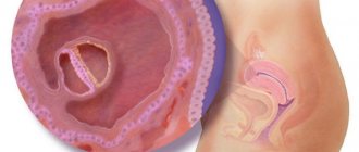
9 weeks after fertilization, 11 obstetric weeks (after the first day of the last menstruation)
1 — Anus 2 — Labial-scrotal tubercles 3 — Legs 4 — Genital tubercle 7 — Urethral recess 8 — Genital folds
The size of the embryo is 45 mm .
At the 9th week there are no noticeable differences between the genitals of a boy and a girl. The genital tubercle and genital folds are surrounded externally by labioscrotal tubercles. The pictures confirm that the boy and the girl are no different in appearance.
Boy (11 weeks post fertilization, 13 obstetric weeks)
Boy, 11 weeks
The size of the embryo is 64 mm .
The development of male external genitalia depends on dihydrotestosterone, which is produced by the testes. The genital tubercle lengthens and grows, forming into the penis, and the urogenital folds on both sides of the urogenital membrane begin to fuse, forming the urethra. The labial-scrotal tubercles grow rapidly and turn into the scrotum, fused along the midline.
In boys, the genital tubercle forms the penis (4) . The body of the penis is formed from the genital folds; at this stage of development, the formation of the penis is not yet complete (7) . The scrotum (6) is formed from the labioscrotal tubercles (2) . The scrotal fusion line (5) is formed by the connection of the labial-scrotal tubercles.
At this stage of development, the testicles are located in the abdomen. They do not descend into the scrotum (6) until 7-8 months of pregnancy.
The foreskin at the 12th week of embryo development is already formed.
Changes in the development of the genital organs of girls at 13-20 weeks after fertilization
Girls have very little testosterone in their blood. Therefore, after the formation of the external genitalia in the 8th week, in the future they practically do not change in appearance.
The genital tubercle turns into the clitoris; it can increase not only while it is in the mother’s stomach, but also after the birth of the girl.
The genitourinary folds form the labia minora. The labioscrotal tubercles enlarge and become the labia majora, while the urogenital groove remains open, forming the entrance to the vagina.
The size of a 13-week embryo is 90 mm , a 17-week embryo is 150 mm , and a 20-week embryo is 185 mm .
- Girl, ultrasound 13-20 weeks
- 1 - Anus 2 - Buttocks 3 - Clitoris 4 - Labia majora 6 - Legs 7 - Labia minora
- Girls' genitals are formed from the same folds and tubercles as boys' genitals.
By the 20th week, the labial-scrotal tubercles and genital folds in girls do not fuse and form the labia minora (7) and labia (4) The clitoris is formed from the genital tubercle (3) .
The ovaries are not identified until the 10th week.
Gender determination during the second planned ultrasound at 20-22 weeks
In boys, you can see a lump between the legs, which is the scrotum and penis. may show a round, raised area within the genital area, which is the scrotum and penis. On the ultrasound machine screen, the boys' genitals in profile look like a small snail.
- Some babies turn around during an ultrasound so that their genitals are not visible even at the third screening ultrasound at 32-34 weeks.
- Determination of the sex of the fetus is influenced by factors such as the position of the fetus, the amount of amniotic fluid and the thickness of the abdominal wall.
- Three-dimensional (3D) ultrasound makes it easier for specialists to determine the sex of the fetus.
Answers to the most frequently asked questions
Question: Can the sex of the baby be determined at the first screening ultrasound at 12 weeks?
Answer: At 12 weeks, the ultrasound doctor can make a guess regarding gender, sometimes it is a little more accurate than 50/50.
- fetal position,
- amount of amniotic fluid,
- thickness of the abdominal wall, etc.
- So, if you are very curious about who will be born, then here are several possibilities to guess about the gender based on the results of an early ultrasound examination.
- Such successful pictures as in the photo on the right are extremely rare.
- If the child turns so “comfortably”, the sex can be determined 12 weeks after conception (14 obstetric weeks).
3 ways to determine sex by ultrasound in the early stages
1. Determination by analyzing the angle between the genital tubercle and the baby’s back .
In the ultrasound screenshots below you can see what it looks like. All screens are of embryos at the 12th (14th obstetric) week of pregnancy, the size of the embryos is about 75 mm.
In boys , the genital tubercle forms an angle of approximately 30 degrees or more with the back (left column in the photo).
In girls, the genital tubercle forms an angle of less than 30 degrees (right column of examples in the picture).
- 2.According to the location of the placenta
- If the placenta is located on the right side of the uterus , the baby is more likely to be a boy .
- If the placenta is located on the left side of the uterus , expect a girl .
Source:
How to find out the gender of the unborn child in early pregnancy using a table?
Future parents are faced with a huge amount of advice on how to find out the gender of their child.
Doctors urge you to undergo an ultrasound examination, the older generation recommends monitoring the mother’s external changes, and forum visitors make numerological calculations.
We will consider further what methods really help to obtain information, how long it takes to find out 100%, and what methods you should not waste your time and money on.
Other options
In addition to assessing the state of the anatomy and physiology of the child, the ultrasound doctor evaluates the embryonic structures, the condition of the uterus, chorion, appendages, cervical length and other indicators. And it indicates whether there is a risk of miscarriage.
- To do this, first of all, the features of the chorion (future placenta) are described. Its location is determined.
- The threat of miscarriage is indicated by the presence of signs of chorionic detachment.
- Subchorionic hematoma is often found in pregnant women even in the absence of complaints.
- The amount and homogeneity of amniotic fluid is assessed.
- Using ultrasound, the tone of the uterus can be recorded.
- A corpus luteum is detected in one of the ovaries. Its function is to produce progesterone to maintain pregnancy.
- The mother's reproductive organs are examined for the presence of pathology and developmental features.
- Saddle-shaped and bicornuate uterus, uterine fibroids, cysts and ovarian neoplasms are diagnosed.
Yolk sac
Table for assessing the parameters of the fetal yolk sac.
Look at the shape, measure the internal diameter and echogenicity. The indicator may indicate a normal course of pregnancy or a deviation.
Chorion position
The position of the chorion is determined - this is the future placenta. Normally, it is attached to the back or front wall of the uterus.
If presentation is determined, then it will be necessary to develop separate pregnancy management tactics. Low placentation at this stage is often a physiological phenomenon. As the uterus grows and the pregnancy progresses, the placenta rises in most cases.
In addition, the structure of the chorion is assessed; normally it is homogeneous.
Condition of the uterus
At twelve weeks of pregnancy, the length of the cervix must be measured; normally it is 30 mm. An indicator of 20 mm is considered critical. In such cases, bed rest is prescribed, and in more complex cases, hospitalization. The muscle tone of the uterus is also assessed; with hypertonicity there is a risk of miscarriage. Treatment is prescribed on an outpatient basis or hospitalization for conservation.
Amniotic fluid
The condition and amount of amniotic fluid is examined. An increase or decrease in amniotic fluid is a sign of pathology. May indicate infection, renal dysfunction, or central nervous system disease. Cloudiness of the amniotic fluid may also indicate pathology, so additional examination is prescribed.
Indications
There are several reasons why an ultrasound examination of a pregnant woman at 12 weeks is of great importance:
- At this time, the age of the unborn child can be determined with high accuracy, and later he will gain weight, and the error in the date of conception will be significant (up to a week).
- At this time, it is important to determine the size of the collar area (the so-called soft tissues of the back of the head and neck). This indicator is key in the early diagnosis of chromosomal diseases, including Down, Patau, and Edwards syndromes. The examination must be carried out before 12 weeks , because after 14 weeks, chromosomal pathologies can no longer be detected and some fetal malformations too.
- Assessing the placenta insertion site, the presence of abruption, the quality and quantity of amniotic fluid are significant indicators of pregnancy that affect the health of the child and the management of pregnancy - they are also important to find out at 12 weeks.
- The doctor assesses the condition of the uterus - there should be no increased tone (that is, tension) or isthmic-cervical insufficiency. All these conditions require timely treatment, so high-quality and timely diagnosis during pregnancy plays a key role.
FAQ
- 3D ultrasound - Three-dimensional volumetric ultrasound is not performed at 12 weeks due to objective reasons, namely the fetus is too small. The optimal period is the 24th week of pregnancy, and it is carried out according to indications, and not at the whim of the parents. Three-dimensional ultrasound is performed for special indications as prescribed by a doctor, and is not recommended for a standard examination for every pregnant woman. Therefore, it’s not even worth talking about 4D ultrasound for standard 1 screening.
There are 2 fetuses on the 3D ultrasound image. Twins. - Ultrasound photo - in the pictures of the first screening, if the picture is successful, you can see the profile of the unborn child, distinguish the arms, legs, and umbilical cord. Naturally, future parents are more concerned about whether they can see the gender of the baby; here everything is much more complicated and you can easily mistake a girl for a boy.
- Twins - ultrasound images always show one of the fetuses better. There are times when one child conceals the second child, and the ultrasound specialist may confuse you and say that you have one fetus. But in addition to visualization on the monitor, twins can be distinguished by their heartbeats.
Ultrasound image shows twins
What tests and studies should I undergo?
Of course, you need to get tested. All examinations that your doctor recommends are aimed at ensuring that you can:
- Assess the current health status of the woman and fetus. This is very important, because it is difficult to recognize the onset of a disease that is hidden behind the symptoms of pregnancy.
- Prevent possible complications. A timely detected violation of hormonal status or hemoglobin level will help to correct pregnancy management and give birth to the baby on time.
- Carry out the necessary treatment.
At 12 weeks of pregnancy, you must visit a gynecologist. If you haven’t done this before, then it’s high time to register with the antenatal clinic! If you have complaints to other doctors, you should also visit them.
If screening in the form of a prenatal double test was not carried out in a timely manner (at 10-11 weeks), it needs to be done now. The level of PAPP-A and hCG, when tested simultaneously, will help in diagnosing fetal development. Although a screening test cannot make a diagnosis, it gives an idea whether an extensive examination is necessary. Find out how the first screening goes in a separate article on our portal.
Ultrasound is performed if necessary.
When prescribing additional research methods, it is necessary to remember that timely diagnosis and treatment lead to better results.
Recommendations at 12 weeks of pregnancy
In 12 weeks, a lot has changed: larger underwear appeared in the wardrobe for enlarged breasts, low-heeled shoes are now much more comfortable than stilettos, smoking and alcohol are a thing of the past.
By the end of the first trimester, she underwent correction and physical activity. After the first classes, the initial complex of yoga for pregnant women has already been mastered. Shaping began to bring pleasure even to absolutely unsportsmanlike ladies. If this has not happened yet, choose a gymnastics club, pool or section, guided by the following principles:
- Non-traumatic. Dizziness and unsteady gait are common during pregnancy. If you add skates or a tennis ball to this, you can cause damage to yourself. We strive to improve health, which means that priority areas will be swimming and sets of physical exercises for pregnant women.
- Close to home. Physical exercise is necessary after going on maternity leave even more than during the working period. Therefore, it will be more convenient to visit a section near your place of residence, regardless of the start of your vacation.
- Personal preference. If you're bored in yoga, choose fitness. If you enjoy the water element, sign up for a swimming pool.
Nutrition
try to choose something that is balanced and healthy.
The diet must include meat, fish, dairy products, vegetables and fruits. This will provide your baby with everything he needs. Proteins, carbohydrates, fats and vitamins will be used to build a growing body.
Avoid lots of mayonnaise, fried foods and fast food. Sex deserves special attention. In the absence of contraindications, it will help the couple develop a special attitude towards childbirth. For some women, sexual sensations during pregnancy are revealed much more clearly. Do not deny yourself and your partner pleasure.