Unlike other human organs, the adrenal glands are rarely examined using magnetic resonance imaging. If this happens, doctors usually do an MRI of the adrenal glands and kidneys at once. This is explained by the fact that these organs are located extremely close.
All tissues in the human body, including those that make up the adrenal glands, respond to physical stress in a specific way. Since the structure and density of different tissues, as well as the properties of their cells, are different, the reaction will also be different. The essence of all instrumental diagnostic methods is based on this phenomenon.
If you take an ultrasound, then in this case the tissues will absorb and push out ultrasonic waves. A similar process occurs during radiography.
As for magnetic resonance imaging, it is based on something else - the reaction of cell nuclei during their excitation in a magnetic field by electromagnetic waves. All these processes are read by the ultra-precise sensor of the MRI machine. Next, the results obtained are processed by a computer and, as a result, a three-dimensional image of the organ under study appears on the monitor screen.
This image is very accurate and clear. This allows the specialist to examine the organ in as much detail as possible, which is extremely important when diagnosing pathologies of such a small organ as the adrenal gland.
Advantages of the procedure and possible risks
The main advantages of MRI include the fact that it provides very accurate data on the condition of organs.
In addition, during the study the patient does not risk his health, because magnetic radiation does not have any effect on the body and does not change the functioning of organ cells.
This makes it possible to diagnose pregnant women, children and people with serious diseases of internal organs.
MRI is not an invasive procedure. During it, the patient does not feel any discomfort.
What can replace MRI?
An alternative to magnetic resonance scanning is scintigraphy and CT scanning of the adrenal glands. They require special training and preliminary laboratory diagnostics.
CT scan reveals benign and malignant tumor processes. It has wide capabilities for recreating images in different planes.
The disadvantage of the procedure is exposure to radiation. In this case, MRI scans the human body using a magnetic field that is safe for humans.
Procedure with and without contrast
At the moment, MRI can be performed in two ways: with and without contrast.
The attending physician chooses exactly how the procedure will be performed for a particular patient. This will depend on the preliminary diagnosis. So, if a specialist suspects a tumor in a patient, then the administration of contrast will be mandatory.
Without this, it will simply be impossible to determine the structure of the neoplasm. And this is very important, because this is how you can distinguish a benign neoplasm from a malignant one. A cancerous tumor has a heterogeneous structure. It is lumpy and has no clear boundaries.
This way you can determine the exact location of the tumor and identify metastases, if any. This will help preserve not only health, but also human life. The contrast agent in this case is made on the basis of gadolinium, magnevist and some other substances.
When performing MRI, contrast agents containing iodine are not used. Thanks to this, the likelihood of an allergic reaction is significantly reduced. The contrast agent is injected directly into the vein. This is done a few minutes before the start of the study.
Contraindications to MRI of the adrenal glands
MRI of the adrenal glands still has some contraindications. To avoid negative consequences, they cannot be ignored. This study is not carried out if:
- the presence of infectious diseases;
- pregnancy;
- epilepsy;
- obesity (more than 120 kg);
- claustrophobia;
- allergies to the dye.
Also, tomography is not allowed for people who have metal implants in their body (pacemakers, bullets, staples, prostheses, etc.).
Using the latest computer technology in MRI, doctors can now create a three-dimensional model of an organ and view it from all sides. In different cases, the frequency of examinations may be different. Thus, for stable tumors, MRI is performed every six months to a year, and if there is suspicion of their growth, twice as often. It all depends on the doctor's prescription.
Attention!
This article is posted for informational purposes only and under no circumstances constitutes scientific material or medical advice and should not serve as a substitute for an in-person consultation with a professional physician.
For diagnostics, diagnosis and treatment, contact qualified doctors! Number of reads: 751 Date of publication: 07/19/2018
Urologists - search service and appointment with urologists in Moscow
Indications for the study
The function of the adrenal glands is to produce hormones that are necessary for the functioning of the entire body.
Various pathologies of the adrenal glands are extremely dangerous not only for health, but also for human life. Even a minor failure in this case will not pass without a trace for a person. This is why diagnosing adrenal gland diseases on time is very important. Doctors say that the cortex of these organs usually undergoes pathological changes.
There are many indications for which you need to contact a medical facility as soon as possible and perform an MRI of the adrenal glands. These include:
- significant and persistent increase in blood pressure;
- sudden weight gain;
- swelling of the face;
- formation of stretch marks on the skin;
- impaired glucose tolerance;
- disruptions in the immune system;
- pathological leaching of minerals and calcium from bones;
- severe weakness in the body;
- sudden mood swings;
- loss of gender characteristics;
- too early puberty;
- tachycardia, disruptions in the functioning of the cardiovascular system.
In addition, diagnosis is recommended for patients whose blood tests showed an increase in the amount of adrenal hormones.
X-ray or MRI, which is better?
X-ray and MRI are fundamentally different examination technologies. If we compare them with each other, then according to the following criteria:
Image quality
. A tomograph scans the human body and builds an image in layers. The image has high resolution, detail, data on soft and bone tissues. An X-ray machine can highlight only one layer and demonstrate the condition of the tissues; the quality of the image will be worse.
| X-ray | MRI of the adrenal glands |
Harmfulness
. X-ray radiation in large quantities is dangerous to health, while MRI can be done at least every day.
Availability of procedures
. An X-ray machine can be found in every ordinary clinic. Equipment for MRI requires large financial costs and the availability of specialists who know how to operate it, so large city and regional medical centers and commercial hospitals can afford a tomograph. You can get an X-ray quickly, but the appointment for an MRI is distributed over weeks in advance.
Price
. An X-ray examination can be done free of charge under the compulsory medical insurance policy in a regular clinic. Only patients from surgery and inpatient departments can get an MRI free of charge.
Main contraindications
MRI of the adrenal glands is strictly contraindicated in patients in the following cases:
- the body has a pacemaker;
- there are any metal objects in the body that cannot be removed;
- there is an allergy to the contrast agent (in this case, the classical procedure can be performed, but not with contrast).
In addition, there are conditional contraindications. This means that the procedure can be performed on patients, but some difficulties may arise.
In most cases, diagnosis turns out to be more important and then the doctor decides to perform an MRI. However, all the nuances in this case will be taken into account.
Relative contraindications include the following:
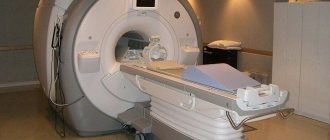
- Claustrophobia . Most MRI machines are closed. This can cause shock, panic, and hysteria in the patient. To avoid this, doctors may suggest that the patient take a dose of a sedative before the procedure. If possible, the diagnosis will be carried out in an open-type tomograph.
- Epilepsy . Sometimes during diagnosis, patients begin another attack of seizures.
- First 2 trimesters of pregnancy . The fact is that it is at this time that the formation of all the organs and systems of the fetus occurs. At the same time, the effect of a magnetic field on an embryo has not yet been studied. MRI is performed extremely rarely in such patients. However, after this, not a single case of adverse effects on the fetus was recorded.
- Inability for patients to remain motionless . The more motionless the patient is during the procedure, the more accurate results can be obtained in the end. It is very difficult for preschool children to remain in one position. It is impossible to control this process for adults who are under anesthesia.
- Extremely serious condition of the patient.
- The patient's weight is more than 120-150 kg . Most devices simply cannot withstand such a load.
If a woman is breastfeeding, then after the procedure she is not recommended to do so for 2 days. During this period, the contrast agent will be completely eliminated from the body and will definitely not pass into the milk.
The essence of MRI, advantages and disadvantages of diagnosis
MRI is a modern and accurate instrumental diagnostic method
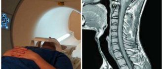
Magnetic resonance imaging is based on the use of magnetic nuclear resonance. The examination uses a tomograph, which is essentially a large magnet. Using a strong magnetic field, you can examine any organ of the human body. The picture is displayed on a computer monitor and recorded dynamically. You can also receive a disc and pictures after the procedure is completed.
MRI of the adrenal glands is often performed to make a diagnosis, since ultrasound and blood tests are less informative. The procedure is quick and painless. It can be carried out either as prescribed or at the personal request of the patient, but in the second case it will be paid.
MRI has many advantages over other diagnostic methods:
- Non-invasive and painless. During the procedure, no foreign bodies are introduced into the body tissues. The patient does not experience any physical discomfort. In some cases, MRI with contrast is used, which means an injection of a contrast agent is necessary. However, there are no strong painful sensations in any case.
- High information content. The MRI procedure is currently considered one of the most informative. A three-dimensional image of the organ is displayed on the screen. You can evaluate both its size and thickness, structure, and uniformity.
- Safety. There is no radiation effect on the body. The procedure is absolutely safe for people of all ages in the absence of contraindications to its implementation. In some cases, side effects may occur from the contrast agent rather than from the magnetic effect.
The disadvantages of MRI include a fairly high price (if the procedure is performed without a doctor’s referral), as well as a large number of contraindications, both absolute and relative. The device is designed for people with a certain weight, so when examining people with a body weight of 120 kg and above, it may be useless.
How to prepare for diagnosis?
Another advantage of MRI is that this procedure does not require prior preparation.
However, if the patient is scheduled for a contrast study, then he should come to it with an empty stomach. This is because the contrast agent can cause nausea and vomiting.
In addition, 3-4 days before the procedure, the patient should exclude soda and foods that cause increased gas formation from the diet. These are, first of all, beans and cabbage. Excess gases can distort the resulting images and thereby make the results less informative.
If an MRI needs to be performed urgently, there is no need to talk about preparation.
MRI scan results: what is normal and what is abnormal
Normally, the adrenal glands are presented in the form of homogeneous formations, up to 10 mm thick. Thanks to the contrast agent, the cortex and medulla are distinguished.
Nodular hyperplasia - thickenings up to 1.5 cm with uneven, bumpy outer contours.
Myelolipomas are round formations with unclear contours, homogeneous structure, and large sizes.
In adrenal tuberculosis, the cortex and medulla are separated, and the glands are affected by calcifications.
In malignant tumors (pheochromocytomas), heterogeneous, clear formations with a high degree of intensity are visualized.
Benign tumors (aldosteromas) are smaller in size, round in shape, and have clear contours. They affect one adrenal gland and are prone to accumulation of contrast media.
Metastases on MR images have a heterogeneous structure, uneven contours, and tend to accumulate contrast agents. Both adrenal glands are affected.
Pheochromoblastoma of the left adrenal gland
Aldosteroma of the left adrenal gland
Malignant corticosteromas appear as unclear contours, a heterogeneous structure with cystic or fatty deposits. They accumulate contrast and are large in size. In a benign course of the disease, an even contour and homogeneous structure are visualized.
How are they diagnosed?
MRI of the kidneys and adrenal glands is performed as follows:
- Before entering the room where the diagnosis will take place, the patient must put away his phone, watch and keys, and also remove all metal objects. If your clothing has metal buttons, these will also have to be removed. This is due to the fact that metal distorts the magnetic field and can affect the information content of the study.
- The patient lies down on the tomograph conveyor table. If necessary, a contrast agent is injected at this time. The patient's limbs are secured using special straps.
- The table moves into the tomograph together with the patient. This is where the research takes place. Scanning is done in two planes: frontal and transverse.
- The patient should lie still. It is very advisable to synchronize breathing with taking pictures.
- During the procedure, the doctor will sit in the next office at the computer. You can contact him at any time through the public address system.
Typically, the patient is alone during the procedure. However, if the diagnosis is being carried out on a child, one of the adults may be with him (before entering the office, he must also remove all metal objects from himself).
The duration of an MRI is usually no more than 50 minutes.
Anatomy and physiology of the adrenal glands
The adrenal gland is a paired gland responsible for the activity of internal secretion. This is the name given to the process of producing and releasing hormones into the bloodstream. The organ consists of 2 morphofunctional endocrine glands - the cortex, the medulla, which have an embryonic appearance.
The cortex is secreted from interrenal tissues. This is a piece of mesoderm. The medulla has an appearance together with the NS. It has an organ of similar origin to chromaffin tissue.
Hormones influence the brain matter. If there is a malfunction of the adrenal glands, the patient experiences unpleasant conditions, ranging from severe irritability to whole body tremors and panic attacks. This is due to the fact that the organ is responsible for the normal functioning of the endocrine system. If it functions incorrectly, the patient's condition will deteriorate sharply.
According to histology, there are 3 zones in the adrenal cortex (it accounts for up to 90% of the organ). Under the capsule is the zona glomerulosa, which is responsible for the secretion of aldosterone. The reticularis acts as the innermost zone and is responsible for the secretion of hormones called androgens.
If necessary, an MRI or CT scan is done - if there is a suspicion of neoplasms or other serious diseases. To better see the structure and boundaries of the organ, the procedure is performed with contrast enhancement. Before the examination, the patient is prepared; below we will look at how this is done. Kidney pathologies are a serious problem due to the fact that the body’s activities are disrupted. It is better to be examined in a clinic that has high-quality equipment. The price of diagnostics is high, but it is justified by the costs that are used during the procedure, as well as the price of a tomograph - a device that plays a key role in the examination.
Feelings during the procedure
During the study, the patient does not feel anything unpleasant. He can hear the knocking and noise that accompanies the operation of the device. Heat may be present directly in the adrenal gland area.
If during an MRI the patient experiences any discomfort or pain, this should be reported to the doctor immediately.
An allergic reaction to a contrast agent can manifest itself in different ways. While some patients simply feel a slight itching of the skin, others may experience a state of shock. Fortunately, this happens extremely rarely and if it happens, the allergy is mild. After just a day, this reaction will disappear.
Ultrasound or MRI, which is better?
Both methods are safe, effective and informative. However, it is necessary to know their capabilities, advantages and disadvantages in order to determine what is more appropriate to apply in a particular case. To do this, let's compare the following aspects:
- Operating principle of the devices
. MRI uses magnetic pulses to visualize internal organs, and ultrasound diagnostics is based on the conversion of ultrasonic waves. People with metal implants, dentures, pins, cochlear devices, and pacemakers are not safe from exposure to electromagnetic fields. The ultrasound technique is suitable for them. - Research speed
. For MRI you need to spend 20-30-40 minutes and at least an hour for description. The ultrasound procedure takes 5-10 minutes, and the result is immediately available. - Device capabilities
. Ultrasound examination does not provide 100% accurate results. The processing of the received data is carried out by a person. The information content and reliability of the examination depends on the experience and skills of the specialist. An MRI machine processes signals, generates images, and interprets data using a computer processor. And only then does the radiologist begin decoding. The likelihood of errors and errors in this case is reduced. - Availability and cost of the procedure
. An ultrasound machine can be found in rural clinics, while an MRI scanner is the prerogative of large regional centers. It is easier to undergo ultrasound diagnostics through compulsory medical insurance. In the case of paid services, the cost of ultrasound is 2-3-4 times lower than MRI.
| Ultrasound | MRI of the adrenal glands |
Alternative Research Methods
If a patient cannot undergo an MRI for any reason, then his doctor should choose another way to examine the adrenal glands. It is desirable that the alternative procedure also gives accurate results and is highly informative.
The choice of procedure in this case will directly depend on the preliminary diagnosis. Thus, an ultrasound can indicate the presence of a tumor, but in this way it will not be possible to identify its exact location. Similar data can be obtained using PET-CT, as well as MSCT.
It is worth noting that these techniques have even more contraindications, so MRI is the most preferred diagnostic method.
What conditions will MRI diagnostics show?
MRI is indicated for patients with endocrine pathologies. Scan shows:
- Addison's disease, which manifests itself as impaired resistance to stress, cravings for salty foods, chronic fatigue;
- psycho-emotional disorders;
- Itsenko-Cushing syndrome;
- adrenocortical cancer;
- hypercortisolism;
- Conn's syndrome, which manifests itself in the form of increased blood pressure;
- congenital hyperplasia.
After confirming the diagnosis, refrain from self-medication and present the scan results to the attending physician.
Adrenal glands. Symptoms of the disease in women
A woman's body is prone to hormonal changes. The main function of the adrenal glands is to produce hormones. The main disturbances in the functioning of organs are associated with insufficient production of these substances.
How do you know when you should have your adrenal glands checked? Symptoms of the disease in women may vary. Since their nature depends on the disease that is present in the body. For example, the signs may be the following:
- bone fragility;
- lack of libido;
- disruptions of the menstrual cycle;
- tendency to high blood pressure;
- obesity;
- drowsiness;
- increased weakness;
- panic attacks;
- increased irritability;
- headache;
- frequent muscle cramps.
- appetite decreases;
- bluish lips;
- unnatural thinness appears;
- tachycardia;
- severe fatigue;
- tremor of the limbs;
- increased gas formation;
- depressive states.
In these and some other cases, the doctor may prescribe an MRI of the adrenal glands.
Which specialist should I contact?
You can undergo an MRI examination of the adrenal glands by receiving a referral from your attending physician or from an endocrinologist, who will indicate in the direction of the presumptive diagnosis and the purpose of the diagnosis. The results of the study are deciphered by the doctor who performs the tomography procedure.
It is very important to choose a highly qualified clinic and doctor. It is worth giving preference to more modern diagnostic centers that are equipped with high-quality equipment and employ qualified medical staff.