In the modern world, any person, even without a medical education, has heard more than once about the important role of hormones and the glands that produce them. One of these endocrine organs is the pituitary gland. This gland serves as a structural unit of the brain and is located in a special depression at the base of the skull, called the “sella Turcica”.
It consists of two parts: the anterior one is the adenohypophysis, the posterior one is the neurohypophysis. The first accounts for about 80% of the volume of the entire gland. The hormones it produces regulate the functioning of other endocrine glands and take part in many physiological processes. Thus, the pituitary gland influences most human systems and organs, and disruption of its function leads to serious problems.
This is why a qualitative study of this organ is so important when alarming symptoms appear. One of the most informative diagnostic methods today is MRI of the sella turcica. This procedure is recognized as the safest and most accurate compared to radiography. The images obtained on the tomograph show a complete picture of even the most minor changes in the organ, which helps to make a diagnosis at an early stage and begin adequate therapy.
Explanation of the term
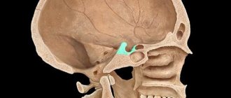
The sella turcica is a bone formation in the form of a dimple located in the sphenoid bone. Its shape resembles the outline of a saddle - hence the specific name.
In the central part of the sella there is a depression where the pituitary gland is localized - a small compaction loosely located on the stalk or stalk of the gland. This element of the endocrine system is responsible for the production of hormones, the functioning of the glands of the endocrine system, and the regulation of metabolism.
When the bone formation is compressed, the gland is injured and partially or completely loses its functionality. Why do X-rays take place in this case?
An x-ray helps to identify defects and dysfunction of the pituitary gland and begin therapy in a timely manner.
In what cases does a gynecologist refer for the procedure?
The entire human body is a system with a complex structure. In it, all parts are interconnected, even if at first glance the connection is not obvious. Therefore, when a gynecologist sends you for an X-ray examination of the sella turcica - a bony depression in the structure of the skull - you should not be surprised.
The condition of the sella turcica directly affects the health of the pituitary gland. The pituitary gland, in turn, is part of the human endocrine system, and in the female body it produces various hormones that are closely related to the sexual and reproductive health of women, including:
- thyroid-stimulating;
- follicle-stimulating;
- luteotropic (prolactin);
- luteinizing;
- gonadotropic;
- adrenocorticotropic;
- oxytocin;
- melanostimulating hormone.
Each of these hormones plays its role in the functioning of the female genital organs. Some are involved in the ovulation process; without others, follicles cannot form in a woman’s body; others are formed during pregnancy and contribute to its normal course.
A gynecologist may send a patient for an X-ray examination of the sella turcica if:
- menstrual irregularities;
- infertility;
- increasing the level of prolactin in the blood.
Disturbances in the functioning of the pituitary gland in women are also manifested by general malaise, increased formation of wrinkles, and deterioration of skin elasticity, which is not associated with age-related changes. In such patients, hair begins to fall out rapidly, it becomes dull and brittle, appetite worsens and frequent constipation appears.
In addition to x-rays of the sella turcica, the gynecologist may recommend visiting an endocrinologist, as well as testing for hormones of the pituitary gland and other endocrine glands.
Indications for the study
X-rays of the bone bed of the pituitary gland are prescribed in a number of the following cases:
- the need to determine the neuroendocrine pathological process;
- high concentration of prolactin in the blood;
- high intracranial pressure;
- abnormal formation of the skull;
- slow or too rapid growth;
- detection of damage to cranial bone elements.
The doctor may refer the patient for examination if there are the following complaints:
- disruptions in the menstrual cycle;
- periodic severe headaches;
- inability to conceive (subject to hormonal diagnosis).
Diagnostic value of examination for empty sella syndrome and similar pathologies
MRI of the sella turcica is prescribed if there is a suspicion of damage to the pituitary gland, which is accompanied by some clinical signs. They appear due to compression, the gland itself and the structures above are damaged.
Physiological disorders occur, which are indications for MRI of the pituitary gland:
- Visual impairment - the optic chiasm located above is damaged.
- Changes in blood pressure, headaches and dizziness are common symptoms of brain damage.
- Hormonal disruptions - weight problems appear, chronic diseases worsen, and the menstrual cycle in women is disrupted.
Sometimes one type of clinical sign is recorded; symptoms may not appear or may occur sporadically.
The described disorders are also observed in other pathologies:
- circulatory disorders;
- increased intracranial pressure;
- oncology of the pituitary gland or neighboring structures.
MRI will help to finally put an end to the issue of pathology. During the study, layer-by-layer images are obtained that help to study the pituitary gland and neighboring structures in detail in a three-dimensional projection.
Contraindications to X-rays
The described procedure has no absolute contraindications. However, this type of diagnosis is not recommended for pregnant women, especially in the first trimester.
If the potential result of an x-ray exceeds the risk of harm to the fetus, the procedure will not be canceled. All of the above is also relevant in relation to diagnosing children. When examining a pregnant woman, the area of the reproductive organ is covered with a protective blanket.
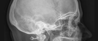
Other radiological signs of sella turcica pathology
X-rays of the skull sometimes reveal an “empty sella turcica.” In the picture, changes appear below the diaphragm of the sella turcica itself and consist of the following signs:
- the bottom in the frontal plane is symmetrical;
- vertical enlargement of a closed-shaped saddle;
- double-contour bottom on a sagittal image.
Clinical signs with such changes in the sella, however, are minimal. The patients do not have any endocrine disorders, but they are subject to dynamic monitoring. Malfunctions of the pituitary gland occur more often in women.
Preparatory stage and methodology
The diagnostic procedure is not preceded by special preparation. On the eve of the x-ray, the patient will have to remove metal objects (jewelry, watches, glasses, etc.): they can distort the results of the examination.
Pictures are taken in the following projections:
- direct frontonasal;
- right and left side;
- straight back.
Often, an additional radiography of the skull is performed. This measure helps determine the size and shape of the sella turcica in comparison with similar characteristics of the skull.
Preparation for the procedure
Before prescribing magnetic resonance imaging of the sella turcica, the doctor directs the patient to undergo general clinical tests, determine blood hormones, and also undergo additional diagnostic methods. If necessary, consultation with narrow specialists is carried out. Magnetic resonance imaging can also be done on your own initiative. The exception is the use of contrast. In this case, a referral from the treating doctor is necessary.
Before starting the diagnosis, you need to remove all metal objects and accessories (dentures, earrings, etc.), as well as put out mobile phones and magnetic cards. They will interfere with the magnetic field and the examination result will be unreliable due to poor-quality images.
If an MRI is performed with intravenous dyes, patients are advised to fast for at least 8 hours before testing.
Interpretation of examination results
The radiologist begins interpreting the resulting radiograph. The specialist evaluates the size, shape, contour line and condition of the bone.
A number of the following indicators have a diagnostic value:
- Sagittal size: standard value - 9-15 mm.
- Vertical size: normal - 7-12 mm;
- Saddle Index: A comparative measure of saddle height and length. In healthy children this figure exceeds one, in adults it is slightly less.
The image is normal.
Based on the examination results, the radiologist may suspect the development of a gland tumor. The sella turcica on an effective x-ray will demonstrate one or more pathological pictures:
- osteoporosis of the walls of the bone formation (without damage to the structural components of other elements);
- wall atrophy;
- thinning of the wedge-shaped processes of the formation;
- unevenness of the internal contour of the walls.
If the doctor assesses the contours of the formation as clear and even, pathologies in the pituitary gland area are highly likely to be excluded. If a blurred contour line is detected, the development of a tumor or empty sella syndrome is likely.
In gynecology, when assessing examination results, signs of neuroendocrine gynecological diseases are sometimes identified. At the same time, on an x-ray, the specialist notes areas of thickening of the occipital and frontal bones, as well as calcifications (calcium inclusions) in the dura mater of the brain.
How does an x-ray work?
There is no need to prepare for the procedure - diet, refusal to take medications, and bowel cleansing are not needed. Immediately before Rg-graphy of the sella turcica, the patient removes jewelry, hairpins, hair clips and other objects from the head, which reduce the information content of the images.
The procedure takes place without pain or discomfort. Duration – 5-15 minutes. How exactly to take an x-ray is prescribed by the attending physician or determined by the radiologist. Pictures can be in lateral, less often in frontal and posterior projections. The patient is in a lying or sitting position. The images are analyzed by a radiologist, and after the procedure a conclusion is drawn up. An accurate diagnosis is made by the attending physician at a subsequent consultation.
Computed tomography as a modern alternative to x-rays
CT is an improved method of radiography. The difference between the methods lies in the diagnostic capabilities: with computed tomography they are immeasurably wider. This fact is explained by the ability of CT to make layer-by-layer sections of the area under study. With traditional x-rays, linear scanning is carried out - the output is images in one plane.
The second point is the safety of the procedures. X-ray is considered a safer diagnostic method compared to CT, although not as informative.
Based on practical data, in the initial stages of diagnosis, an x-ray is often sufficient. If the examination does not provide the necessary information, they resort to computed tomography.
An alternative procedure helps to examine the area under study in detail (from different angles) and determine the size and depth of the pathology focus. As an alternative imaging method, MRI of the sella turcica is also used.
The most harmful diagnostic method
X-ray of the sella turcica is used in diagnosing the condition of the pituitary gland, a gland in the human skull. In addition to the named element, the bone formation itself acts as an object of study. Upon completion of the procedure, the radiologist evaluates the condition, contour line, shape and size of the bone tissue.
Analysis of indicators helps determine the likelihood of developing a tumor, empty sella syndrome, neuroendocrine gynecological diseases and other pathological conditions. If there is a lack of information obtained, they resort to an improved method of x-ray examination - computed tomography.
MRI of the pituitary gland and sella turcica
Targeted MR imaging clarifies the diagnosis or features of the pathology identified using alternative research methods: X-ray or CT.
Pathological processes in the sella turcica, pituitary gland and blood vessels are difficult to visualize using other techniques, as they are small. For example, the sella turcica does not exceed 1.2 cm in height and 1.5 cm in width.
Oncological and benign tumors in the structure of the pituitary gland are almost impossible to notice using x-rays. During an MRI, the specialist is able to magnify suspicious areas and look at the smallest details in detail. The study not only diagnoses small tumors, but also allows us to determine their nature - oncological or benign.
MRI with contrast to examine the pituitary gland and sella turcica is prescribed in complex cases. The contrast is evenly distributed throughout the human body and accumulates in pathological areas. Due to this, the size and location of the tumors are determined. One of the indicators for MRI with contrast is a suspicion of secondary Raynaud's syndrome.
Contrast drugs rarely cause allergies or other adverse reactions. The substances are completely excreted by the kidneys after 24 hours. Drugs leave the body faster when drinking large amounts of water.
Treatment of empty sella syndrome
The prescription of a course of therapy depends entirely on the underlying cause against which the development of sella turcica syndrome began. As a rule, the underlying disease is treated and the symptoms are suppressed. Treatment options can be divided into two main groups – medications and surgery. Therapy with folk remedies is not carried out for this type of disease.
Medication
When a sella turcica is detected in the brain during examination for another disease, as a rule, no treatment is prescribed. In these cases, the pathology does not manifest itself in any way and does not cause discomfort. All you need is regular examination by a doctor so as not to miss the worsening of the condition. In other cases, they are guided by the following principles:
- In case of hormonal imbalances, when there is a deficiency in the production of specific hormones, replacement therapy is prescribed. It consists of maintaining the missing elements from the outside.
- Asthenic, vegetative problems are solved with the help of symptomatic treatment. Your doctor may prescribe sedatives, pain relievers, or medications to lower your blood pressure.
Surgical
Surgical intervention for leakage of cerebrospinal fluid due to thinning of the bottom of the sella turcica into the nasal cavity. The same treatment method is necessary for sagging of the optic chiasm into the diaphragm and compression of the optic nerves, which leads to visual field impairment. After the procedure, the patient must undergo a course of radiation and hormone replacement therapy. There are two techniques for performing surgery:
- Through the frontal bone. This type is used when there is a large tumor, which does not allow removal through the nose. This method is more traumatic.
- Through the nose. A more common option for the operation. An incision is made in the nasal septum, through which all other manipulations are carried out.