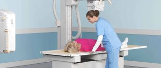
Radiography is a method of obtaining images of internal organs based on the use of x-rays . X-rays passing through the body are partially absorbed by tissues and, therefore, are attenuated. The attenuated radiation is then captured on x-ray film and an image is formed. The image is obtained due to the fact that the degree of absorption of most tissues is different, and therefore, the intensity of the rays passing through them will also differ, which can be seen on the x-ray.
Currently, there is a fluoroscopy method, which is also based on the use of X-rays. It differs from radiography in that the study can be carried out in real time. This allows you to evaluate parameters such as the displacement and distensibility of organs.
Ultrasound of the stomach or FGDS - which is better?
It is impossible to give a definite answer to this question:
- some pathological processes can be noticed only with the help of an endoscopic examination, others, on the contrary, will only be shown by ultrasound of the organ;
- Both types of research should be carried out if the digestive system causes serious concern to the patient, and the gastroenterologist finds it difficult to make a diagnosis.
In any case, the decision must be made by a qualified doctor.
Previously, there was an opinion that gastroscopy is much preferable to ultrasound, since the latter does not make it possible to see the smallest details and give an accurate assessment of the condition of the stomach and intestines.
However, modern technologies have made a great leap forward, and today ultrasound of the stomach deserves no less attention than gastroscopy. In some cases, ultrasound can serve as a full-fledged alternative to FGDS, which makes it possible to study organs much more comfortably and painlessly.
Benefits of stomach ultrasound
Ultrasound examination is certainly a more comfortable and gentle method for the patient, and has the following advantages:
- Absence of pain not only during the study, but also after (unlike FGDS).
- No equipment penetrates inside, which avoids any microtrauma and ensures psychological comfort for the patient.
- The structure of the affected organ can be examined from any convenient angle.
- The contrast method is physiological (sometimes the patient is asked to drink a liquid containing a contrast agent).
- With ultrasound, it is possible to take photos and videos of the organ, which can be useful in further treatment and in the medical history.
- Quite high resolution.
- It is possible to examine the blood flow of an organ and examine the vascular network.
- The research doesn't take much time.
- You can examine neighboring organs, whereas gastroscopy allows you to study only the condition of one organ.
- A high level of comfort for the patient during the procedure makes ultrasound of the stomach painless.
Disadvantages of stomach ultrasound
Like any method, ultrasound also has a number of disadvantages, but they are relatively few.
Here are the main ones:
- The inability to see the full picture of the stomach cavity, since it is a hollow organ.
- There is no way to collect physiological fluids in the stomach. In some cases, there is a need to examine them to make a correct diagnosis.
- Does not allow for a biopsy (taking part of the stomach tissue for further research).
Benefits of gastroscopy
Fibrogastroduodenoscopy has always been considered a more accurate method of examining the stomach, allowing the patient’s diagnosis to be determined with the highest accuracy.
FGDS of the stomach - what it shows and what advantages it has:
- Excellent resolution.
- It is possible to take a biopsy.
- Allows you to collect physiological fluids from the stomach for analysis.
- Possibility to take photos and videos of the organ being examined.
- The ability to view the stomach cavity and its walls in their natural form, and not through an ultrasound screen.
Disadvantages of gastroscopy
FGDS has a number of disadvantages, which are slightly greater than those of the ultrasound method:
- Severe discomfort during the procedure, which can manifest itself in the form of nausea, gagging, vomiting, lacrimation, discomfort in the esophagus and stomach, as well as sore throat. Some symptoms may persist for some time after the procedure is completed.
- Gastroscopy does not make it possible to examine the organ being examined from all sides.
- The study cannot be classified as physiological procedures.
- There is no way to determine gastric blood flow.
- FGDS does not provide access to other organs.
- The presence of foreign objects in the body during the examination.
- Relatively long research time.
- Hollow organs are filled with air in order to obtain good examination results. This causes additional discomfort and is not physiological.
X-ray of the stomach: preparation
X-ray of the stomach - preparation, how it is done, why interpretation of gastrography is not a diagnosis, but only a presumptive conclusion that should be analyzed with other tests
X-ray of the stomach: preparation, how it is done, indications, contraindications
X-ray of the stomach is one of the leading methods for identifying the pathology of the organ. An alternative is FGDS (fibrogastroduodenoscopy), which studies the internal lining of the stomach, detection of erosion, ulcerative defects, inflammatory changes, and cancer. The choice of examination depends not only on diagnostic purposes, but also on the tolerability of the examination to patients.
The insertion of a probe during FGDS can provoke a gag reflex and pain, so gastroscopy is prescribed. X-ray of the stomach with contrast is characterized by human radiation exposure, and therefore is not an alternative to fibrogastroduodenoscopy.
Preparation for an X-ray of the stomach and FGDS is carried out according to similar principles, but must be carried out correctly. The presence of mucus, food particles, and hydrochloric acid in the gastric cavity greatly complicates diagnosis.
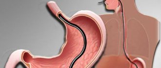
The stomach is a hollow organ that, when at rest, contains a gas bubble. X-rays pass through the gastric walls without reflection, so organic changes cannot be visualized without the use of contrast.
Gastric contrast is performed to study the following characteristics of the organ:
• Pathological changes; • Form; • Size; • Position; • Structure;
• Relief.
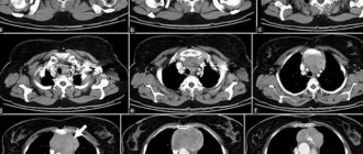
The contrast is a suspension of barium, which is not absorbed into the blood, which allows the substance to be freely excreted naturally through the gastrointestinal tract. During the examination, the doctor looks at the changes on a special X-ray fluorographic screen and takes a series of images that display the distribution of contrast. During one procedure, 8-12 images are taken. Series of radiographs show not only static data, but also the dynamic distribution of contrast as it passes through the stomach.
X-ray of the stomach: preparation, indications, contraindications
Preparing for a stomach x-ray is not difficult. All manipulations are performed at home. If you follow the procedure, 2 important conditions must be met:
• The study is carried out on an empty stomach, so do not eat anything 8 hours before the procedure; • Avoid alcoholic beverages, smoking, and medications the day before gastrography.
Some difficulties in preparation arise for people with diseases of the gastrointestinal tract. The formation of gas makes it difficult to perform FGDS and contrast gastrography. To eliminate gas formation, you should exclude the following foods from your diet:
• Sweets; • Sparkling water; • Fermented milk drinks; • Cabbage;
• Bread.
The patient's diet should include low-fat fish, meat, and water porridge. The rest of the menu is prescribed by the doctor. Before the procedure, the patient is required to remove metal objects that make it difficult to create high-quality radiographs.
The rest of the preparation depends on the type of stomach x-ray.
With the standard contrast technique, the patient is given a glass of barium sulfate to drink 15 minutes before the procedure, supplemented with several Aeron tablets under the tongue. The drug promotes relaxation under the tongue. Tablets are prescribed to relax the intestines. The double contrast technique requires thorough cleaning of the gastric lining for good visualization of the relief, where folds need to be analyzed in order to identify ulcerative, cancerous or other organic defects.
For older people, we recommend using Fortrans to thoroughly cleanse the stomach of toxins. The calculation of the drug is one sachet per 60-70 kilograms of weight.
To prevent allergic reactions when taking barium, a preliminary provocative test should be performed. To do this, a person should first drink one or two sips of barium sulfate. If the patient does not experience redness of the skin or changes in the feeling of health, the examination can be continued.
According to the standards, all alternative examinations must be carried out before performing an x-ray of the stomach and other organs.
Preparation for gastrography with double contrast
Double contrast - the use of barium with a gas-forming substance. The technique helps to identify not only large “plus shadows” and niches, but also changes in relief in chronic gastritis and tumors.
Diet for colon cleansing:
• Exclusion of gas-forming products - potatoes, bread, milk, peas; • A glass of semi-sweet tea with bread.
Immediately before gastrography, you should do a cleansing enema or take Fortrans.
Indications for stomach x-ray:
• Suspicion of peptic ulcer of the duodenum and stomach; • Suspicion of a tumor process in the stomach; • Gastroesophageal or duodenogastric reflux; • Comprehensive diagnosis of gastroduodenitis, gastritis; • Diverticula, gastric malformations; • Control after gastrectomy; • Indigestion, heartburn, belching, pain in the left hypochondrium;
• Intestinal obstruction.
Contraindications to stomach radiography:
• Continued gastric bleeding; • The patient's serious condition;
• Intolerance to iodine preparations.
How to do a stomach x-ray
X-rays of the stomach are done not only if organic lesions of the gastric wall are suspected. The study allows us to study the evacuation function of the organ. With stenosis of the pylorus and antrum, the contrast agent remains in the cavity for a long time. Physiologically, the contrast is cleared from the stomach after 45 minutes. A contrast X-ray of the stomach is done after a survey X-ray of the abdominal cavity, in which there should be no accumulation of gas along the right dome of the diaphragm. The presence of free gas indicates perforation of the organ (through destruction of the wall) with air escaping. If the condition is present, barium contrast is prohibited, since contrast is a water-insoluble substance. If it enters the abdominal cavity, it will provoke a fatal condition - peritonitis.
We described above how to prepare for an x-ray of the stomach. Next, we will consider the principles of gastrography. First, let us remind you once again that esophagoduodenoscopy (EGD) is done before gastrography.
The first image is taken during the passage of the contrast agent through the esophagus, which is necessary to identify organic formations and analyze the nature of peristalsis in the upper parts of the digestive system. For these purposes, the patient drinks a sip of barium. If there is no allergy to the drug, the person drinks the remaining amount (250 ml glass). Barium does not have specific taste characteristics, so manufacturers add various flavoring additives to it.
The duration of fluoroscopy is about 40 minutes, which is necessary for a thorough analysis of not only all the walls of the stomach, but also the condition of the mucous membrane. After the procedure, older people may experience constipation for several days. Gray stools are caused by the release of suspended barium. To speed up the elimination of these symptoms, we recommend drinking about 2 liters of water per day.
What does the interpretation of a stomach x-ray describe?
The results of gastrography are interpreted by a radiologist. If questions arise when reading the x-ray, the specialist consults with a gastroenterologist or oncologist.
When interpreting radiographs, it is important to consider the following features:
1. Movement of contrast through the esophagus and stomach; 2. Time of entry into the duodenum; 3. The nature of intestinal motility; 4. Contrast evacuation through the pylorus; 5. Uniform distribution of barium suspension over the relief; 6. Changes in the esophagus, stomach;
7. State of the lumen.
Particular attention is paid to the pylorus and duodenal bulb, where ulcers, scars, and cancerous formations are often localized.
In conclusion, let us recall that the interpretation of the radiograph is not a diagnosis, but only a preliminary conclusion, which the attending physician must compare with other diagnostic information. Pathology can be diagnosed only by analyzing all the information. Sometimes additional examinations are prescribed to confirm the presumptive conclusion.
The results of radiography are evaluated taking into account objective research, laboratory, instrumental diagnostic methods, and complaints. X-ray scanning can be done for a fee or free of charge, in public and private clinics, but its conclusion is only conjectural and must be analyzed with a complex of all diagnostic methods!
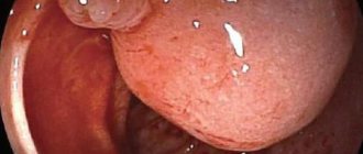
Diagram of a gastrogram of the stomach - an ulcer on the lesser curvature is indicated by an arrow
Sight radiograph after double contrast - small erosions are indicated by arrows
- Send your research data and receive qualified assistance from our specialists!
secondopinions.ru
What does ultrasound and FGDS allow you to see?
Ultrasound of the stomach: what does it show?
Ultrasound examination makes it possible to diagnose diseases such as:
- diaphragmatic hernia;
- gastroesophageal reflux;
- swelling of the stomach walls;
- acquired pyloric stenosis;
- malignant and benign tumors;
- dilation of gastric varicose veins;
- mesenchymal hereditary neoplasms;
- neoplastic thickening of the stomach walls, etc.
In rare cases, cystic formations can be detected. In children, ultrasound often reveals hypertrophic pyloric stenosis.
What does fibrogastroduodenoscopy allow you to see?
FGDS shows:
- the presence of scars and narrowing in the stomach;
- cancerous nodes;
- diverticula;
- polyps;
- changes in the lining of the stomach during gastritis.
FGDS also provides information:
- about the patency of the stomach and esophagus;
- determines the degenerative and inflammatory nature of the mucosa;
- gives an idea of the degree of reflux - backflow of contents.
Advantages of the method
Gastroscopy using swallowing a probe is an old and reliable way to examine the cavity of the stomach and duodenum for the presence of diseases. With this method, the endoscopist has the opportunity to visually examine cavity structures and determine the degree of tissue damage. Also, using an endoscope, you can perform a mini-operation to remove pathologies and take tissue samples for a biopsy.
Despite the versatility of the method, it is not suitable for every patient. What is the alternative, and what can replace the examination of the gastrointestinal tract with a probe? Modern medicine offers several diagnostic options:
- ultrasound examination;
- magnetic resonance imaging MRI;
- by X-ray irradiation.
What is better - ultrasound or FGDS of the stomach, magnetic resonance imaging or x-ray? What is the difference between the methods, which one is the best?
What is magnetic resonance imaging? This is a modern system for studying the internal structures of the body, with which you can get a detailed and detailed picture of the clinical condition of any organ. MRI diagnostics do not make mistakes, since the data obtained is processed by a computer program. An MRI study completely ruled out medical error.
However, this technique cannot replace gastroscopy in two cases:
- detection of helicobacteriosis;
- tissue sampling for biopsy.
The MRI method cannot characterize the condition of the mucous membrane, like a probing method. Also, this diagnosis is impossible if there are iron implants in the patient’s body - pacemakers, dentures, etc.
Gastric juice
Contraindications for ultrasound and FGDS
Ultrasound of the stomach has no contraindications as such. Relative contraindications may include skin damage in the area being examined (burns, open wounds, infections, etc.)
FGDS has a more expanded list of contraindications, namely:
- Tumors of the digestive system: stomach, duodenum, biliary tract, esophagus.
- Malformations of the digestive organs.
- Acute intestinal and gastric bleeding.
- Vomit.
- Intestinal obstruction.
- Foreign body in the intestines or stomach.
- Bowel perforation.
- Stroke.
- Heart failure stage 3.
- Central nervous system instability, extreme emotional sensitivity.
Which method should you choose in a particular case?
Due to the nature of the procedures, X-rays and FGDS have some contraindications. In the case of radiography, the main stumbling block is the relatively high ionizing radiation. It is for this reason that this diagnostic method is undesirable during pregnancy, as well as for children. In addition, this procedure should not be performed in case of through formations in the organ, since the ingress of contrast agents into them can cause complications.
Gastroscopy is considered a safer method, however, there are some contraindications, and their number is quite impressive. Absolute contraindications include heart failure, atherosclerosis, and aortic aneurysm.
Some structural features of the esophagus may also be a reason to refuse FGDS. There are also relative contraindications, i.e. indications for which special precautions must be taken. These include angina pectoris, diseases of the pharynx, larynx and esophagus, stomach ulcers, etc. That is why in each specific case a consultation with a gastroenterologist is necessary.
How are the examinations carried out?
Ultrasound
Ultrasound goes like this:
- It is carried out exclusively on an empty stomach.
- During the procedure, the patient lies on his back or takes a semi-sitting position.
- The doctor places the sensor on the area being examined so that an image of both the anterior and posterior walls of the stomach can be seen. In addition, the doctor needs to be able to set the parameters of the greater and lesser curvature. Normally, a small amount of fluid is found in the stomach cavity.
- A special gel is applied to the patient's skin to facilitate the sliding of the sensor and ensure good contact of the scanner with the surface.
- The doctor moves the sensor over the skin, pressing it at the required angle.
- The monitor displays an ultrasound image of the organ being examined.
Sometimes it becomes necessary to use contrast agents. The special product is diluted in 500 ml of water with gas. The patient drinks it in one gulp. Upon completion of the procedure, the patient is given a conclusion with the results of the study.
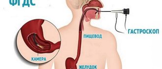
FGDS
FGDS goes like this:
- It involves the patient swallowing a special flexible hose - an endoscope. Its diameter is about 1 cm. The probe is equipped with a video camera, which, penetrating into the stomach, gives the specialist the opportunity to see the condition of the organ in its natural form.
- A few minutes before the examination, the patient is given local anesthesia - the throat is treated with ice caine. Sprays are usually used. In some cases, general anesthesia is used, however, side effects can have negative consequences, and therefore general anesthesia is used extremely rarely if the patient (usually a child) is particularly agitated.
- The patient lies on his left side. A towel is placed under the head to catch the saliva that will inevitably be released during the examination.
- The patient clamps a plastic ring resembling a mouthguard with his teeth, into the hole of which an endoscope is inserted to the root of the tongue.
- The person being examined is asked to swallow several times, as a result of which the probe moves into the stomach.
- After the endoscope has reached the stomach cavity, air is supplied to straighten the walls of the organ.
- The liquid is removed with an electric suction (gastric juice, bile, mucus). Next begins the examination of the mucous membrane of the stomach and duodenum.
- At the end of the procedure, the probe is carefully removed.
Tears and saliva are natural secretions during FGDS. These phenomena cannot be avoided, and they are normal and physiological. Therefore, you should not be embarrassed or worry about this.
Purpose and features
Magnetic resonance imaging is a method for diagnosing the gastrointestinal tract, which is based on the interaction of magnetic fields and radio frequency pulses with biological tissue. MRI is characterized by:
- high information content;
- painless implementation.
It is carried out using a tomograph, which is a large metal capsule with a computer attached to it; a three-dimensional image of the stomach is transmitted to the latter’s monitor. Based on the results of MRI, accurate images are obtained showing the area under study in an enlarged view and in projection.
Indications for ultrasound of the stomach
Ultrasound is prescribed for suspected diseases such as:
- gastritis;
- ulcer;
- stenosis of the pyloroduodenal type;
- abnormalities in the structure of the stomach;
- intestinal obstruction;
- oncology;
- frequent pain in the abdominal area of unknown origin;
- chronic heartburn;
- vomit;
- burping and other digestive problems.
Sometimes an ultrasound of the stomach is prescribed:
- patients with acute bronchial asthma;
- patients with coughing attacks;
- with severe regurgitation in children.
How is gastroscopy performed?
Gastroscopy refers to endoscopic examinations. It is carried out using a special device - a gastroscope. It is a long flexible probe, inside of which there is a fiber-optic system. In fact, the instrument is a soft, long tube with a camera.
During the procedure, a probe is inserted through the mouth into the esophagus, from where it enters the stomach. During the study, the surface of the organs is displayed on the monitor, and video and photographic recordings are made. If necessary, other procedures can be performed during gastroscopy:
- biopsy;
- pH-metry;
- removal of polyps;
- stopping bleeding;
- administration of necessary drugs.
In most cases, the patient does not need anesthesia, but some clinics offer temporary “freezing” of the throat. This is due to the unpleasant side effects of gastroscopy: unpleasant painful sensations occur in the throat, and the gag reflex is acute.
If additional procedures are not performed during gastroscopy, the procedure takes 1-3 minutes. Discomfort in the throat goes away after a few hours, and at first there are problems with speech.
Preparing for an ultrasound
The main point of preparation is the diet, which should be switched to 2 days before the examination. The diet is aimed at reducing gas formation.
It is necessary to exclude products that provoke flatulence:
- legumes;
- dairy products;
- rye products;
- fresh fruits and vegetables;
- carbonated drinks;
- alcohol.
The last meal should occur no later than 8 pm before the examination day. On the day of the ultrasound, you should not eat or drink anything. Smoking is also prohibited.
What common
The inspection methods described above are the most common in our time. With their help, complete visualization of the internal state of the body is achieved, after which it will be very easy for the doctor to make a diagnosis.
They allow you to see the following:
- Condition and relief of the mucous membrane.
- Presence of foreign objects or formations.
- The presence, location and degree of development of various deformations and destructions, such as ulcers or pre-ulcerative conditions.
- Amount of bile.
- Tendency to heartburn.
- Hemorrhages on the inner membrane.
Preparation for FGDS
Rules:
- Do not eat on the day of the test.
- The last meal should be 12 hours before the examination.
- It is advisable not to consume coffee, chocolate and alcohol for 2 days.
- You need to give up liquids 2 hours before.
- Smoking is not strictly prohibited, however, it can distort the results due to the fact that nicotine increases gastric secretion and constricts blood vessels. Therefore, it is still better not to smoke on the day of the study.
Possible risks
Side effects are very rare - in 2-4 patients out of a hundred. They are associated with the use of contrast, which sometimes causes an atypical reaction in the body.
In this case, the radiology office always has a set of antihistamines that will help take emergency measures. Another risk is radiation, which in some cases provokes the growth of malignant tumors.
However, the dosage used during CT is so small that it is not capable of harming health in any way. You just need to take into account that radiation can accumulate in the body, so certain intervals are taken between examinations or replaced with non-radiological techniques.
Conclusion
So what to choose, ultrasound of the stomach or FGDS? Both methods of examining the stomach have their pros and cons; the choice of method must be approached individually in each case. It should be noted that a contraindication for ultrasound of the stomach is the recent intrusion of an endoscope into it.
This means that if it is advisable to use both methods, you first need to undergo an ultrasound, and only then an FGDS. Particular attention should be paid to preparation for the examination, since up to 60% of the success of the procedure and the accuracy of the diagnosis depend on it.
What is the difference?
These are two completely different methods , which have their own characteristics in preparation, conduct of the procedure and the number of results collected.
Their main similarity is that X-rays of the stomach and FGDS help the doctor assess the general condition of the organ. FGDS has one big advantage - the ability to perform a biopsy. In addition, the doctor can install a clip or administer the necessary medication. It is not possible to carry out these manipulations when performing x-rays.
The next difference is the quality of the information received. Even the most modern X-ray machine is inferior to FGDS . The fact is that the image transmitted by a gastroscope is color and has very good quality, which increases the chance of seeing various damage to the stomach that an x-ray will not show. And the last disadvantage of x-rays is the radiation that the patient receives.
But radiography also has its advantages, namely the absence of damage that may occur during FGDS and complications after the procedure.
X-rays are not accompanied by unpleasant sensations and during this procedure no allergic reaction to an anesthetic develops, because it is not used. But an allergy may occur to the contrast agent.
Where can I get tested? Price
To undergo this type of diagnosis, you need to consult a gastroenterologist. This is a rather expensive procedure, and it is currently possible to carry it out in private clinics or regional hospitals.
The gastroenterologist will also recommend additional types of tests that this procedure involves.
This diagnostic method has a high price, but it varies in different cities and regions of Russia:
- In Moscow, such a procedure will cost from 15,000 to 70,000 rubles. It all depends on the clinic you visit.
- In St. Petersburg, its average price will be from 25,000 to 30,000 rubles.
- In Krasnodar, such a procedure will not exceed 22,000 rubles.
- An economical option can be considered the city of Minsk. This type of diagnosis will cost no more than 20,000 rubles.
Do they do MRI of the stomach and intestines?
Based on complaints and objective examination data, the gastroenterologist makes a preliminary diagnosis. To clarify the type of pathology and localization of the process, the specialist resorts to a number of auxiliary studies.
Depending on the level of damage, diagnostic methods for the stomach and intestines are prescribed, presented in the table.
Table 1. Diagnostic methods for assessing the gastrointestinal tract
Diagnostic option | Digestive Tract Imaging Department | Features of the technique |
| FGDS | Stomach, duodenum | Clarifies the condition of the mucous membrane, the presence of ulcerative defects, bleeding, indirect signs of malignant neoplasms |
| Ultrasonography | Pancreas, gallbladder, liver, spleen | Detects organ enlargement, structural changes, stones, space-occupying formations, functional changes, congenital defects |
| Colonoscopy | Colon | Detects changes in the mucous membrane, tumors, ulcerative disorders, bleeding, developmental abnormalities |
| Sigmoidoscopy | Rectum, distal sigmoid colon | Determines the presence of inflammation, dilation of hemorrhoidal veins, neoplasms |
| Irrigoscopy | Large intestine, partly small intestine | It is possible to determine indirect signs of tumors, ulcerative-necrotic changes, functional disorders |
| CT scan | All parts of the gastrointestinal tract | Visualizes malignant neoplasms, including metastases to regional lymph nodes and other organs, structural disorders of various departments |
An alternative diagnostic direction is the method of layer-by-layer imaging of the digestive system, which is not associated with x-ray radiation. Specialists perform MRI of the stomach and intestines, taking into account the safety and high information content of the examination.
Types of stomach radiography
An X-ray examination of the stomach involves the introduction of a contrast agent (barium suspension) into the organ. Without contrast, the information content of the study in this case will be practically zero. Depending on the characteristics of contrast, there are two types of radiography of the stomach:
- Radiography with conventional contrast.
- Double contrast radiography. Using this technique, both barium and air are injected into the stomach, which straightens the folds of the organ so that the doctor can more carefully examine the relief of the mucous membrane and evaluate the elasticity of the muscle layer.
X-ray images allow us to evaluate only the structure of the organ, which, of course, is not enough when studying the digestive tract. Therefore, a full X-ray examination of the stomach usually includes fluoroscopy (monitoring the progress of contrast through the esophagus, stomach, small intestine in real time) and radiography in different positions.
As a result, the specialist can judge how the organ under study functions and what changes there are in its morphology.
Computed tomography of the stomach
Before the advent of computed tomography, the main diagnostic tests for identifying various gastric diseases were endoscopy and x-rays. An endoscopic examination allows you to examine the mucous membrane of the esophagus, stomach, the beginning of the small intestine (examination of the upper part of the gastrointestinal tract), as well as the large intestine (examination of the lower part of the gastrointestinal tract). Examination of the stomach is carried out using gastroduodenoscopy, the following pathological conditions are detected:
- Gastritis.
- Gastroesophageal reflux disease.
- Portal hypertension.
- Stomach ulcer.
- Polyps.
- Cancer of the esophagus, stomach.
- Gluten intolerance.
- Anemia.
An endoscopic examination allows for therapeutic measures to be taken - to stop bleeding, remove foreign bodies, and also during the examination a biopsy of stomach tissue is performed.
X-ray of the stomach is a dynamic research method that allows you to take a picture of the stomach and select the desired projection of the organ. An X-ray with barium is also performed; the study allows you to observe the passage of contrast through the gastrointestinal tract using images. X-ray examination allows us to identify the following pathological conditions:
- Varicose veins of the esophagus and stomach walls.
- Polyps.
- Stomach ulcer.
- Malabsorption syndrome.
- Gastrointestinal motility disorder.
- Benign and malignant tumors of the gastrointestinal tract.
- Narrowing of the duodenum.
- Hiatal hernia.
Computed tomography is most often performed to detect stomach tumors. Indications for use include suspected ulcers, Crohn's disease, colitis, cancer and many other diseases. Computed tomography allows you to determine the presence of foreign bodies, injuries, polyps, and make a quick decision on whether to perform surgery or prescribe a specific treatment method.
- Indigestion.
- Heartburn.
- Nausea.
- Blood in urine or stool.
- Pain in the stomach after eating.
- Feeling of heaviness in the stomach after eating or having a small snack.
- Loss of appetite, loss of body weight.
- Digestive disorders.
CT with contrast is used to diagnose stomach diseases. Before a computed tomography scan with contrast, preparation is carried out. The patient undergoes tests for allergies to contrast agents, and kidney function is determined. CT scans are performed on an empty stomach; before the study, the patient abstains from eating for 6-7 hours.
A computed tomography scan of the stomach takes an average of 20 minutes and causes virtually no discomfort to the patient. Computed tomography very well allows you to see the walls of the cavity filled with gas or liquid. The contrast agent is administered intravenously or drunk by the patient. The patient is then placed on a rotating CT scanner table, which moves under the scanner.
The contrast agent is drunk by the patient in several stages, allowing soft tissues and blood vessels to be more clearly visible. Using contrast, tumors of the stomach and intestines are detected in the early stages of development - contrast highlights the smallest malignant and benign tumors in images of the circulatory system.
Computed tomography helps determine the location of the tumor, size, type, circulatory characteristics and other features of the tumor. CT helps to identify the onset of tumor metastasis, the boundaries of the lesion, and the stage of development of the tumor is very accurately determined. The doctor can see signs of perforation of the stomach walls, obtain accurate data on the condition of other abdominal organs, data on the functionality of the organs, and the peculiarities of the functioning of the organs.
Differences
These two methods, although used for the same purpose, have significant differences:
- There are no contraindications to ultrasound, unlike FGDS, which is strictly prohibited in some cases.
- You need to prepare much more carefully for the fibrogastroduodenoscopy procedure than for an ultrasound examination.
- Ultrasound does not bring any discomfort at all, which cannot be said about FGDS.
- Using an ultrasound procedure, you can examine not only the stomach, but also its circulatory system, in addition, you can examine other organs of the abdominal cavity.
- Fibrogastroduodenoscopy allows you to see a much larger number of pathologies and determine them much more accurately than ultrasound.