Pain, itching, discomfort that interfere with your usual lifestyle is a sufficient reason to visit a doctor. But why waste time and money on a medical examination if nothing hurts?
The reason is simple: in most cases, noticeable symptoms appear when the pathological process has gone far enough. First, cholesterol levels rise in a person’s blood, then cholesterol appears on the walls of blood vessels.
What is atherosclerosis? atherosclerotic plaques, hypertension develops and stroke occurs. A minor and imperceptible violation can cause dangerous consequences. It is possible to break the chain leading to lifelong dependence on drugs, disability and even death. It is enough to regularly examine the entire body.
A full medical examination is carried out to identify disturbances in the functioning of the body, even if they are asymptomatic. This diagnostic method is useful primarily for people who do not notice their health problems and want to make sure that there are no diseases.
What it is?
CT is a modern study based on x-rays , during which the scanned organs are displayed in images. Unlike a typical X-ray machine, a tomograph allows you to obtain more informative images, from which you can not only detect a pathological focus, but also identify its exact location and determine its size.
CT principle
Computed tomography capabilities
During scanning, X-rays pass through tissue and are captured by special sensors and transformed into an image. A computed tomograph is more complex than a conventional x-ray, which greatly expands its diagnostic capabilities.
Advantages of CT:
- Three-position scanning - this allows you to obtain detailed images of the pathological focus without “overlapping” the images on top of each other, as with an x-ray. When viewing in a special program, you can change angles along three axes, which cannot be done with ultrasound.
- Great diagnostic capabilities - CT produces layer-by-layer sections, from which it is easy to determine the size of the pathological focus. When working in the program, you can select the measurement option and obtain data on the width of the cavity or channel down to a hundredth of a millimeter.
- Possibility of studying hollow organs - the use of contrast allows you to obtain a clear image of soft tissues and blood vessels.
Safety of the procedure
CT scanning is often prescribed to diagnose abdominal diseases because this method:
- does not require special preparation;
- carried out quickly;
- painless;
- informative;
- accessible.
Help Unlike X-rays, CT scans expose the body to more radiation.
But single dosages are safe, and clear images are often enough to make a final diagnosis after a single examination. more about the harm caused to the body during a CT scan and the radiation dose received by a person from this article.
What's new? Modern tests that will tell you even more about your health
MICROBIOTA ANALYSIS
Our health largely depends on the species and quantitative composition of intestinal microflora. Friendly bacteria help absorb microelements and vitamins and synthesize useful substances, such as serotonin. Opportunistic bacteria, if they are in excess, can affect intestinal permeability, exacerbating allergic reactions. The composition of the microbiota largely determines the functioning of the immune system, the speed of metabolic processes, the state of the gastrointestinal tract, the appearance of the skin, general well-being and even mood. A complete picture of the microbiome can be obtained by studying the inhabitants of the small and large intestines. In the first case, blood is donated, in which the metabolic products of microorganisms are determined and conclusions are drawn about the composition of the microbiota. In the second case, feces are examined.
Price: gas chromatography-mass spectrometry method - from 5 thousand rubles, analysis of microbiota (feces) - from 1,200 rubles.
A number of experts recommend including ultrasound of the abdominal cavity (liver, pancreas, gallbladder, kidneys), thyroid gland, pelvic organs and mammary glands in the annual medical examination.
GENETIC ANALYSIS
The test is done once - once in a lifetime. Saliva or blood are suitable for testing. You can examine your entire genome and ultimately get a complete genetic portrait. But you can study only some genes that are responsible for certain processes in the body or a tendency to certain ailments.
A genetic test allows you to determine:
- Features of the metabolism of proteins, fats and carbohydrates. This allows you to choose the optimal diet. The need for vitamins and microelements, as well as the characteristics of their absorption. Knowing the characteristics of your genetics, it is easier to choose a preventive complex.
- The functioning of the detoxification system. Shows how much the body is able to neutralize harmful substances.
- Tendency to develop certain diseases. A suitable option for physical activity. The effects of medications on the body, their effectiveness, the likelihood of side effects.
Price: the cost of individual panels is from 5 thousand rubles, complex genetic analysis is from 15 thousand rubles.
5 vitamins that women usually lack by age 40
TESTS FOR FOOD INTOLERANCE
Difficulties with losing weight, problems with the gastrointestinal tract, chronic fatigue are often associated with the presence of food intolerance to a particular product. This condition is not a true allergy, but nevertheless provokes constant stress on the immune system. IgG, which perceives the remains of undigested food as “strangers,” is involved in the development of food intolerance. As a result, food is not digested properly, which leads to the development of a chronic inflammatory process both in the gastrointestinal tract and throughout the body. The manifestations of food intolerance are very vague. As a rule, its symptoms include: increased fatigue, gastrointestinal disturbances, skin rashes, stool disorders, and weight gain. These symptoms do not appear immediately. The most common food intolerances are dairy products, grains (wheat, rye, barley), peas, mushrooms, and strawberries. To diagnose food intolerance, blood is tested for antigens of foods that enter into an immune reaction. The test allows you to check from 20 to 300 products.
Price: from 18,000 rub. (definition of 95 antigens)
A number of experts recommend including ultrasound of the abdominal cavity (liver, pancreas, gallbladder, kidneys), thyroid gland, pelvic organs and mammary glands in the annual medical examination.
Natalya Grigorieva anti-age expert, nutritionist, general director of the aesthetic medicine clinic “Premium Aesthetics”
What internal organs are checked?
The images clearly show all the organs of the abdominal cavity:
- stomach;
- duodenum;
- jejunum and ileum;
- colon;
- liver, gall bladder and their ducts;
- pancreas and spleen.
The examination also includes organs of the retroperitoneal space - kidneys, adrenal glands, cellular spaces, vessels and nerves, and with advanced diagnostics, the gastrointestinal tract, chest and pelvic organs can be examined, which can provide additional information for making a diagnosis.
Differences between men and women
There are slight differences in abdominal scans depending on gender. During an extended examination, when the boundaries of the pelvis are captured, the following images are visualized:
- In men , the prostate gland and vas deferens. The urethra passes through the prostate, which can be compressed due to inflammation. When a contrast agent is introduced, developmental anomalies of these organs, tumors and the consequences of injuries are visible.
- In women - the uterus, fallopian tubes and ovaries. In this case, CT is an excellent method for the prevention of gynecological diseases.
The pelvic organs are always evaluated, since the symptoms of pathologies of the reproductive system are often similar to intestinal diseases. This is an important step in differential diagnosis.
How is the diagnosis carried out?
The medical examination plan is established by the therapist according to the patient’s age and gender, as well as based on a survey about well-being and complaints. After receiving all the results of tests and appointments with specialized specialists, the patient undergoes a second consultation with a general practitioner, who makes diagnoses and writes a final report. The conclusion is accompanied by a list of recommendations for improving health or, if deviations are identified, a treatment regimen is described if the prescriptions have not previously been prescribed by a cardiologist, neurologist, ophthalmologist or other specialists.
Where does the research begin, and is preliminary preparation necessary?
Clinical examination begins with a preventive examination by a therapist. The doctor conducts a questionnaire, examines the patient - his skin, mucous membranes of the lips and mouth, palpates the thyroid gland and lymph nodes. He finds out the patient’s complaints, the fact of aggravated heredity, asks questions about the presence of symptoms characteristic of diseases of the cardiovascular, bronchopulmonary, and digestive systems. Notes are made about bad habits, sports, and diet.
The therapist determines the body mass index based on height, weight, and waist circumference. The doctor measures blood pressure, writes out directions for laboratory tests and fluorography of the lungs.
Next, the patient is sent to specialized doctors and undergoes hardware-type tests.
The procedure for preparing for ultrasound examinations depends on the area being examined.
- When preparing for an ultrasound of the abdominal organs, it is necessary to abstain from food several hours before the procedure. A few days before the appointed date, you should exclude gas-forming foods, including flour, from your diet. In case of severe flatulence, carminative medications should be taken. Gastro- or colonoscopy cannot be performed on the day of the procedure.
- To improve visualization when examining the organs of the urinary system, it is necessary to fill the bladder as much as possible.
- Before an ultrasound of the gallbladder, the day before the procedure, exclude flour, dairy products, fresh fruits and vegetables. It is important to exclude gas-forming foods, namely legumes, carbonated drinks, and sweets.
- Preparation for transrectal ultrasound of the prostate gland involves using a cleansing enema the day before or on the day of the procedure.
Preparing for an x-ray of the peritoneum involves avoiding heavy foods on the eve of the procedure. In the morning, the procedure is done on an empty stomach. It is strictly forbidden to smoke and take medications on this day.
Preparation for endoscopic examinations goes like this.
- Esophagogastroduodenoscopy. On the eve of the procedure, it is necessary to avoid heavy meals and limit yourself to a light snack until 7 pm.
- Colonoscopy. For three days, you need to eliminate plant fiber from your diet. Activated carbon and preparations containing iron should not be used. The evening before the study, you must start taking the laxatives recommended for this procedure.
- Bronchoscopy. If the patient has chronic diseases, allergies or intolerance to any drugs, it is necessary to inform the attending physician about this in advance. Smoking is strictly prohibited before the study. Bronchoscopy is performed on an empty stomach (the last meal should take place on the day before the procedure, no later than 19:00). You can drink, but no later than an hour before the procedure.
How long does the diagnosis take?
Medical diagnosis begins with making an appointment with a therapist. The duration of the entire set of diagnostic procedures is usually one day (if no deviations are detected).
What does the patient receive after completing all procedures?
After completing the Check-up, the patient receives a document called a “health passport.” This is a detailed report containing indicators of all analyzes and expert opinions.
Indications
Your doctor may order a CT scan if you have the following symptoms:
- abdominal or epigastric pain;
- flatulence;
- increase in abdominal size;
- prolonged absence of stool;
- change in color of stool;
- rapid weight loss ;
- nausea and vomiting;
- constant belching “rotten”;
- change in skin color.
Before scanning, the patient undergoes blood and stool tests, ultrasound or endoscopy. CT is prescribed when the information content of these methods is weak or to clarify the diagnosis.
Where to begin
Today, the choice of foreign clinics where you can undergo an examination of the body is quite large. It can be difficult to choose on your own. We will try to answer the question: how to undergo a complete examination of the body?
First, you should decide whether a full comprehensive examination of the body in a hospital is required, assess your health status, whether there are already identified serious pathologies, the assessment of which requires certain equipment or qualified medical personnel. If there is, then the choice should be limited to more specialized clinics, or undergo a full examination of the body in a sanatorium.
If there are no specific health complaints, then you can plan the examination in such a way as to combine it with a business trip or vacation in a certain country.
Well, if you can’t decide yet, then contact travel agencies, study the level of service, prices, and then choose a clinic.
Having a medical record will allow the doctor to track the dynamics of your condition.
Today you can reserve a place in many clinics by phone or directly on their websites. Also, a travel company can take care of the entire organization of your survey, including accommodation and entertainment.
Do not forget to prepare your medical record, because it may contain valuable information for the doctor about previous illnesses, test results or examinations. This will allow you to track some of the dynamics of your condition.
What does it show?
Computed tomography helps to recognize any changes in the abdominal organs and retroperitoneal space - inflammation, bleeding, damage to their walls and blood vessels. When scanning, a clear display of the boundaries and structure of anatomical units in the studied area is obtained.
A detailed list of organs and possible pathologies that the tomograph will show is shown in the table below:
| Organ | Possible pathologies |
| Stomach | Ulcer, gastritis , perforation, bleeding, foreign bodies, abscesses, tumors. |
| Intestines | Adhesions, fistulas, developmental abnormalities, ulcerative lesions, bleeding, suppuration, polyps , foreign bodies, oncology. |
| Liver | Cirrhosis, hepatitis , parasites, abscesses, anomalies, duct blockage, hypo- and hypertrophy. |
| Gallbladder and ducts | Sediment, stones, dyskinesia , the presence of parasites, perforation and oncology. |
| Spleen | Damage to the parenchyma, changes in size in systemic diseases. |
| Kidneys | The presence of stones , signs of infection by changes in the renal pelvis. |
| Adrenal glands | Changes in size due to endocrine diseases. |
| Vessels and nerves | Damage, developmental abnormalities, atherosclerosis in the arteries and thrombosis in the veins. |
Please note CT is a valuable method in diagnosing gastric ulcers . To increase information content, the use of contrast , thanks to which perforation can be determined and peritonitis can be prevented .
What blood tests should I take to check my health?
Blood circulates throughout the body, transporting necessary substances from one organ system to another. That is why blood is the best indicator of the body’s condition during a medical check-up examination. A general blood test allows you to determine the level of hemoglobin and the number of blood cells (erythrocytes, leukocytes, platelets). A deviation in the number of blood cells from the norm indicates to the doctor the nature of the disease and further direction of diagnosis.
A biochemical blood test gives a more detailed picture of the state of various body systems:
- measuring glucose levels allows you to assess the risk of developing diabetes;
- deviations from the norm in the level of urea and creatinine indicate disturbances in the functioning of the kidneys;
- measuring cholesterol helps predict the risk of developing atherosclerosis and heart problems;
- the level of bilirubin indicates the condition of the bile ducts, liver and gallbladder;
- the level of total protein content may indicate nutritional disorders, digestive problems, liver disease;
- A pathological change in the blood level of albumin, the main plasma protein, is characteristic of malnutrition, chronic renal pathologies, and diseases of the digestive tract.
Also, in a biochemical blood test, the concentrations of certain types of enzymes are checked - complex proteins responsible for the synthesis and breakdown of substances necessary for the body. Knowing the functions of enzymes and their physiological norm in the blood, doctors can determine in which organ the dysfunction has occurred:
- pathological levels of alkaline phosphatase in the blood indicate bone diseases (including cancer), disruption of hormonal secretion of the thyroid and parathyroid glands;
- an increase in the level of AST (aspartate aminotransferase) in the bloodstream indicates the destruction of myocardial cells or hepatocytes and dysfunction of the heart and liver;
- deviation from the normal level of the ALT enzyme (alanine aminotransferase) in the blood is characteristic of liver and kidney diseases;
- An abnormal level of GGTP (gamma-glutamyl transpeptidase) indicates damage to the liver, pancreas, thyroid or prostate glands.
General and biochemical blood tests during a comprehensive “Check-up” examination are sufficient to detect almost all major disorders in the functioning of the body. Comparing various indicators allows you to determine which organ has failed and begin to look for its cause. In some cases, the therapist may prescribe additional tests to measure the level of thyroid or other hormones (if there are risk factors), antibody tests to diagnose helminthiases, etc.
Decoding the results
After the scan, the patient must wait 40-60 minutes to receive the examination results. The decoding is carried out by a radiologist and in the conclusion indicates the location and size of organs, features of their structure. When pathological changes are detected, the doctor characterizes them in detail, notes their location and makes a full description. If there is a suspicion of oncology , the specialist will definitely mark the boundaries of the neoplasm , describe the features of the parenchyma and the nature of vascular growth.
You will be given a description, photographs on film, flash media (disk) . Copies must be shown to the attending physician, since the doctor may order an additional consultation with a radiologist or transfer the patient to another institution where a re-analysis of the scan will be required.
What is included in a comprehensive examination of the body?
The advantage of a comprehensive “Check-up” examination over a regular medical examination is the understanding that a potentially healthy person does not need to visit all doctors to detect health problems. To identify pathology, it is enough to pass a basic set of tests to check the functions of the liver, kidneys, pancreas and thyroid glands, do an electrocardiogram, and undergo an ultrasound examination of the digestive and genitourinary systems.
The results of the body check-up do not allow a final diagnosis to be made, but are the basis for further additional research if deviations from the norm are detected and the patient is referred for treatment to a specific specialist. Examining the body using the Check-up service is recommended for early diagnosis and prevention of complications:
How to increase hemoglobin in a child? anemia;- hypertension;
- cervical or breast cancer in women;
- adenoma or prostate cancer in men;
- diabetes mellitus;
- hyperfunction of the parathyroid glands;
Can hypothyroidism be cured? hypothyroidism;- a number of viral (hepatitis, HIV), venereal (syphilis, chlamydia, mycoplasmosis, etc.) and parasitic (opisthorchiasis, echinococcosis, anisakidosis, etc.) diseases.
Early diagnosis and initiation of treatment for hypertension significantly reduces the risk of complications and disability, reduces the need for hospitalization, and increases life expectancy. According to the results of medical studies, the likelihood of stroke in patients with diagnosed asymptomatic hypertension who receive the necessary treatment is reduced by 25%, coronary insufficiency by 19%, and other heart diseases by 17%.
Statistics among breast cancer patients also show a significant increase in survival rate for women who were diagnosed with the disease at the first stage (85%) compared to patients who sought help at the second or more stages of cancer (53%). For breast screening examinations in young women, safe ultrasound is now used, while mammography is prescribed only if there are special indications.
Early recognition of cervical cancer and treatment reduces the risk of further tumor growth. The number of women with tumors that have spread beyond the cervix among those who underwent recommended medical examinations is 6 times lower than among patients who did not undergo preventive examinations.
Studying the amount of sugar in the blood, hormone levels, and enzyme activity makes it possible to notice disturbances in the activity of various organ systems even before the first symptoms appear. At the initial stages, many dangerous diseases can be prevented by giving up harmful habits, adjusting the diet and normalizing the level of physical activity.
Contraindications
A detailed list of restrictions on the use of abdominal CT is given in the table:
| Contraindications for adults | Contraindications for children |
| · pregnancy and lactation in women; · overweight; · mental disorders; allergy to contrast; · chronic diseases that can be aggravated by contrast. | · age up to 5 years; hyperactivity or mental disorders; Contrast intolerance or systemic diseases that react to it; · severe obesity. |
Behavioral or mental disorders are a relative contraindication; if necessary, scanning can be performed under medicated sleep conditions.
Indications for examination
A complete medical health diagnosis is an effective preventive measure. Medical examination is necessary for people who want to make sure that there are no diseases and who lack time for a more in-depth examination. Moreover, the method is necessary for those who have health complaints or for a long time will not be able to see a doctor for any reason.
Are there any contraindications
There are no contraindications for undergoing a comprehensive medical examination. There are restrictions on prescribing individual procedures, but the doctor will select alternative methods.
Kinds
When examining the abdominal cavity, three types of scans can be performed - survey, native and enhanced CT. When choosing a method, the doctor is guided by the characteristics of the suspected disease and the localization of the pathological focus.
Survey
During a general CT scan the entire abdominal cavity is scanned - the intestines, organs of the biliary system, blood vessels and nerves. This diagnosis is prescribed for general symptoms when the suspected disease is unknown.
The doctor studies all anatomical structures, pays attention to their location, boundaries, shape and condition of soft tissues.
With a survey scan, it is possible to detect damage to the abdominal part of the aorta and its branches - atherosclerosis, thrombosis, embolism, ruptures due to trauma.
Native
Targeted CT is prescribed to clarify the diagnosis - this is a scan of one area or organ . The study produces layer-by-layer images in a three-dimensional projection, from which the exact location and size of the pathological focus can be determined. The images also show the topography of the organ and the condition of its membranes.
With gain
This is the use of a contrast agent that stains the soft tissue or walls of hollow organs, as a result of which they are accurately visualized in photographs.
Contrast can be entered:
- orally - “through the mouth” when examining the intestines;
- intravenously - when scanning other organs.
The doctor prescribes enhancement for the examination of soft tissues, which are poorly displayed during a regular scan. Diagnostics is also appropriate when identifying tumors - cancer tends to accumulate such substances.
How to check the entire body at once?
To check the entire body, a person has to go to many doctors’ offices and undergo a whole list of tests. Visits to doctors, instrumental and laboratory diagnostics take time and financial resources and are not always justified.
Unlike outdated medical examinations, a comprehensive Check-up examination involves passing a basic list of diagnostic procedures to identify the most common and life-threatening diseases. The doctor determines how to check the entire body, taking into account the gender, age, and family history of the patients. The results of tests and instrumental diagnostics obtained as a result of the examination are assessed by the therapist. After this, he gives an opinion on the state of the human body, a list of recommendations for maintaining health, or a referral to a specialist to clarify the diagnosis and prescribe treatment if the research results deviate from the norm. A well-thought-out scheme for taking tests and undergoing diagnostic procedures allows you to undergo an examination of the body in 1 day.
Preparation
Before an enhanced examination, the patient undergoes a compatibility test - usually a contrast agent is placed under the skin and the body’s reaction is assessed. If there is no allergy, a CT scan is prescribed.
Preparation for the study includes:
- within three days, products that contribute to gas formation are excluded;
- Adsorbents are taken the day before the scan;
- It is recommended not to eat in the morning; you can drink unsweetened tea.
Contraindications to the procedure
Complex tomography of the whole body, like its other types, has a standard set of contraindications. Thus, magnetic resonance imaging scanners do not test people with implanted medical devices:
- hearing aids;
- neurostimulants;
- insulin pumps;
- pacemakers.
It is not recommended to look at an MRI of the entire body in patients with installed metal crowns, fixed dentures, plates, staples and screws that hold skeletal bones. Patients recently operated on in whom metal staples were used to fasten soft tissues (for example, after surgery for hemorrhoids using the Longo method) are not studied using a magnetic field.
How do they do it?
The study is carried out on a tomograph - the patient is placed on a platform that extends into the tunnel of the device. The doctor then leaves the office and the scan begins.
X-rays pass through the body of the subject, which are captured by sensors and converted into an image. Communication with the patient is carried out through an intercom.
The procedure is completely painless. There can only be two things that make you feel uncomfortable:
- Closed spaces scare you . Try to concentrate on the tips of your toes, feeling each one individually. Then gradually work your way up your body. Try to feel your heels. Then the whole feet. Then the shins. Are they cold? Do they touch the surface? This meditation will allow you to escape from the feeling of being confined in space. And by the time you reach the sensations of the scalp, the examination will already be over.
- The tomograph makes a very loud sound , which is completely tolerable for an adult (in the absence of a headache), but can frighten a child. Therefore, you should prepare the child in advance and explain that there is nothing dangerous or frightening behind this sound.
How often can the study be done?
CT is a harmful method that causes radiation, and when scanning the abdominal cavity, many vital organs fall into the affected area. Therefore, you can be examined at certain intervals so as not to provoke complications. The optimal period is no more than once every 4 months .
How long does the procedure take?
The procedure itself takes 5-7 minutes, with contrast it takes a little longer. This time is enough to scan the abdominal cavity and obtain detailed images.
When can I expect results?
The time to obtain a report and images is about 20-30 minutes. During this period, the doctor looks at the images and writes conclusions. If the patient ordered a disc recording, the wait may be a little longer.
How often should I undergo a full body diagnostic?
The recommended frequency of body check-ups depends on a person’s age, gender, lifestyle, past pathologies, and hereditary predisposition to life-threatening diseases.
Women under the age of 40 should have their reproductive system examined at least once every three years, have a smear from the cervix, and check the condition of the mammary glands (self-examination to detect lumps should be carried out every month after the end of menstruation). After reaching the age of 40, the chances of developing gynecological diseases and breast cancer increase significantly, so do not forget about annual screening.
Men's bodies are less subject to hormonal changes as they age, so fewer visits to the doctor are acceptable for them. Men under 40 years old need to have their whole body checked at least once every five years, until they reach the age of 55 - once every 2-3 years, and then annually.
WHO recommends that all patients undergo a preventive medical examination annually, including those who feel healthy or have only general complaints about their health (fatigue, drowsiness, apathy, etc.), to prevent the development and early diagnosis of diseases, when their treatment is most effective, and The prognosis for recovery is favorable.
Photo
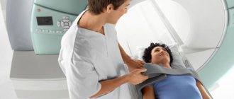
Fig.1. Tomograph image
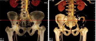
Fig 2. CT image of the abdominal area
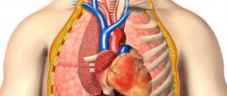
Fig 3. What the doctor can see in the cross-section
What deviations are detected
CT is necessary to detect pathological processes both in the initial and advanced stages of treatment. When diagnosing, you can identify:
- kidney stones and their number;
- malignant formation and probable metastasis;
- signs of thrombosis;
- foreign bodies in the abdominal cavity;
- purulent formations;
CT scan can detect the presence of kidney stones
- hepatitis;
- cirrhosis;
- damage to the lymph nodes;
- congenital pathologies;
- cysts;
- blood diseases;
- gallstones.
Computed tomography is a universal procedure due to the high detail of the resulting images.
Alternatives
If it is not possible to perform an abdominal CT scan, the doctor may prescribe:
- Ultrasound is a cheaper examination; scanning produces images with a two-dimensional image. It shows pathologies of hollow organs well, but during analysis you can “overlook” some injuries, perforations, and bleeding.
- MRI uses magnetic waves to produce three-dimensional images. Such a study is informative, is not inferior to CT in price, but is longer and requires mandatory immobility.
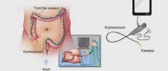
- Endoscopy is known among patients as “swallowing the lightbulb.” During diagnosis, a device with a camera is used, which can be used to study the condition of the mucous membrane of the stomach and duodenum. The lower parts of the large intestine are examined with a colonoscope.
chooses MRI as an . Our separate article will tell you about the differences between the two procedures and help you choose what suits you best.
Comprehensive programs
The all-Russian public organization “League of Nations” has been conducting comprehensive health screening programs in Russian cities for several years now. True, such events are temporary, and it is worth carefully monitoring information about where and when they will take place. But at the same time, during them you can completely check your health, since the programs include such actions and projects as “Check your heart”, “Check your spine”, “Check your cholesterol levels”, “Check your hearing”, “Rinse” the nose is a barrier to viruses”, “Mobile”, “Diabetes: time to act”, etc. They all take place within the framework of one comprehensive program.
Anyone can take part in the survey.
Article on the topic
“Express” diagnosis: what tests are done at Health Centers
Where to get tested
Generally recognized, trustworthy medical clinics and diagnostic centers are primarily European medical institutions. Switzerland, Germany, France, a complete examination of the body in Israel - these are the most popular places for medical tourism.
But today, the same medical clinics equipped with the latest technology have appeared in Korea, Thailand and other countries. In order not to go blind, you can go through the websites of these organizations; many of them have Russian-language versions of their websites or provide information on Russian-language medical portals.
