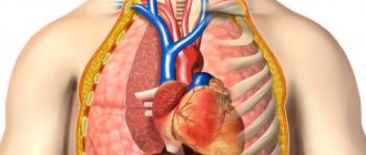
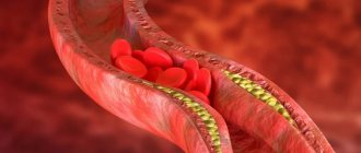
Tumor markers are special substances that are detected in blood or urine tests in patients with malignant neoplasms. At the beginning of the study of this area, it was assumed that tumor markers would be used for early diagnosis and screening of cancer.
In reality, everything turned out to be much more complicated, for example, an increase in the level of many tumor markers is not associated with malignant processes, and, moreover, can manifest itself normally in healthy people. It was also found that in many patients with an already established diagnosis of malignant neoplasm, the level of tumor markers remains normal or increases slightly.
To date, the determination of tumor markers for early diagnosis is used for a limited number of nosologies. They are mainly used for additional diagnostics and dynamic monitoring of the effectiveness of treatment and disease progression.
- What tumor markers are used to diagnose stomach cancer?
- What do tumor markers show for stomach cancer?
- Indications for testing for gastric tumor markers
- Preparation for analysis for tumor markers of stomach cancer
- Reliability and validity of tumor marker analysis results for gastric cancer
- Interpretation of tumor marker results in gastric cancer
- Causes of stomach cancer
What tumor markers are used to diagnose stomach cancer?
CA-19-9
This marker is produced by epithelial cells of the gastrointestinal tract. Accordingly, in tumors originating from these cells, the level of CA19-9 increases significantly. The indicators change most with pancreatic cancer, but can also increase with cancer of the colon, liver, bile ducts and gallbladder, pancreatitis, and cystic fibrosis. Thus, given the low sensitivity and specificity, this indicator is not used for self-diagnosis and monitoring.
REA
CEA (carcinoembryonic antigen) is a protein, a tissue marker of oncopathology. It is used in the diagnosis of colon tumors, but can also be effective in stomach cancer. Normally, its levels are extremely low, but in the presence of a malignant neoplasm, its level increases sharply.
SA 72-4
CA 72-4 is a glycoprotein that is located on the surface of the epithelium of the gastrointestinal tract during human intrauterine development. In adults, it appears with colorectal cancer, stomach cancer and other malignant tumors. However, in 6.7% of patients, its increase was also detected in the presence of benign tumors.
Sensitivity for stomach cancer is 40-46%, and the higher the tumor marker, the more widespread the malignant process, i.e. there is a correlation with stage. After radical removal of cancer, the CA value of 72-4 returns to normal within 3-4 weeks. As for metastases, this tumor marker for stomach cancer is more sensitive than CEA or CA 19-9. The determination of CA 72-4 is carried out to assess the chances of survival in patients with an established diagnosis of gastric carcinoma. The higher the tumor marker, the higher the stage of the disease.
Gastrointestinal tumor markers
Recently, there has been a persistent trend towards an increase in the number of cases of primary diagnosis of gastrointestinal cancer, with an increasing share of young patients.
In light of this, many, alas, know the answer to the question: “what are tumor markers?” Based on these sad statistics, timely, highly effective diagnosis becomes of great importance. These requirements are fully met by the analysis of serum blood for the content of tumor markers.
The numerous markers of the gastrointestinal tract determine the advisability of conducting one or more tests, based on family history, age, symptoms, laboratory data and other indicators.
What do tumor markers show for stomach cancer?
In general, tests for tumor markers for stomach cancer are not highly sensitive or specific. This means that normal results do not guarantee the absence of a tumor, and elevated results do not mean the presence of cancer. However, when the tumor marker level increases tenfold, the likelihood of this increases.
The diagnostic value of the study of tumor markers increases with the simultaneous study of several indicators, for example, CA 19-9 and CEA, or CA 72-4 and CEA. But they still cannot be used for self-diagnosis of stomach cancer.
SA 19-9
Reference values for the CA19-9 tumor marker are in the range of 0-34 U/ml. Such indicators are observed in 95% of healthy people.
An increase in tumor marker levels may indicate the following diseases:
- Pancreas cancer. The higher the level of CA 19-9, the more widespread the pathological process. The highest concentration is observed in the presence of distant metastases, i.e. at the 4th stage of the disease.
- Stomach cancer.
- Colon cancer.
- Liver carcinoma.
- Cancer of the bile ducts and gallbladder.
- Ovarian cancer.
In addition, an increase in the level of this tumor marker is observed in some benign diseases:
- Hepatitis.
- Cirrhosis of the liver.
- Pancreatitis.
- Cholelithiasis.
- Cystic fibrosis.
REA
The REA reference values are as follows:
- For non-smokers below 3.8 ng/ml.
- For smokers - 0-5.5 ng/ml.
These are normal levels of CEA, characteristic of healthy people, but this result is also possible in the presence of a tumor that is insensitive to this test.
An increase in the level of this tumor marker can be observed in both malignant and benign diseases. Malignant tumors:
- Colon cancer.
- Stomach cancer.
- Lungs' cancer.
- Breast and pancreatic cancer.
Non-oncological diseases:
- Cirrhosis of the liver.
- Hepatitis.
- Intestinal polyps.
- Chronic inflammatory bowel diseases, such as ulcerative colitis.
- Some lung diseases, including tuberculosis.
- Autoimmune pathologies.
SA 72-4
Reference values for tumor marker CA 72-4 0-6.9 U/ml. A normal analysis indicator is typical for healthy people; an increase in its value is observed in the following cases:
- Stomach cancer.
- Some forms of ovarian cancer.
- Colorectal cancer.
- Lungs' cancer.
- Hepatitis.
- Cirrhosis of the liver.
- Ovarian cyst.
- Inflammation of the gastrointestinal tract.
CT, MRI
These diagnostic methods are used quite rarely, which is directly dependent on their high cost and the lack of necessary equipment in many clinics. But in all complex cases, when there are inaccuracies in the results of the studies performed, which do not allow the leading oncologist to select an adequate treatment for esophageal cancer, or the clinical picture of the tumor process occurring in the esophageal canal is blurred, the patient is recommended to undergo computed tomography or magnetic resonance imaging. The most relevant method in each specific case will be selected by the attending physician, taking into account the individual characteristics of the tumor and the general condition of the cancer patient.
The specific need for their use comes down to the following:
MRI (magnetic resonance imaging). Thanks to it, an experienced oncologist can easily determine the location and size of a malignant tumor structure, the degree of prevalence of the abnormal process, as well as disturbances in the structure of the tissues that make up the internal organs, indicating the appearance of metastases in them.
CT allows you to determine at what stage of development, primary or secondary, a malignant neoplasm is located, assess the metastatic essence of the pathological process and analyze the condition of regional lymph nodes.
Important! The decision to use one or another diagnostic method should be made directly by a qualified specialist who can take into account all the nuances of a particular situation. It is the attending physician who is guaranteed to identify any contraindications that a cancer patient has for undergoing an examination using a certain method.
Reliability and validity of tumor marker analysis results for gastric cancer
The reliability and validity of the results will depend on the following aspects:
- Features of collecting material and manipulating it before delivery to the laboratory.
- Diagnostic systems and equipment used for analysis.
- The presence of concomitant diseases that can lead to an increase in the concentration of the tumor markers being studied.
To obtain reliable results, it is recommended to take tests in accredited laboratories that have established pre-analytical and analytical stages of research, as well as internal and external quality control.
What can distort the result
Sometimes it happens that the data obtained may be somewhat distorted. Most often, this happens when all recommendations regarding preparation for a blood or feces test are not followed.
Also, unreliable data may occur if a specialist makes mistakes during the examination of the obtained biomaterial.
In addition, a false positive result may occur when taking the test at the time of development of acute inflammation or in chronic pathologies at the acute stage. Antigens can increase with diseases such as flu, bronchitis or dental problems.
Interpretation of tumor marker results in gastric cancer
A doctor who has experience in diagnosing and treating stomach cancer should prescribe and interpret the results of tumor marker tests. Interpretation is carried out on the basis of examination data, anamnesis and clinical picture. The isolated use of tumor markers for screening or detection of gastric cancer is unacceptable.
Normal values of stomach tumor markers do not guarantee the absence of malignant tumors. Such results can occur in early stages of cancer, when the level of the marker has not yet increased, or there are tumors that do not lead to an increase in its level at all.
Increased results also require additional examination, since they may indicate the presence of a tumor of another location or a non-oncological disease. In doubtful cases, testing for tumor markers of stomach cancer is prescribed over time. An increase in its level indicates the presence of a progressive disease, a decrease in the level of the marker indicates recovery.
Initially, very high levels of tumor markers, especially if their value is exceeded several times, with a high degree of probability indicates the presence of a malignant process, but does not guarantee this 100%, and even more so does not allow one to determine the type of neoplasm.
The most informative value for gastric cancer is the dynamic study of tumor markers. A decrease in their level indicates that the treatment was effectively carried out; normalization of indicators is noted after a radical operation.
If after treatment the tumor marker level begins to increase, this indicates either a relapse of the disease or its progression. If the indicators increase tenfold, this may indicate distant metastases.
Decoding the results
The interpretation of the obtained data should be carried out exclusively by a highly qualified specialist.
Antigens can enter the biological fluid of the human body from healthy or mutated cells. Changes in a specific type of protein will indicate cancer.
Thus, when deciphering the analysis, doctors use the following meanings:
- + – additional tumor marker;
- ++ – significance is of medium degree;
- +++ – antigen is the main one.
Causes of stomach cancer
How and why stomach cancer develops is not completely clear. But the influence of certain factors has been proven in which the likelihood of a tumor increases. These include:
- Chronic atrophic gastritis, especially against the background of hyperplastic processes of the epithelium. This disease was found in 60% of patients with stomach cancer. In this case, the localization of the process was important. For example, when atrophy is located in the antrum of the stomach, the risk of developing cancer increases 18 times, and with total damage to the organ - 90 times.
- Helicobacter Pylory infection. The likelihood of developing stomach cancer in such patients is 3-4 times higher than in the general population.
- Nutritional features. Containing a large amount of hot, spicy, pickled, salty foods, fast food, smoked meats and fats in the diet statistically significantly increases the likelihood of developing tumors of the gastrointestinal tract, including the stomach.
- Presence of adenomatous polyps. Such neoplasms have a relatively high risk of malignancy, so they are recommended to be removed in a timely manner.
- Smoking.
- History of gastric ulcer. When an ulcer is located in the body of the stomach, the risk of developing a malignant neoplasm increases by 2 times.
- History of gastric surgery. Increases the risk of developing cancer by 4 times.
- Menetrier's disease, which is characterized by hypertrophic gastropathy or hyperplastic giant-fold gastritis.
- Drunkenness and alcoholism.
- Pernicious anemia. Anemia with a malignant course, which develops due to the inability to absorb vitamin B 12. It is also accompanied by immunodeficiency conditions, which increases the risk of developing a malignant tumor by 10%.
- Hereditary predisposition. The presence of malignant stomach tumors in close blood relatives increases the risk of developing cancer in this location by 5-20%. It is believed that Napoleon Bonaparte and his father died from this disease.
- Immunodeficiency states.
- Working with carcinogenic substances.
Book a consultation 24 hours a day
+7+7+78
Leading clinics in Israel
Assuta
Israel, Tel Aviv
Ikhilov
Israel, Tel Aviv
Hadassah
Israel, Jerusalem
Science now knows more than 200 types of gastrointestinal tumor markers, but only 20-30 of them are of practical importance for medicine, which have proven effective in detecting early cancer.
Each type of tumor marker corresponds to one type of malignant neoplasm. For example, a diagnosis such as intestinal cancer is more often made to people over 50 years of age, so during medical examinations, determining the appropriate tumor marker is mandatory. The question of which tests to take in order to obtain more reliable information is entirely within the competence of the doctor.
Symptoms of esophageal cancer
The most important symptom of the disease is a violation of the passage of food through the esophagus, the so-called dysphagia, which is caused by a narrowing of the lumen of the esophagus by a growing tumor. As a rule, it increases gradually. First, problems appear with swallowing solid foods, then liquid ones. Sometimes, as a result of spontaneous destruction of the tumor, the patency of the esophagus is partially restored and the passage of food improves, but this is again followed by deterioration. Esophageal vomiting and regurgitation of food just eaten appears. The early stages are characterized by belching, nausea, and heartburn. As a protective reflex, increased salivation may begin. A putrid odor appears from the mouth.
A number of symptoms indicate complications of esophageal cancer: changes in the timbre and sonority of the voice, coughing fits, whistling breathing, constriction of the pupils, drooping eyelids, sunken eyeballs.
Constant companions of the disease are complaints of general weakness, fatigue, and loss of body weight.
What are tumor markers and why do they need to be taken?
Answering this question, experts note that tumor markers are specific substances of various natures, proteins with a lipid or carbohydrate complex, hormones, enzymes that begin to be intensively produced when malignant tumors appear in the body. More than 20 types of biomolecules are used in clinical practice.
Most of them have low specificity, for example, many patients with problems of the digestive organs believe that the CA 19-9 antigen or ACE are tumor markers for cancer of the stomach, rectum or sigmoid colon, although this is not entirely true. Any marker can appear in the plasma in increased quantities during the onset of a malignant process in various systems of the body, therefore gastroenterologists do not recommend undergoing such a study on their own and prescribe it only in a few cases:
- as a regular diagnostic screening method for people at risk and patients with a history of precancerous diseases;
- after alarming symptoms appear that may indicate the onset of a malignant process;
- to monitor therapeutic measures for the purpose of timely course correction;
- after treatment to predict possible relapse.
Important! Only in these cases, passing a test to identify specific tumor markers is justified and can help to timely diagnose the onset of development or activation of a malignant process.
Indications for testing
The need for such research does not always arise. According to experts, these specific proteins serve only to confirm or refute the information available to the attending physician, and can also indirectly indicate the development of a malignant process.
The main indications for undergoing this study are:
- the appearance of suspicious symptoms in a person, indirectly indicating the emergence of a cancerous tumor in any part of the gastrointestinal tract;
- monitoring the effectiveness of radical surgery or drug antitumor therapy;
- the need to obtain prognostic information about the course of the malignant process;
- annual screening of patients with precancerous diseases.
Treatment of esophageal cancer
The goal of treatment is one - to achieve complete recovery of the patient. If it is unattainable, then a set of measures is carried out aimed at maximizing the extension of the patient’s life while maintaining its acceptable quality.
The surgical method is the main one, its essence is to remove a significant part of the esophagus or even the entire esophagus, and replace it with the colon or a tube cut from the stomach wall. An important condition for a successful operation is the absence of metastatic damage to other organs.
Radiation therapy techniques are widely used in the treatment of esophageal cancer, both in inoperable situations and in the pre- and postoperative period. Its goals are to reduce the activity of tumor cells and prevent metastasis.
Chemotherapy in the treatment of esophageal cancer can be used as an independent type of treatment or in combination with radiation therapy, having the same goals. Provided that patients with stages 1 and 2 of cancer are treated promptly and adequately, the prognosis is quite encouraging, but for stages 3 and 4 it is extremely unfavorable.
Essential drugs
There are contraindications. Specialist consultation is required.
- Cisplatin (antitumor agent). Dosage regimen: IV, at a dose of 75–100 mg/m2 on the 1st day. 4 courses are conducted with an interval of 28 days.
- Fluorouracil (antitumor agent). Dosage regimen: IV, at a dose of 1000 mg/m2 (750 mg/m2) on days 1–4. 4 courses are conducted with an interval of 28 days.
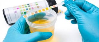
Preparation and delivery
In order for a study on tumor markers of the digestive organs to show accurate results, it is necessary to properly prepare for it. Preparatory measures do not present any difficulties and are similar to those for any venous blood sampling.
Experts recommend that patients comply with the following list of requirements:
- refusal of alcoholic drinks of any strength 72-48 hours before visiting the laboratory;
- stopping taking medications (with the exception of vital medications, information about which is available from the attending physician) one day before blood sampling;
- correction of the diet with the exclusion of spicy, fatty and fried foods, 24 hours before the analysis.
Following these simple rules will allow you to obtain the most reliable results. But even in the case when all the requirements for preparing for the study were fulfilled by the patient with accuracy, the analysis can give false positive indicators, since completely different reasons, often independent of the person, lead to changes in the level of specific proteins. That is why doctors, to exclude medical errors, always prescribe a repeat examination.
How is the analysis carried out?
If a person is scheduled to take tumor markers for laryngeal cancer, he should prepare for the familiar blood draw from a vein and come to the laboratory on the appointed day from 8 to 11 am. At the same time, a specialist can determine up to 3 specific biomolecules, so the SCC tumor marker and the cyfra 21-1 protein are tested on the same day. The collected biomaterial is transferred to a laboratory assistant, who must immediately obtain serum from it and begin studying it under a microscope - calculating the concentration of tumor cells per 1 ml of peripheral blood.
Important! It is not recommended to store the collected blood frozen for some time, so if the laboratory conducts research not daily, but once or several times a week, you should visit it on the day when laboratory technicians are studying the biomaterial.