Cancer is a very common disease these days and it is truly alarming. The relevance of identifying cancer in the early stages is beyond doubt, because it is at the beginning of the development of cancer that it can be successfully defeated. Diagnostic methods, including tests for stomach cancer, make it possible to identify the risks of this disease, and if the disease has already occurred, they make it possible to determine its stage. In addition, thanks to the results of tests and other studies, it is possible to monitor the treatment process and evaluate its effectiveness.
General blood analysis
Many patients are interested in the question: will OAC show specific changes? It is known that a blood test for stomach cancer cannot confirm or refute the diagnosis. Early stages of the tumor may not manifest themselves, so there will be no change in the overall analysis.
With constant blood loss, chronic posthemorrhagic anemia develops. The second mechanism for the development of anemia is impaired iron absorption due to damage to the glands of the gastric mucosa. Cancer intoxication also plays a role in disrupting iron metabolism.
Other indicators also change - the number of leukocytes increases (with tumor disintegration) and ESR, a decrease in leukocytes occurs in the initial stage.
An increase in the number of monocytes is characteristic, which is associated with a tissue reaction. ESR is an important indicator, since its high numbers in a person’s overall normal state of health can lead the doctor to think about a possible cancer.
ESR in cancer
The level of ESR is especially important when diagnosing pathology. If there is a systematic increase in the indicator, and there are no visible changes in the patient’s well-being, the formation of a cancerous tumor should be excluded.
When a tumor forms in the stomach area, high ESR levels are observed. There are many reasons for this. Starting from intoxication and ending with impaired hematopoietic function. In any case, a constant increase in this indicator is a direct indication of the appointment of a cancer screening.
A low ESR level does not exclude the presence of a tumor and cannot be considered as a reliable diagnostic sign.
The development of a tumor in the stomach is accompanied by an increase in ESR levels
All people who lead a healthy lifestyle and provide themselves with adequate nutrition should be wary of the fact that the ESR begins to increase without any reason. In addition to this test, you can donate stool, which can reveal the presence of occult blood.
Study of gastric juice
It was revealed that as the tumor develops, there is a decrease in gastric secretion: hydrochloric acid and pepsin.
Gastric juice acquires a specific odor and contains an admixture of lactic acid. An increase in gastric secretion can be observed in the ulcerative form of cancer. This is especially pronounced in young patients.
The oncological process that arose against the background of atrophic gastritis is characterized by achlorhydria. When analyzing the components of the juice, you can determine: mucus, red blood cells, atypical cells.
When is an analysis for tumor markers of stomach cancer necessary?
It is necessary to do a study if any digestive problems often arise against the background of chronic fatigue and fever.
The analysis is also indicated for those who are over 40 years old or have a family history of cancer. In addition, regular screening during and after treatment is necessary. In the first case, the level of tumor markers informs about the effectiveness of treatment, and in the second, it signals an impending relapse long before its manifestation.
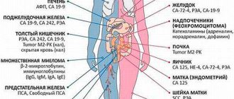
Determination of tumor markers
Markers are specific compounds formed during the cancer process in the body. They can be detected in small quantities in healthy people.
Each type of tumor has its own specific markers. In 50% of cases, CEA – carcinoembryonic antigen – can be detected in the blood. If the stomach is affected, a blood test is prescribed for CA 19-9, CA 72-4.
The diagnostic value of tumor markers is that they allow cancer to be detected at early stages. It is important to observe changes in these indicators over time, since their normal concentration is individual for each patient. The study is carried out on an empty stomach. The material for analysis is venous blood.
Biochemistry
A biochemical blood test allows you to evaluate the activity of internal organs. Deviations of certain indicators make it possible to identify in which system the failure occurred and how much the disease has progressed.
Since tumor cells can spread and affect neighboring organs, when checking for stomach cancer, attention is paid to the functioning of the liver, pancreas, and gallbladder.
Blood for analysis is donated on an empty stomach, taken from a vein. 2-3 days before the test you should refrain from drinking alcohol or visiting a bathhouse or sauna. It is not advisable to take hormonal drugs, antibiotics, diuretics, or statins, as they can distort the results.
The following indicators help identify dysfunction of the digestive system that occurs with stomach cancer:
- amount of total protein. If there are malignant tumors, then its concentration becomes less than 55 g/l. Cancer cells reduce the amount of albumin (less than 30 g/l), and increase the level of globulins;
- increase in lipase. If cancer cells have spread to the pancreas, the amount of fat-digesting enzyme increases;
- Alkaline phosphatase levels increase if a tumor develops in the body;
- increase in gamma GT (glutamyl transpeptidase). This enzyme is involved in the process of amino acid metabolism. An increased amount of this compound in the blood indicates stagnation of bile, which occurs as a result of dysfunction of the liver or biliary system. It is considered normal if the indicator does not exceed 71 units/l for men and 42 units/l for women;
- increased aminotransferase activity indicates liver necrosis or myocardial infarction. The enzyme activity is less pronounced in cirrhosis, skeletal muscle injury, myositis, heat stroke, some liver tumors, and hemolytic diseases;
- cholesterol levels different from normal. The indicator may be low or high, depending on the location of the cancer;
- decrease in glucose;
- increase in bilirubin. The pigment is formed as a result of the breakdown of hemoglobin and is excreted from the body by the liver. An increase in its concentration in the blood indicates pathology of the gland.
Tests for malignant tumors of the stomach
Stomach cancer occurs with multiple pathological symptoms, ulcerations, tumor disintegration, and general intoxication of the body. In addition, the patient has numerous digestive and metabolic disorders. Laboratory studies are based on identifying such processes.
When there is a suspicion that a patient has developed stomach cancer, a blood test makes it possible to make an initial diagnosis quite clearly. The specialist studies the results, comparing the obtained indicators with the normal level. The degree of their change can indicate the development of a tumor.
As a rule, a clinical and biochemical study is first prescribed, and based on the degree of change in their data, the doctor determines the need for other tests or the use of hardware diagnostic methods.
Based on the totality of the results obtained, the specialist makes an expert opinion on the use of a particular treatment method.
According to laboratory tests, it is impossible to clearly judge the presence or absence of stomach cancer, but they can answer basic questions about the cause and nature of the disease observed in the patient.
Very often, studies of blood plasma and its formed elements are used to monitor the effectiveness of the treatment used.
Laparoscopic diagnosis of the stomach
If a tumor is suspected of spreading throughout the abdominal cavity, diagnostic laparoscopy and laparoscopic ultrasound may be performed, allowing for a detailed study of the process in close proximity.
This method allows you to examine the surfaces of the liver, the anterior wall of the stomach, the parietal (lining the wall of the abdominal cavity) and visceral (covering the organs) peritoneum, performing a biopsy if necessary. In some cases, this data is fundamentally important for choosing treatment.
Differential diagnosis of stomach cancer
Due to the fact that identifying a malignant neoplasm in the main digestive organ is always difficult due to the similarity of its clinical signs with some diseases of the internal organs, a differential diagnosis of gastric cancer should always be carried out. It allows you to exclude some precancerous diseases, which include ulcers, polyps, atrophic and chronic gastritis. This is necessary due to the fact that they all have similar characteristics.
For the correct identification of a pathological condition, an adequately collected anamnesis and a complete examination of not only the gastrointestinal tract, but also other organs are of primary importance.
Differential diagnosis of stomach cancer is carried out using the following examination methods:
- endoscopy with simultaneous biopsy;
- gastrobiopsy;
- X-ray examination;
- detailed blood test.
After a specialist has diagnosed the pathology affecting a person, he selects adequate treatment tactics. This disease is undoubtedly very dangerous and the percentage of complete cure for patients is quite low, but all unfavorable prognoses relate directly to those people who trust their health and lives to obvious charlatans or specialists with minimal experience and low qualifications.
It is worth remembering that malignant processes occurring in the main digestive organ can either be completely eliminated, or the life of a cancer patient can be prolonged and made easier. To do this, you need very little - to find an experienced oncologist who is able to provide effective assistance at any stage of the disease.
Methods for diagnosing stomach cancer
As already mentioned, early detection of the development of a malignant neoplasm in the main digestive organ is very important, since only in this case do 70 out of 100 patients have a real chance of a full recovery. That is why experts recommend that people at risk undergo screening. In case of stomach cancer, such a planned annual examination, consisting of gastroscopy, can save a large number of lives.
The procedure itself does not require any special preparation, is performed on an outpatient basis and takes no more than 15 minutes. At the same time, its importance in identifying precancerous and cancerous conditions of the main organ of the gastrointestinal tract is invaluable. If, based on its results, a specialist suspects that a person, even one who does not currently have suspicious symptoms, is developing a malignant tumor, he will be assigned a special diagnosis of stomach cancer.
It consists of a whole range of measures that are aimed at not only identifying the underlying disease, but also identifying the causes that provoked its occurrence.
This examination of the stomach consists of 4 main methods:
- Clinical. It consists of collecting the patient's medical history and compiling a medical history.
- Physical. Includes auscultation (listening to sounds arising in the stomach) and palpation (palpation of the diseased organ). In the early stages of development of a pathological condition in the main digestive organ, using this method it is possible to identify distant signs of the disease. It is worth noting that palpation is carried out in four positions: standing, lying on the right side, on the left side and on the back.
- Laboratory. The first step is to conduct a blood test for tumor markers in a sick person. The material for tumor markers (tumor markers are specific proteins that only cancer cells produce) is blood serum from a vein. The procedure is performed on an empty stomach, the last meal should be no later than 8 hours before blood sampling. For patients who have undergone radical therapy, this study must be repeated every three months. Based on its results, the specialist can confirm or deny the presence of malignant cells.
- Instrumental. It is prescribed last and includes x-ray examination, fibrogastroduodenoscopy with biopsy for a detailed examination of the mucosa and taking a tissue sample for histology, magnetic resonance and computed tomography.
The use of these methods to identify the initial stage of malignancy of the main digestive organ makes it possible to detect stomach cancer at the earliest stages. This gives patients a chance for a full recovery or prolongation of life for the maximum possible period for this disease. That is why experts recommend that all people who are at risk or have a precancerous condition of the gastrointestinal tract undergo them.
Diagnosis of gastric cancer with metastases, stage 4
The advanced stage of disease development is characterized by tumor growth into all layers of the digestive organ, as well as the spread of mutated cells throughout the body. In order to detect malignant gastric lesions at a late stage, in addition to the main ones, additional diagnostic methods are also necessary. Among them, the main one is laparoscopy, performed under direct ultrasound guidance.
This diagnostic test is a minimally invasive operation performed under anesthesia. It is performed through punctures in the abdominal wall into which a camera is inserted. Using this method, a specialist has the opportunity to detect the growth of a tumor into nearby tissues and the spread of metastases to the liver and peritoneum.
Typically, such a diagnosis of stage 4 stomach cancer allows the specialist to identify the following unpleasant signs in the patient:
- mutated cellular structures are in close proximity to neighboring organs;
- the tumor has spread to nearby lymph nodes;
- the tumor process begins to develop in nearby internal organs.
Metastases at stage 4 of this pathological condition can spread not only through the lymph, affecting the lymph nodes, but also by hematogenous (through the bloodstream) or implantation (with close contact of internal organs) routes.
First symptoms of the disease
Cancerous damage to the walls of the stomach in the initial stages of its development, just like any other oncology, does not cause pronounced changes in a person’s well-being. Certain symptoms of stomach cancer begin to appear in the second stage of the cancer process.
Stomach cancer examination
Experts note the following very first signs that suggest that a malignant tumor is forming in the main organ of the gastrointestinal tract:
- gastric dyspepsia, expressed by such negative sensations as constant and causeless bloating, belching or heartburn, periodic nausea, interspersed at times with vomiting;
- loss of appetite, expressed in intolerance to any product, usually meat;
- depression, constant lethargy, pronounced decrease in performance and sleep problems;
- an unprovoked rise in body temperature to subfebrile (37–38°C) levels;
- sudden weight loss.
But pain with stomach cancer does not appear immediately. At first, cancer patients experience only minor pulling sensations in the epigastric or pancreatic region (slightly above the navel), which occur regardless of food intake, but pass fairly quickly.
If the size of the malignant tumor becomes large enough, that is, the tumor begins to occupy almost the entire internal surface of the digestive organ, its volume decreases, which causes a rapid onset of saturation in the sick person. When a tumor develops in the immediate vicinity of the exit sphincter into the intestine, the patient suffers from constant heaviness in the abdomen due to the inability of a bolus of food to pass through it, and the tumor, blocking the connection with the esophagus, leads to difficulty in the swallowing reflex.
All of the above symptoms of stomach cancer associated with digestive disorders contribute to changes in the natural functioning of the digestive tract. This in turn causes a deterioration in metabolism, resulting in the appearance of some external signs. The main ones are an unpleasant, pungent odor from the mouth and a constant coating of the tongue with a dense coating of yellow or grayish color.
Must remember! You should not immediately panic when such symptoms appear, as they can accompany other, less dangerous gastrointestinal pathologies. First of all, you should contact a specialist and undergo appropriate diagnostic tests that will help identify the true root cause of anxiety symptoms. It is categorically not recommended to delay visiting a gastroenterologist in such a situation, since possible oncology of the main digestive organ is always prone to rapid development.
Early diagnosis of stomach cancer
It is very important to recognize the development of a malignant tumor process in the main digestive organ as early as possible. This is of fundamental importance due to the favorable prognosis of this disease - 90% 5-year survival rate is observed only when gastric cancer is detected and operated on in a timely manner. At later stages, based on statistical data, it does not rise above 40%.
There are no specific symptoms accompanying a stomach tumor that is just beginning to develop. A pathological condition that develops directly against the background of ailments occurring in the gastrointestinal tract, chronic gastritis or ulcers that are benign in nature, retains their main manifestations for a long time. Very often it is impossible to diagnose stomach cancer at the initial stage of the disease. This is due to the latent course of the disease, so its development is very slow. In the rarest cases, the occurrence of the disease may be indicated by involuntary internal bleeding from the lower gastrointestinal tract.
Early diagnosis of stomach cancer is possible with direct fluoroscopy. Due to its simplicity and accessibility, this technique is currently used for preventive research. To obtain the most accurate results, large-frame gastrofluorography is used, and the images taken with its help are analyzed by two independent specialists.
The main warning signs considered suspicious at the initial stage of stomach cancer are:
- thickening of the mucous layer and restructuring of its relief in small areas with limited area. Their folds are always located chaotically;
- a barium depot (accumulation of the suspension the patient drank before the examination) repeated repeatedly on an x-ray image between thickened folds. This picture is noticeable even when there is not yet a clearly defined depression between them;
- partial smoothness of the protruding elevations of the mucous membrane, the roughness of their surface, noted in small areas, as well as jaggedness in these places of the contour of the stomach.
If specialists detect such suspicious signs on an x-ray, the patient undergoes a gastroscopy, which is necessarily performed with a targeted biopsy.
Endoscopic diagnosis of stomach cancer in the early stages is a rather difficult task, but it also gives good results. In 18% of observations, using only this study, doctors were confidently able to detect malignancy of the gastric mucosa at the initial stage, in 59% they suspected it, and in 30% they identified a macroscopic picture that was more likely to be characteristic of a benign process.
When assessing the results obtained during an endoscopic examination, early gastric cancer is classified according to the following picture presented in the table:
| MAIN TYPES OF TUMOR IN THE STOMACH | FORMS | CHARACTERISTIC |
| Bulging or protruding | Polypoid | Stomach cancer with this shape looks like a polyp sitting on a broad base. The defect is detected when the main organ of the gastrointestinal tract is completely filled with barium suspension. Typically, such polyps in the stomach have a diameter of no more than 1 cm and are round or irregularly oval in shape. Its contours are unclear and in places with uneven edges. The mucous membrane surrounding the polyp, with an area of approximately 5 cm, has a changed relief, which is represented by uneven elevations. |
| Plaque-like | Defects discovered after tight filling of the stomach with barium suspension appear round, without a protrusion structure, located on the relief of the mucosa. In rare cases, a single defect is discovered that has clear and even boundaries. In the center of it, a more or less deep depot of barium is usually noticeable, indicating ulceration of the tumor surface. Plaque cancer rarely reaches 2 cm in diameter. | |
| Invasive | saucer-shaped | This form of malignant neoplasm occurs most often in the main digestive organ. The main reason for its appearance is the progressive manifestation of the tumor. The defect in this case is a rounded growth, in some cases reaching quite large sizes and capable of growing into neighboring organs |
| Surface | Ulcerated (ulcerative-infiltrative) | Ulcerated stomach cancer is also diagnosed very often, in more than half of cases of malignancy of the digestive organ. It unites tumor-like ulcerated pathologies of the stomach of different genesis, related to the primary ulcerative form. They are a consequence of the progression of a chronic ulcer or saucer cancer |
Methodically correctly performed endoscopic and x-ray examinations allow 40-50% of patients to suspect gastric cancer at a very early stage.
Study of the parameters of the coagulation system
The blood coagulation system is a complex system consisting of:
- The actual coagulation system. Its components are responsible for coagulation, that is, blood clotting when necessary.
- Anticoagulation system, components of this system are responsible for anticoagulation.
- The fibrinolytic system ensures the dissolution of already formed blood clots. This process is called fibrinolysis.
With the development of gastric cancer of various forms, increased thrombus formation occurs. This is expressed by an increase in blood values such as APTT, TV, PTI.
When hypercoagulation occurs, compensatory mechanisms trigger the activation of fibrinolysis, which is necessary for the dissolution of blood clots. Therefore, in gastric cancer, an increase in antithrombin and antithromboplastin levels is detected.
Types of analyzes
The most common test is a simple finger prick. This type of examination is recommended for various pathologies. With the help of a general analysis of biological fluid, it is possible not only to identify how the disease develops, but also to evaluate the effectiveness of the selected treatment.
When malignant processes occur in the human body, the composition of the blood changes. However, to clarify the etiology of atypical changes, one general blood test is not enough.
To establish an accurate diagnosis, the following types of tests are required:
- general analysis;
- biochemical examination;
- detection of tumor markers.
Based on the data obtained, the person’s condition is analyzed and assessed. If the readings are elevated, the specialist suspects the presence of an inflammatory process. However, to make a definitive diagnosis, a biopsy is required. It is this instrumental method that allows you to determine the type of cancer and its stage.
Reasons for diagnosis
Despite the fact that such a dangerous pathology as stomach cancer has been detected more and more often in recent years, many people are interested in the question of why experts recommend undergoing scheduled annual examinations, called screening in medical terminology. This is explained quite simply. Any oncological diseases in the early stages are practically asymptomatic or have vague signs that in no way indicate the appearance of a malignant neoplasm.
Only through early diagnosis is there a high probability that an emerging tumor will be detected in the main digestive organ, and treatment of stomach cancer detected in the early stages gives positive results in 90% of cases. It is also necessary to remember that the basis for such studies as gastroscopy, endoscopy and radiography of the digestive organs, which allow timely detection of dangerous gastrointestinal pathologies, is the appearance of dyspeptic gastric symptoms.
Important! If you suddenly begin to suffer from unexplained discomfort and pain in the epigastrium, loss of appetite, a temperature that often rises to subfebrile levels and constant weakness, you should immediately consult a specialist. Do not forget that such symptoms are a direct reason for undergoing diagnostics, as they may indicate the development of a malignant tumor in the stomach.
Blood test for stomach cancer: indicators and their meanings, cost
Stomach cancer, like any other organ, cannot be diagnosed based on the symptoms of the disease alone.
To confirm the diagnosis, the doctor prescribes a series of examinations; a blood test is also required. Based on changes in normal blood parameters, a specialist determines the likelihood of developing a malignant process.
https://www.youtube.com/watch?v=ycZGO-BiJrI
The most common blood test is a complete blood count.
This examination is prescribed for various diseases, and it allows you to determine not only how the disease progresses, but also serves as a control for the effectiveness of treatment.
When a malignant lesion of the body occurs, certain changes occur in the composition of the blood, but a general analysis alone is not enough to identify them.
A presumptive diagnosis of stomach cancer can be established by conducting several types of blood tests at once, these include:
- General analysis.
- Biochemical research.
- Detection of certain tumor markers.
Assessing the totality of all changes in the composition of the blood allows the doctor to suspect a particular disease, to confirm which instrumental examinations are also necessary. It must be remembered that cancer can only be accurately identified when cancer cells are detected, which requires a biopsy.
A general analysis is a study of blood taken on an empty stomach from a finger, or less often from a vein. If gastric cancer is suspected, special attention is paid to such indicators of a general blood test as ESR, the number of leukocytes in the blood and hemoglobin level.
- ESR almost always increases with malignant neoplasms. The normal erythrocyte sedimentation rate should be no more than 15 mm/h. A sharp increase in ESR indicates that there is an active inflammatory process in the body. SLE indicators characteristic of cancer change little during antibacterial therapy.
- White blood cells in the initial stages of cancer either remain normal or decrease slightly. As the disease progresses, the number of leukocytes increases markedly, and many young forms are found in the blood.
- With stomach cancer, in most cases, hemoglobin falls below 90 g/l. This happens due to the fact that a person consumes less nutrients; the tumor interferes with the complete absorption of food. In the last stages of cancer, anemia is associated with the disintegration of the tumor and bleeding from it.
- The number of red blood cells drops to 2.4 g/l.
The listed changes also occur in other diseases, most of which are successfully treated. Therefore, you should not evaluate the results of a blood test yourself.
A biochemical blood test is performed to check the functioning of internal organs. Changes in some indicators directly indicate in which organ pathological changes occur and which body systems are affected.
Using this analysis, the likelihood of developing cancer can be determined.
Blood for biochemistry is taken from the cubital vein. This is usually done in the morning, since a person should not eat anything for at least 8 hours.
In case of gastric cancer, a number of changes are detected in a biochemical blood test, these are:
- Decrease in total protein. With malignant neoplasms, the level of this blood component drops below 55 g/l. Proteins consist of globulins and albumins. With the development of cancer cells, the albumin content also decreases significantly, it becomes less than 30 g/l. On the contrary, globulins increase.
- An increase in lipase, an enzyme necessary for the breakdown of food, occurs if a malignant tumor from the stomach penetrates into the pancreas.
- An increase in alkaline phosphatase indicates tumors developing in the body.
- Increased glutamyl transpeptidase (gamma GT).
- Increased activity of aminotransferases – AlAT, AST.
- Changes in cholesterol levels. Depending on the location of secondary foci in gastric cancer, cholesterol decreases or, on the contrary, increases.
- Decreased glucose levels.
- Increased bilirubin levels. This pigment usually indicates the functioning of the liver, but with gastric cancer, damage to this organ is also possible.
At the initial stages, any oncological process has almost no effect on the biochemistry of the blood, but as the cancer progresses, the indicators of blood components deviate more and more from the norm. Usually, if there is a change in the biochemical analysis indicating a possible malignant process, the doctor orders a repeat test.
The blood coagulation system is a complex system consisting of:
- The actual coagulation system. Its components are responsible for coagulation, that is, blood clotting when necessary.
- Anticoagulation system, components of this system are responsible for anticoagulation.
- The fibrinolytic system ensures the dissolution of already formed blood clots. This process is called fibrinolysis.
With the development of gastric cancer of various forms, increased thrombus formation occurs. This is expressed by an increase in blood values such as APTT, TV, PTI.
When hypercoagulation occurs, compensatory mechanisms trigger the activation of fibrinolysis, which is necessary for the dissolution of blood clots. Therefore, in gastric cancer, an increase in antithrombin and antithromboplastin levels is detected.
If the examinations carried out suggest that a person has developed a malignant lesion of the stomach, then he may be prescribed a blood test for tumor markers.
In case of gastric cancer, an abnormal tumor marker, designated CA 125, is detected. This is a high molecular weight glycoprotein, which is essentially an antigen. It can also be detected in a certain concentration in the blood of a healthy person, in this case it is approximately 35 units/ml.
The antigen is not elevated during the formation of both malignant and benign tumors. But in cancer, the indicator of this tumor marker increases quite strongly and amounts to more than 100 units/ml.
In case of gastric cancer, the CA 19-9 antigen is also determined. This tumor marker is often used as an indicator of the effectiveness of treatment. Normally, the concentration of C 19-9 ranges from 10 to 37 units/l; with the development of a malignant tumor in the stomach, the antigen value reaches 500 units/l.
If an increase in this type of tumor marker is observed after surgical treatment of cancer, this indicates the formation of secondary foci of a malignant tumor.
Cancer is a very common disease these days and it is truly alarming. The relevance of identifying cancer in the early stages is beyond doubt, because it is at the beginning of the development of cancer that it can be successfully defeated.
https://www.youtube.com/watch?v=fmbe5svRrUw
Diagnostic methods, including tests for stomach cancer, make it possible to identify the risks of this disease, and if the disease has already occurred, they make it possible to determine its stage.
In addition, thanks to the results of tests and other studies, it is possible to monitor the treatment process and evaluate its effectiveness.
Gastric cancer is a malignant neoplasm, that is, a tumor that originates in the epithelial layer of the gastric mucosa.
Under the influence of various unfavorable factors, healthy epithelial cells are modified and degenerate into a malignant tumor, which over time spreads through metastases to other organs and tissues.
Another name for this type of cancer is gastric adenocarcinoma.
Typically, the cells that make up our body die over time, and new ones take their place. But it happens that this process is disrupted, and new cells are formed when the body does not need them at all. At the same time, the old cells remain in their places. As a result, the tissue grows and a tumor forms. It can be benign or malignant.
In contrast, a cancerous tumor does not have any membrane, so its cells easily and quickly penetrate into neighboring tissues and organs, and through the flow of blood or lymph, pieces of a malignant neoplasm can reach very far, forming new foci of the disease.
Doctors have still not been able to establish the exact causes of the occurrence and development of stomach cancer, but some factors have been identified that can increase the likelihood of this disease. These include:
- gender: there are twice as many stomach cancer patients among men as among women;
- race: representatives of the Negroid and Mongoloid races are more susceptible to this disease compared to Caucasians;
- genetic predisposition: if close relatives have had cancer, the risk of developing stomach cancer and other types of cancer is higher;
- geographical location: in the countries of Eastern Europe, Central and South America, as well as in Japan, there is a higher percentage of patients with stomach cancer;
- blood type: the risk of developing this disease is higher in people with blood group O;
- age: older people are more likely to suffer from this cancer: men after 70 years, women after 74 years;
- unhealthy diet: if the diet includes salty, spicy, fatty, fried, sour foods, but is characterized by a lack of fresh vegetables and fruits, then there is a high probability of developing stomach cancer;
- sedentary lifestyle;
- excess weight: obesity increases the risk of tumor formations in the upper part of the stomach;
- Helicobacter pylori bacterium: leads to inflammation and stomach ulcers, and can also increase the likelihood of cancer;
- gastrointestinal diseases: prolonged inflammatory processes and swelling lead to an increased risk of stomach cancer; dangerous diseases include gastric polyps, pernicious anemia, chronic gastritis, intestinal metaplasia;
- smoking: nicotine increases the risk of developing cancer, including this;
- professional factor: if the work is related to the extraction of minerals such as nickel, coal, processing of wood, rubber, or work with asbestos, then this leads to an increased risk of cancer.
The problem is that the presence of the above factors does not always lead to stomach cancer. But at the same time, their absence does not mean that a person will not get sick. There were many cases where the causes of the disease were never clarified.


