Comparison of MRI of the intestine and colonoscopy
The advent of magnetic resonance analysis in medical diagnostics has significantly improved the quality and level of diagnosis of many pathologies and increased the number of detected pathologies several times. With the help of an MRI machine, such complex conditions as tumors of the brain, spine, and bones of various locations began to be accurately diagnosed. The diagnostic method makes it possible to assess the size, location of the formation, its relationship and relative position in relation to neighboring organs and tissues.
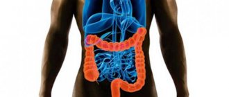
To identify intestinal pathologies, the magnetic resonance analysis method is not used so often. Most appointments are for diagnosing neoplasms. This is explained by the peculiarity of the target organ. The intestinal loops are located tightly to each other and can be layered on top of one another, which reduces their visualization for magnetic waves. It is more suitable for examining pathologies of the small intestine, since it is not possible to reach it during a colonoscopic examination.
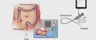
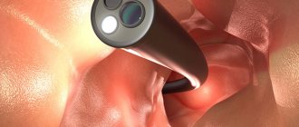
Colonoscopy involves an invasive examination. This means that the mucosal imaging device is inserted directly into the intestine through the anus. On the monitor screen, the doctor sees exactly what the surface of the intestinal mucosa looks like and can assess the presence or absence of pathological formations. Using video colonoscopy, you can not only examine the intestines from the inside, but also take material for biopsy examination.
Indications and contraindications
It’s unlikely that anyone would even think of going through a colonoscopy procedure just like that. It is prescribed only by a doctor if the patient has indications for it. When seeking advice, there is usually a standard set of complaints:
- pathological discharge from the anus;
- painful act of defecation;
- stool disorder;
- constipation;
- flatulence;
- weight loss;
- manifestations of anemia: pallor of the skin;
- diagnosing a history of intestinal neoplasms.
Such subjective complaints can be made by patients in whom the doctor suspects intestinal tumors of various locations, intussusception of an intestinal loop, Crohn's disease, colitis of various origins and clinical manifestations.
It is important to consider when choosing which is better: MRI or colonoscopy of the intestine, the subjective state of the patient, a list of concomitant diseases and an anamnesis of the previous medical history.
Contraindications to colonoscopy examination are as follows:
- diseases of the cardiovascular system;
- mental disorders;
- pregnancy with gestosis;
- intestinal damage;
- tendency to bleed;
- post-infarction state;
- pelvic joint injuries.
If possible, it is better to conduct a magnetic resonance examination in such cases. The diagnostic method is no less informative, but not always realistic for several reasons:
- lack of a tomograph;
- inability to ensure patient immobility during the procedure;
- pregnancy, especially the first three months;
- presence of a pacemaker;
- metal prostheses;
- claustrophobia - fear of closed spaces;
- deep drug or alcohol poisoning;
- general serious condition of the subject.
It is impossible to say unequivocally whether it is possible to do an MRI of the intestine instead of a colonoscopy. The indications for both procedures are similar, but the result cannot always be obtained from any one of them.
CT scan
CT is a painless procedure and is to some extent similar to magnetic resonance imaging. But unlike MRI, a computer analogue scans organs using X-rays, transferring information about the object under study to a digital medium.
The CT machine is highly informative, just like MRI, it shows a three-dimensional image of the area under study. But the presence of radio waves limits its use: the procedure cannot be performed repeatedly, they try not to prescribe it to children, and it is strictly not performed on pregnant women.
If it is impossible to perform a colonoscopy of the intestine, a double diagnosis may be prescribed: CT and MRI. This method will provide clearer information about the patient’s condition. Just like MRI, computed tomography is performed using contrast agents to better visualize the area being examined.
Preparation and performance of MRI
In order to obtain the maximum diagnostic result and prescribe adequate treatment for the disease, it is necessary to properly prepare for the study. The intestines should be practically free of feces. To do this, a few days before the scheduled examination, you need to start following the following diet:
- pureed or slimy soups with water;
- homogeneous porridges from neutral cereals;
- Drinks allowed include water, herbal tea, and choleretic infusions;
- limit meat and eat only boiled;
- the last meal no more than 10-12 hours before the study.
Alcoholic drinks, smoking, so-called fast food products, pickled dishes, dairy products, confectionery, mushrooms, and nuts are strictly prohibited these days. All of the listed products cause increased gas formation and make it difficult to stool, which is unacceptable for obtaining a good result of magnetic resonance imaging. On the eve of the appointed time, do several cleansing enemas to completely cleanse the intestinal walls of feces.
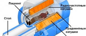
An MRI takes from 40 minutes to one hour. At this time, the patient must remain motionless inside the chamber; to ensure this condition, he can be additionally fixed with special fasteners. The method is absolutely painless; its negative property for the patient’s subjective perception may be psychological discomfort. The doctor evaluates the data obtained from the images, which remain in digital form.
Preparing and performing a colonoscopy
In order for a colonoscopy to give the most informative results, you need to prepare for it in much the same way as for an MRI. The intestines must be completely freed from feces, follow a diet, take laxatives the day before and do several high cleansing enemas.
During the examination, the patient experiences discomfort because the colonoscope tube is inserted directly into the intestine through the anus. During the examination, the patient is on his side, his legs are bent and brought to his stomach. A complete examination lasts approximately 20–30 minutes. The doctor visually assesses the condition of the intestinal mucosa, the presence of tumors, their shape, appearance, location directly during a colonoscopy, since he sees the picture transmitted by the camera on the monitor screen.
Which is better: CT scan or colonoscopy?
Despite the modernity and relative safety of CT, examination of the intestinal mucosa using standard colonoscopy remains an important need.
The traditional method of examining the intestines with an endoscope is characterized by unpleasant sensations, but provides maximum information about any pathology and allows the doctor to evaluate the condition of the epithelium. Anesthesia is often required to make this procedure easier.
A comparison of CT and colonoscopy helps determine which is better for a person:
- Endoscopy allows you to identify the disease by changes in the shade of the mucous membrane, characteristic darkening and spots. The technique detects even the smallest polyps, adhesions or erosions. Computer technology often misses tumors up to 0.5–1 mm.
- When performing tomography, one of the contraindications is the patient’s weight over 150 kg. There is no such limitation with colonoscopy.
- CT excludes damage to the mucous membrane, accidental ruptures and other painful moments. When the endoscope operates with severe spasms or inappropriate behavior of a person, in rare cases, perforation occurs, requiring urgent surgical intervention.
- During colonoscopy, any radiation is excluded, which significantly reduces the number of contraindications.
- With the help of a modern endoscope, during the procedure the doctor has the opportunity to collect biological material. He often uses a special loop to remove benign polyps.
- With CT, you can examine organs located near the tumor to exclude metastases and proliferation of the mediastinum. Endoscopic equipment limits the view only to the intestinal cavity.
In severe conditions, the doctor may recommend a combination of two techniques simultaneously to prepare the patient for a complex operation.
Features of CT examination
CT allows you to examine and study large intestinal tumors in combination with the condition of the surrounding lymph nodes, blood vessels and bone tissue. But it cannot show the shade of the mucous membrane, which is important when determining the location of inflammation in colitis, ulcers and other diseases. Therefore, after a diagnosis, a regular colonoscopy is often recommended to clarify the results and exclude dangerous conditions.
Features of traditional colonoscopy
When carried out, a special tube-probe is inserted into the anus, which moves along the loops of the intestine. This creates discomfort and pain. With large tumors, it is almost impossible to examine the intestinal cavities without the risk of injury and damage. But the information received is highly reliable. In addition, during work, the surgeon can stop the bleeding and take a polyp for a biopsy.
Comparative advantages and disadvantages of methods
How to make a choice: MRI of the intestines or colonoscopy, which is better to do, what are the reviews, all this worries those who are faced with the issue of examining the intestines for the first time.
The indisputable advantages of magnetic resonance diagnostics include:
- digital accuracy of the obtained data;
- the ability to assess the spatial location of detected tumors;
- safety of data in the form of images and computer material;
- non-invasive, painless for the patient.
The method has the following disadvantages: the ability to detect inflammatory changes in the mucosa is very limited, when intestinal loops are superimposed on each other, pathology may not be detected, the price of the procedure is quite high.
The advantages of colonoscopy are the following:
- obtaining visualization of the pathological formation;
- During the procedure, it is possible to take material for histological examination;
- colonoscopy makes it possible to immediately cauterize the ulcer and cut off the polyp;
- objective assessment of the condition of the mucous membrane.
The disadvantages of a visual examination of the large intestine are quite significant: pain, psychological discomfort, the likelihood of injury to the intestinal mucosa, and the potential danger of bleeding.
Indications and contraindications
At the initial stages of the pathological condition, when changes in the intestine are subtle, MRI is not so informative. But during pregnancy, as well as in seriously ill patients, the procedure is the only possible way to diagnose gastrointestinal disorders.
Indications for MRI:
- diverticulosis of the sigmoid or colon;
- intestinal obstruction;
- Crohn's disease;
- ulcers;
- presence of stones;
- congenital organ pathologies;
- neoplasms;
- polyps;
- bleeding.
The procedure is strictly contraindicated if the following is present in the body:
- artificial metal bones, teeth, joints;
- pacemakers;
- pacemaker;
- hearing device;
- iron fragments, bullets;
- ferromagnetic clips.
Tomography is also not prescribed for contrast agent intolerance, hyperkinesis, claustrophobia, and children under 7 years of age. The examination is relatively contraindicated in the presence of:
- severe renal failure (only with the permission of a nephrologist);
- implants in the middle ear;
- pregnancy (up to 12 weeks);
- insulin pumps and nerve stimulators;
- prosthetic heart valves;
- hemostatic clips.
MRI is considered insufficiently informative in the following cases:
- for small areas of inflammation;
- minor changes in the organ mucosa;
- very active peristalsis;
- in emergency situations.
Colonoscopy is the gold standard for bowel examination. Prescribed for:
- suspicion of the presence of tumor formations;
- narrowing of the intestinal lumen;
- bowel prolapse;
- ulcers and polyps;
- proctitis.
The procedure is not performed when:
- the patient’s refusal to undergo it;
- toxic megacolon;
- perforation of the large intestine;
- peritonitis;
- heart attack;
- heart and pulmonary failure;
- shock states;
- colds;
- blood clotting disorders;
- fulminant, ischemic, ulcerative colitis.
Relative contraindications for colonoscopy are:
- acute diverticulitis;
- unstable hemodynamics;
- myocardial infarction that occurred less than a year ago;
- early postoperative period;
- aneurysm of the abdominal aorta.
Which method is more informative?
It is not possible to give an unambiguous answer to this question. Medicine is not an exact science, so diagnostics must complement each other.
The data obtained from MRI or colonoscopy are equally valuable, it all depends on how much they helped to make the correct diagnosis. The doctor has education, experience, clinical thinking, so the assessment of how much more he needs this or that method is only up to him.
Reviews from doctors and patients
The doctor has a specific task: to make a diagnosis and carry out competent, adequate treatment. In order to achieve this goal, a specialist must understand the details of instrumental diagnostics and prescribe exactly the type of study that will facilitate the path to diagnosis.
Reviews from doctors about the advantages or disadvantages of intestinal MRI and colonoscopy are different in each individual case, it all depends on whether the planned information was received or not enough.
Patients are afraid of the discomfort that a colonoscopy can bring. This is understandable and justified, but you should not categorically refuse it if the doctor insists on conducting a colonoscopy examination.
Distinctive subtleties of the procedures
The machines for MRI and colonoscopy are completely different, so the technique is radically different.
MRI
MRI scanning is performed while holding your breath. Therefore, the patient wears headphones, and a microphone is installed in front of the doctor, through which he gives commands.
The study consists of a native and a contrast part. The total duration is at least 45 minutes, during which time you must lie as still as possible. The tomograph is sensitive to changes in body position.
At the end of the procedure, the doctor receives an image of the abdominal organs and the dilated small intestine. The introduction of a contrast agent makes it possible to correlate the detected changes with a specific group of diseases.
Colonoscopy
The patient changes into a disposable gown, goes into the endoscopy room, and lies down on the table on his left side. The anesthesiologist puts you into a state of medicated sleep or the doctor administers local anesthesia.
Who chooses
The choice in this case should be a priority for the attending physician. The patient has the right to control his own health; he may be subject to subjectively unpleasant, invasive diagnostics, but this is not always justified. MRI, despite being painless, is a fairly serious test for the nervous system.
The doctor must choose because he is the one to diagnose and treat the patient. Without complete confidence in the clinical picture and pathogenesis of the disease, it is impossible to prescribe treatment and choose the right tactics. It is necessary to take into account the patient’s subjective attitude, his somatic diseases, and the state of the nervous system, but one should only be guided by the achievement of the result.
CT bowel
Virtual intestinal colonoscopy can be considered a complete analogue of a conventional examination. However, for all its maximum information content, it is slightly inferior to colonoscopy. Simply because the study does not allow direct contact. However, CT became a real lifeline when mortality rates from untreated bowel cancer began to rise alarmingly. The reason was simple: the reluctance of the patients themselves to be diagnosed using colonoscopy. According to statistics, intestinal cancer was in 3rd place in terms of mortality. With the advent of CT, the figure began to decrease, patients undergo timely examination without fear.
CT has many advantages, despite some disadvantages:
- painlessness;
- extreme visualization of tumors;
- intestinal stenosis does not become an obstacle;
- it is possible to check neighboring pathologies;
- The pictures are clear, even contours.
CT scan allows you to better see tumors in the intestine.
An important point is that colonoscopy is not permitted if the body is weakened, including for older people. For CT there are simply no such restrictions; diagnosis can be carried out even by the weakest and elderly. Additionally, there is no need to resort to anesthesia or sedative, the patient feels comfortable and calm.