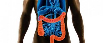
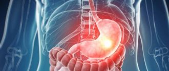
CT scan of the intestine is most often used as a diagnostic tool in the gastroenterological area. This method is non-invasive and is based on the use of x-rays. Allows you to obtain very clear and detailed images of internal organs. Often, computed tomography of the intestines is also called “virtual colonoscopy,” since the effectiveness of the procedure allows you to examine the intestines no less accurately than invasive methods.
Features of virtual research methods
In recent years, doctors have diagnosed a sharp increase in intestinal diseases associated with the appearance of tumors and polyps. Until a certain time, the pathology does not make itself felt, there is no pain or signs of inflammation. Many patients seek help at a late stage, so the role of early diagnosis of diseases with a high degree of information content is increasing.
Virtual methods have relatively recently begun to be used for intestinal research. The most popular and effective for doctors are CT and colonoscopy. From different angles, they allow you to examine the intestinal cavities, carry out diagnostics quickly and efficiently, and resolve controversial issues. Many patients do not fully understand the difference between the methods and their features.
When choosing which is better: CT or colonoscopy, the doctor in each specific situation considers the technique from several angles:
- the presence of tumors in the cavity, their size and shape;
- the need to collect biomaterial;
- patient's age and weight;
- condition of the rectum;
- concomitant diseases and pathologies.
Despite the popularity of computed tomography, virtual colonoscopy in many respects occupies one of the first places in demand. Sometimes an examination provides data for a correct diagnosis that other newer studies are not capable of.
Comparison of the cost of CT and FCS
Multispiral tomography of the abdominal cavity in different institutions is estimated at 2-5 thousand rubles, you will have to add for contrast, in the end the amount reaches 7-10 thousand. According to the compulsory medical insurance policy, tomography is done on a first-come, first-served basis according to the quotas provided. As a rule, only beneficiaries and seriously ill people can actually take advantage of this opportunity, and only after a long wait.
Colonoscopy is provided for 1000-5000, but with the use of sedation the costs will increase to 7-18 thousand. Removal of polyps, taking a biopsy, and histology are also calculated separately. It can be done free of charge, but without anesthesia.
It turns out that the price of the procedures is almost equal, but if on MSCT the radiologist finds something similar to a polyp, then the patient will have to go to the FCS.
Attention! The cost also varies depending on where the colonoscopy is performed. Often large numbers are just an overpayment for the brand of the clinic and the name of the diagnostician.
What is MSCT colonoscopy
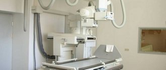
Multispectral computer or MSCT colonoscopy is one of the newest ways to examine the human intestines without the use of an endoscope. But the method is just beginning to be actively used in diagnostics, and such equipment is presented only in large medical centers where treatment and diagnosis of diseases of the digestive tract are carried out.
Colonography is based on the ability of X-rays to penetrate to different depths, delivering information in the form of signals of different lengths. They seem to be repelled from mucous membranes or hard tissues, recreating the outlines in the form of a three-dimensional picture. When passing through the study area, information is removed layer by layer, so the results are accurate and realistic.
Using special equipment, doctors can perform the following manipulations: measure the density of the tumor to identify its structure;
- examine the coronary and large vessels feeding the peritoneal organs;
- confirm aortic aneurysm;
- diagnose liver or kidney failure;
- detect metastases or benign polyps.
The addition of a contrast agent in some situations only improves the picture, helps to find blood clots and blocked vessels, the cause of rectal bleeding.
What is CT colonoscopy
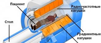
Virtual colonoscopy is used to identify various intestinal diseases associated with changes in the mucous epithelium, the appearance of tumors and polyps. A special field of electromagnetic radiation, created by the phenomenon of nuclear magnetic resonance, forms many thin image slices, each of which has a thickness of no more than 1 mm.
It is based on a combination of X-ray work and the acceleration speed of the smallest particles. Unlike MSCT, virtual colonoscopy only works with hollow organs filled with air, which makes it similar to the standard operation of an endoscope. In the hands of an experienced diagnostician, a computed tomography machine can provide a large amount of information about the condition of the large intestine and internal organs located in the abdominal cavity.
The main advantages of CT technology for the patient:
- absence of pain and discomfort;
- there is no need to use anesthesia for medicated sleep;
- minimum preparatory period;
- Possibility of use for patients of almost any age;
- the opportunity to evaluate the operation performed on the rectum.
CT colonoscopy is not used as a diagnostic procedure due to the risk of radiation exposure to patient tissue.
Indications for diagnosis
CT scan of the small and large intestines is performed in the following cases:
- suspicion of cancer and polyps;
- persistent constipation of unknown etiology;
- intestinal obstruction;
- causeless weight loss;
- the appearance of blood or pus in the stool;
- monitoring the state of the gastrointestinal tract during treatment;
- postoperative monitoring;
- constant pain in the lower abdomen and bloating;
- skin rash with accompanying gastrointestinal problems;
- loss of appetite;
- definition of appendicitis.
The main importance of CT in proctology is the early diagnosis of malignant neoplasms. For this purpose, it is recommended to undergo an MRI or CT scan of the intestine with contrast and a virtual colonoscopy every 5 years.
Features of the methods
Diagnosis of the intestines in the minds of many patients is associated with a painful colonoscopy using an endoscope. But the emergence of new techniques has opened up unique opportunities for early detection of negative phenomena. Indications for such an inspection:
- rectal bleeding;
- suspected tumors and polyps;
- severe pain in the intestines of unknown etiology;
- preparation for surgery.
MSCT differs from CT colonoscopy in the operating principle of the device. The latest multispectral tomographs rotate around a table on which the patient is located. Its sensors work with high precision, so spasms or any muscle contractions do not affect image quality and do not produce erroneous distortions or defects. It is more suitable for studying hard and soft tissues and identifying secondary signs of oncology.
A CT scan helps the doctor examine the structure of the mucous membrane, highlighting irregularities and compactions characteristic of polyps. When carried out correctly, it becomes possible to obtain dynamic data and understand the cause of contraction of the intestinal walls, bleeding and other complications. In fact, both methods are important for high-quality diagnostics and in some issues duplicate each other.
Differences between MSCT and standard CT colonoscopy:
- reduced distortion;
- thinner sections;
- improved contrast of images, allowing you to see the smallest details from 0.5 mm;
- a huge amount of material to study;
- reduced radiation exposure to the patient’s body (up to 25–30%);
- minimum duration.
A serious difference is the cost of the methods: the price of a CT scan using the latest equipment is 4–6 times higher than an examination with an endoscope.
Enterography with contrast
Used for diagnostics:
- Crohn's disease,
- oncological pathologies,
- ulcerative colitis.
The contrast agent is based on iodine. Contrast is injected through a vein. The substance is distributed in the body, which makes it possible to visualize tumors and metastases in detail and determine the spread of the disease with high accuracy.
The study allows you to detect pathology of the walls of blood vessels. Often in old age, diverticulitis (inflammation caused by infection that can cause rupture of the intestine) is detected, which is characterized by swelling of the wall. Wall thickening in cancerous tumors is often difficult to distinguish from changes caused by colitis.
If intestinal obstruction exists, fluid is visible at a horizontal level. Obstruction caused by adhesions or gallstones is visible when the intestine is swollen.
Does CT scanning with contrast affect the body?
If there are no contraindications to CT of the intestine, there is no doubt that the contrast agent is harmless. After the first day, 10% of the drug remains in the body. Soft tissues do not absorb the contrast agent. But the components of the drug in some cases cause allergies. Before the examination, it is necessary to undergo tests to identify the risk of complications. If side effects occur in the form of dizziness, headaches, nausea, you need to wait a while - the symptoms will pass.
Which is better: CT scan or colonoscopy?
Despite the modernity and relative safety of CT, examination of the intestinal mucosa using standard colonoscopy remains an important need.
The traditional method of examining the intestines with an endoscope is characterized by unpleasant sensations, but provides maximum information about any pathology and allows the doctor to evaluate the condition of the epithelium. Anesthesia is often required to make this procedure easier.
A comparison of CT and colonoscopy helps determine which is better for a person:
- Endoscopy allows you to identify the disease by changes in the shade of the mucous membrane, characteristic darkening and spots. The technique detects even the smallest polyps, adhesions or erosions. Computer technology often misses tumors up to 0.5–1 mm.
- When performing tomography, one of the contraindications is the patient’s weight over 150 kg. There is no such limitation with colonoscopy.
- CT excludes damage to the mucous membrane, accidental ruptures and other painful moments. When the endoscope operates with severe spasms or inappropriate behavior of a person, in rare cases, perforation occurs, requiring urgent surgical intervention.
- During colonoscopy, any radiation is excluded, which significantly reduces the number of contraindications.
- With the help of a modern endoscope, during the procedure the doctor has the opportunity to collect biological material. He often uses a special loop to remove benign polyps.
- With CT, you can examine organs located near the tumor to exclude metastases and proliferation of the mediastinum. Endoscopic equipment limits the view only to the intestinal cavity.
In severe conditions, the doctor may recommend a combination of two techniques simultaneously to prepare the patient for a complex operation.
Features of CT examination
CT allows you to examine and study large intestinal tumors in combination with the condition of the surrounding lymph nodes, blood vessels and bone tissue. But it cannot show the shade of the mucous membrane, which is important when determining the location of inflammation in colitis, ulcers and other diseases. Therefore, after a diagnosis, a regular colonoscopy is often recommended to clarify the results and exclude dangerous conditions.
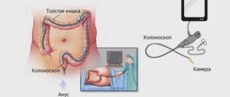
Features of traditional colonoscopy
When carried out, a special tube-probe is inserted into the anus, which moves along the loops of the intestine. This creates discomfort and pain. With large tumors, it is almost impossible to examine the intestinal cavities without the risk of injury and damage. But the information received is highly reliable. In addition, during work, the surgeon can stop the bleeding and take a polyp for a biopsy.
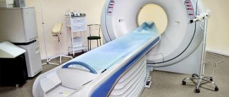
CT scan of the intestines: preparation for the procedure
Preparation for a CT scan of the intestines is usually the same for everyone, but you should always consult with your doctor in advance and follow all his instructions to achieve the best results during the examination.
Purgation
Since the study is carried out on an empty stomach, doctors usually prescribe the procedure in the morning. Computed tomography of the intestine requires preparation, namely, cleansing measures should be carried out. It is necessary to get rid of accumulated gases in the intestines; for this you should take Espumisan, as well as an enterosorbent such as activated carbon. The doctor should also prescribe an osmotic laxative, which is taken the day before the examination. It is advised to completely cleanse the intestines with an enema, and drink half a liter of still water an hour before the CT scan.
Diet
Of course, it is necessary to completely cleanse the intestines of feces; to do this, you should switch to a special diet for 2-3 days, which excludes from the diet foods that contribute to the formation of gas. These are fermented milk products, legumes, raw vegetables and fruits, sweets, and fermented foods. You should also not eat foods that promote constipation to avoid constipation, but it is also not recommended to eat foods with a laxative effect to prevent flatulence. The day before a CT scan of the intestine, it is best to consume liquid food, water, and broth.
Manipulative diagnostic algorithm
Colonoscopy does not require prior hospitalization. The patient first takes a laxative and tries to begin active cleansing of feces one day before.
The manipulation is carried out according to the following scheme:
- the patient is placed on a retractable table;
- after wiping the anus with an anesthetic, a probe is inserted to pump air;
- the table is placed in the middle of the scanning device;
- the patient follows the specialist’s recommendations (holds his breath, turns around).
Preparation and execution takes on average no more than an hour. In rare cases, with rectal pain, air injection is accompanied by an anesthetic injection.
How is the procedure done?
A CT scan of the intestine may be slightly uncomfortable because it requires filling the rectum with air or carbon dioxide. To do this, the patient should lie on the examination table in the desired position as recommended by a specialist. Afterwards, the limbs are secured with special belts to prevent unnecessary movements for clear diagnosis. A thin tube is inserted into the patient's anus, through which gas or air is pumped into the rectum. This is done in order to straighten the intestinal walls, since in its normal state it has many folds. The rectum allows for better diagnosis of the organ along its entire length, even in hard-to-reach places.
The procedure itself takes 15-20 minutes. During the images, the patient will need to hold their breath for 3-4 seconds.
Contrast enhancement
In some cases, contrast enhancement is required to diagnose the intestine. To do this, the patient must take the necessary medication several hours before the examination. Sometimes contrast is administered immediately before the procedure using an enema. This allows you to better visualize the contours of the intestines in the images.
Contraindications
It should not be forgotten that during a CT scan the human body is exposed to x-ray radiation. Therefore, the procedure is contraindicated for pregnant women up to 12–14 weeks. The list of prohibited conditions includes:
- open sores;
- enterocolitis;
- intestinal infection in the acute stage;
- renal or liver failure;
- severe heart disease.
Virtual colonoscopy is ideal for examining the intestines without pain or discomfort. But the choice of technique should be made by a doctor who understands how beneficial the method will be for the patient’s health.
We compare the safety of methods and possible complications
Advantages of CT:
- painless procedure (the patient does not need anesthesia);
- the diagnostic information content is not inferior to the results obtained during colonoscopy;
- there is no possibility of intestinal perforation;
- the device is not inserted into the body;
- the effectiveness of diagnosis in the presence of strictures and polyps in the intestine, which do not allow inserting a colonoscope and thoroughly studying the features of the intestinal wall;
- availability of clear photographs;
- no need for anesthesia;
- Diagnosis of a patient of any age.
Disadvantages of CT:
- cannot be used during pregnancy;
- it is impossible to take a biopsy sample for analysis;
- it is impossible to evaluate the fine mucous structure;
- not very informative in obese patients;
- it is impossible to evaluate the shade of the mucous membrane.
Complications during CT scans are rare, last a short time and disappear almost the next day.
Among them:
- general weakness;
- allergy to contrast injection;
- abdominal pain.
Advantages of using colonoscopy:
- the ability to identify the slightest pathological foci;
- taking a biopsy;
- reliability of research information;
- simultaneous implementation of therapeutic measures (removal of polyps, elimination of the source of bleeding).
Possible complications from colonoscopy:
- pain in the abdominal area after manipulation;
- reaction to drugs administered for anesthesia;
- risk of bleeding;
- intestinal perforation is possible.