Once upon a time, X-ray diagnostics, discovered by the physicist Roentgen, became a real breakthrough in diagnostic, and later in therapeutic medicine. Many years have passed, new methods of visual hardware diagnostics have appeared. Computed tomography and MRI have proven their advantages. But X-ray diagnostics remains today an informative, easiest to perform, and cheapest diagnostic method. It is used in the diagnosis of pathologies of various diseases: pathologies of the lungs, heart, blood vessels, musculoskeletal system, gastrointestinal tract...
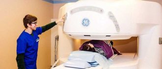
X-ray room
For a long time, only radiography was used. It had its pros and cons. The main disadvantage was the inability to see the pathology of the organ under study during its functioning and thus determine the exact location of the pathological focus and its changes during the operation of the organ.
Therefore, radiography was improved, and, over time, a new method of using X-rays became in demand - fluoroscopy, which is distinguished by the ability to see the organ under study during its physiological movement.
The essence of an X-ray examination is that X-rays, passing through a person, have different degrees of absorption by different tissues of his body. The rays, passing through the human body, are projected onto film, a film cassette, an electronic matrix, or a fluorescent screen. Depending on the structure and density of the tissue, there are X-ray positive and X-ray negative. This is how an x-ray image is obtained. But in the case of radiography, this is a one-time image, and with fluoroscopy, it is an image that changes with functional movement.
What is fluoroscopy
Fluoroscopy is a type of medical examination using x-rays, which results in an image of the internal organ of interest on a fluorescent screen. This method is also called transillumination.
Fluoroscopy and radiography should not be confused. Radiography produces a static (still) image. Fluoroscopy allows you to study the functioning of internal organs in dynamics. Confusion in these concepts arises due to the fact that both research methods are based on the use of X-rays to obtain a picture of internal organs and entire body systems, for example, respiratory, circulatory or digestive.
How is the fluoroscopy procedure performed?
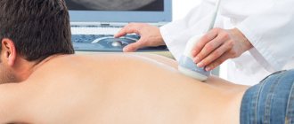
The examination is painless for the patient. You must follow your doctor's instructions. After the equipment is adjusted (an X-ray tube is installed) to the height and position of the patient’s body, the room with the equipment and the patient is closed. The operation of modern X-ray machines is almost silent. The image is transmitted in digital format to the monitor to the doctor, who analyzes the radiological syndromes on the spot and selects an acceptable angle for maximum visualization of the pathological focus. At the end of the study, the results are known immediately; if necessary, the patient receives an electronic version of the study.
Types of fluoroscopy
There are different types of fluoroscopy based on the position of the patient and the direction of the x-rays.
- With the patient in an upright position.
- With the patient in a horizontal position:
- lateroscopy – horizontal direction of rays;
- trochoscopy – vertical direction of rays.
Depending on the area of study, the following types of fluoroscopy are distinguished:
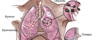
- examination of the lungs and respiratory tract;
- heart examination;
- diagnosis of diseases of the gastrointestinal tract (GIT).
When examining the lungs, it becomes possible to assess the state of the lung tissue in motion.
The transillumination method is especially effective for identifying congenital heart pathologies, acquired diseases, as well as diseases with an unclear etiology (origin). With this examination, you can find calcified areas of the cardiovascular system - places where inflammatory processes occurred and then calcium salts were formed. This can be the myocardium, coronary arteries, valves.
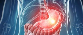
When examining the organs of the gastrointestinal tract, the condition of the duodenum, stomach, and, less often, the esophagus is assessed. Fluoroscopy helps to identify various pathological changes in the digestive tract.
X-ray of the lungs
So, we have already decided what fluoroscopy is. Now let's look at what it is used for. Among pulmonary diseases, lung fluoroscopy is one of the main diagnostic methods. This is due to the ease of implementation and minimal material costs.
The study makes it possible to study the organ shadow not in one position, but in movement, that is, during breathing, heartbeat. This facilitates differential diagnosis between diseases of the pleura and lungs and diagnosis. Fluoroscopy allows one to distinguish inflammatory infiltration from a malignant focus.
By analyzing the movement of foreign bodies in the chest, it is possible to clarify their localization and damage to surrounding structures. Also, x-ray of the lungs in motion evaluates the functional capacity of the respiratory organs, detects atelectasis, pleurisy, inflammatory foci, hydrothorax, emphysema, cancerous tumors, pathology of the lymph nodes, abscesses.
Indications for use
Indications for prescribing a fluoroscopic examination may be:
- from the respiratory system - suspicion of lung pathology, intrapleural bleeding (the pleura is the serous membrane covering the lung, diaphragm, inner surface of the chest wall), pneumothorax (entry of air into the pleural cavity), fractured ribs with damage to the respiratory system;
- from the cardiovascular system - suspicion of heart disease, pathology of the coronary vessels, impaired functionality of individual organs or the entire system;
- from the gastrointestinal tract - diagnosis is needed to identify ulcers, tumors, stenosis (narrowing of the lumen of hollow structures in the body), scars and other painful processes in the organs of the digestive tract.
In addition, the procedure is indicated for the following symptoms:
- dysphagia – difficulty swallowing accompanied by pain;
- abdominal pain – pain arising from stretching of hollow internal organs, tumors, excessive muscle contraction;
- heartburn;
- decreased appetite;
- belching;
- bloating;
- causeless weight loss;
- liver enlargement;
- ascites (accumulation of fluid in the abdominal cavity);
- the presence of blood in the stool;
- detection of compactions in the abdomen upon palpation;
- anemia of unknown origin;
- violation of secretion-forming and acid-forming functions of the stomach.
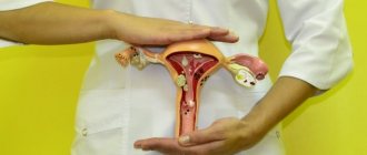
In some cases, the study is also used in gynecology, urology and traumatology.
Contraindications
Contraindications for the procedure are:
- pregnancy;
- serious condition of the patient;
- internal gastric or esophageal bleeding;
- reduced immunity.
For children, such examination is carried out with extreme caution.
Indications, contraindications
The method is used in cardiology, gynecology, traumatology, and urology. Changing the patient's position helps to identify only a developing tumor. Fluoroscopy is used when the lungs are subject to heavy load or it is necessary to monitor puncture during pleurisy and pericarditis. Other indications:
- diverticula;
- esophageal erosion;
- intestinal obstruction;
- intestinal inflammation;
- search for heart valves with limescale deposits;
- esophageal stenosis;
- neoplasms of any nature;
- hiatal hernia;
- ulcer of the duodenum or stomach;
- achalasia cardia.
Contraindications include pregnancy and childhood. The examination is not done if:
- low immunity;
- active tuberculosis;
- contrast intolerance;
- thyroid pathologies;
- kidney and heart diseases;
- internal bleeding.
Since the patient drinks a barium mixture before the examination, the examination is prohibited for allergy sufferers. They may experience immediate anaphylactic shock.
Patient preparation
Often, fluoroscopy of the gastrointestinal tract requires special preparation of the patient. This includes compliance with the following rules.
- Special diet. This involves avoiding foods that cause increased gas formation a few days before the procedure. It is allowed to eat porridge cooked in water, eggs, fish, and lean meats. You should not eat sweets, flour, dairy products, cabbage. It is recommended to avoid chewing gum. On the day of the examination, you should not eat or consume any liquid before the examination.
- It is necessary to stop smoking, drinking alcohol, and postpone taking medications.
- For bloating, flatulence or constipation, gastric lavage or enemas may be prescribed.
- It is necessary to conduct allergy tests when administering a contrast liquid, in order to avoid the development of severe allergic reactions (up to anaphylactic shock).
Precautionary measures
You need to make sure that there are no contraindications to the procedure. Before the study, the patient’s health status is checked to make sure that there is no individual intolerance to the contrast liquid (which contains barium salts). In chronic diseases, a necessary condition is the onset of a period of remission.
Digital fluoroscopy of the stomach
Modern medicine has computer technologies and new highly sensitive sensors.
Digital fluoroscopy of the stomach has numerous advantages:
- the patient receives a low dose of radiation;
- research is carried out quickly and simply;
- the specialist receives an image of the highest possible quality;
- X-rays can be sent by mail, printed, enlarged, or saved digitally.
The low cost and environmental safety of digital gastric fluoroscopy is explained by the fact that during the study, medical workers do not use laboratories or chemical reagents.
How is fluoroscopy done?
X-ray of the lungs is performed to assess their function in different phases of respiratory activity. So that the doctor can evaluate the functioning of the lungs and the respiratory system as a whole, the patient, at the request of the laboratory assistant, inhales, and then holds his breath and exhales.
When examining the digestive organs, contrast liquid is often used. The procedure itself takes about half an hour, but sometimes it takes a little longer.
The examination takes place in two stages. First, the patient is in a standing position, and the contrast fluid flows down the gastrointestinal tract, while internal organs are displayed on the screen. The patient holds his breath and exhales when he is told this. Then the subject takes a lying position so that the doctor can examine the organs from a different angle.
When you can't do without contrast
Contrast liquid is used to improve image quality. For example, in order to examine the work of the heart muscle in more detail, contrast is introduced, since the colored muscle is better visible against the background of the relatively transparent lung.
When examining the gastrointestinal tract organs, their images may overlap each other, so the use of contrast leads to a gradual highlighting of individual objects in color.
A procedure using a contrast method contributes to a more accurate diagnosis and helps make a decision on surgical intervention or alternative therapy.
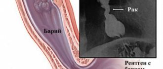
Barium sulfate is used as a contrast fluid. It does not dissolve in gastric juice, is not absorbed into the blood, and is excreted from the body practically unchanged.
Using this method, elevations, depressions, and other anatomical features of the mucous membranes can be examined in the gastrointestinal tract.
If you add more barium sulfate, you can obtain data on the shape, size, functioning, and position of the gastrointestinal tract.
There is also double contrast. In addition to the coloring liquid, air is introduced. This allows you to increase the size of the organ being examined, stretch it and make the image more clear.
A contraindication to this method is suspected perforation. When perforation occurs, a through hole appears in the wall of a hollow internal organ; such a pathology occurs, for example, with an ulcer.
To view the condition of the colon, barium sulfate is also administered, only the intestines are first cleansed, and the drug is administered by enema. This procedure is called irrigoscopy.
When examining other organs of the gastrointestinal tract, the patient drinks liquid. To prevent the appearance of a gag reflex, the drug is combined with various flavoring additives.
Although fluoroscopy involves a fairly large amount of radiation, many doctors often use this examination method due to the availability of fluoroscopic equipment. Patients are attracted by the relatively low price.
The administration of a contrast agent intravenously is contraindicated in cases of severe heart disease, renal failure, dehydration and diabetes mellitus.
How it works
There are several options for where this type of testing can be done. These are private clinics or public hospitals with an inpatient department. But private medical institutions are increasingly abandoning such studies of the stomach in favor of modern, safer analogues, so it is easier to find working devices in public hospitals. They use outdated technology that allows them to study the internal parts of the body based on the resulting black and white “photographs.” All bone structures, together with possible foreign objects, are painted in dark tones on visualization, while the soft tissues of the abdominal cavity have lighter shades.
Content:
- How it works
- When you can't do without contrast
- Indications for use
- Trendelenburg study
- Contraindications for
The main advantage of the analysis is the ability to conduct assessments in real time, which guarantees the provision of fresh sections. This is relevant for situations when, during the examination, it turns out that the victim needs emergency surgery.
A popular method helps to determine not only the structure of the organ being studied, but also to understand in detail its distensibility, contractility, and displacement. When the patient is injected with barium sulfate, aimed at improving the final detail, it is possible to consider patency, as well as filling.
By controlling the flow of the contrast agent, it is possible to see even the localization of the changes occurring. This became possible thanks to rotation during direct manipulation.
Sometimes candling is a mandatory phase, which is typical of the preparatory stage if there is a need to carry out any instrumental procedure in the future. We are talking about:
- angioplasty;
- fistulography;
- catheter installation.
Initially, analog devices were used to obtain the necessary information, which required that the results be printed on film material. Today, the mechanism of how fluoroscopy is done is no different from the version ten years ago, but it displays the image directly on a computer screen. A digital installation can do this. It is believed that innovative analogues, while maintaining the same data collection algorithm, load the body with a lower percentage of radiation. Against the backdrop of this advantage and the ability to immediately see what is happening in the lungs, people prefer to use digital equipment. The visualization received from it can be stored on a disk or flash drive, or printed through a printer on paper.
The updated approach allowed diagnostics to get rid of several significant shortcomings, the main one of which was a reduction in radiation dosage by approximately 90% compared to previous machines.
Another difficulty was the low spatial resolution. Modernized units, which have a copy mode, are practically not inferior in clarity to the standard radiography mode. Previously, the problem was the inability to properly monitor the functional state of some internal organs that were dynamic. It was especially difficult to recognize heart parameters. Now, thanks to enhanced stabilization, it is possible to record the state of the organ during its active activity.
The final difficulty was the inability to properly document the information received. Thanks to digital detectors, doctors have learned to save examination results not only frame by frame, but also in short video format.
Despite the fact that survey diagnostics of this kind are a tool for studying soft tissues, they are occasionally used to evaluate the skeletal structure. The unusual direction of use is explained by the urgent need to consider the localization of complex traumatic injuries. And based on the data obtained, a decision is made on which area to conduct a classic X-ray, or even send the victim for an MRI.
But if the sternum area lends itself well to examination, then studying the characteristics of the duodenum, as well as other components of the peritoneum, is a real problem. Here you will have to use a special solution with barium, which will “illuminate” the organs of the retroperitoneal space. The effort will pay off in differentiation, as in the traditional black-and-white “photo” of a bone structure.
What can you find out after the procedure?
The result of the procedure is a series of images. On them you can see the condition of the internal organs, including the gastrointestinal tract - the esophagus and stomach. You can study the shape, structure, size of the objects under study. Darkening in the images may be evidence of various diseases or pathologies. Knowing the normal position of the organs and taking into account the anatomical features of a particular person, a diagnosis can be made.
How the research is carried out
Diagnosis is carried out in any position of the patient: sitting, standing, lying down and even in an unconscious state of the patient. In some cases, the laboratory assistant or radiologist asks the patient to turn around, take a deep breath or exhale, and bend over. This is necessary for better visualization of the pathological focus and differential radiological diagnosis.
Complications after diagnosis
Complications that may occur after fluoroscopy include:
- the occurrence of bleeding after hysterosalpingoscopy - fluoroscopy of the uterine cavity;
- manifestation of allergic reactions of varying severity to contrast liquid - from minor redness to anaphylactic shock. Factors contributing to the manifestation of allergic reactions are the presence of food allergies or asthma;
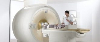
- during pregnancy, in order to avoid severe consequences of the procedure for the fetus, it is recommended to conduct other studies, such as ultrasound or MRI (magnetic resonance imaging).
Interpretation of the results obtained
X-ray of the stomach is an informative examination that will allow the gastroenterologist to establish an accurate diagnosis and select the most effective treatment for the patient.
The results of the study help identify various diseases:
| Name | Description |
| Stomach deformity | The pathological condition can be congenital or acquired. A characteristic feature in the image is the irregular shape of the stomach. A certain section of it narrows, forming an “hourglass” shape. |
| Peptic ulcer | The disease can be detected by fluoroscopy with contrast. The examination allows you to determine signs of pathological processes in the area of the lesser curvature or lower part of the stomach. |
| Stomach cancer | The disease leads to deformation of the walls of the organ. The mucous membrane thickens, the boundaries in the image are blurred. More often, changes are detected in the area of the old ulcer. |
| Gastritis | Fluoroscopy can reveal compaction or deformation of the mucous layer on the inside of the stomach. |
| Pyloric stenosis | In the photographs, the body of the stomach is expanded, and in the area of the pylorus there is a sharp narrowing. The results of the study will also show the absence of contrast transition in the duodenum. |
Fluoroscopy helps to identify an inflammatory process in the digestive organs, varicose veins, disruption of the lumen, the formation of polyps or changes in the size of the gastric cavity.
Advantages and disadvantages of fluoroscopy
The advantage of fluoroscopy compared to radiography is the ability to examine internal organs in motion, during their functioning, and to track their displacement and extensibility.
It also becomes possible to accurately determine the sites of organ damage, since objects can be viewed from different angles. With radiography, multiple images would be needed to obtain such images.
X-ray examination is used when installing a catheter, angiography (contrast examination of blood vessels), and examination of fistulas (pathological openings in the body).
The disadvantages of this method are:
- significant exposure to x-rays;
- the need to view images in a dark room, since the glowing screen is not bright enough to view images in a bright room;
- getting an image that is not sharp enough. Because of this, it is impossible to detect diseases in the initial stage. Additional x-ray examination will be required, which means the patient will be exposed to additional radiation.
Radiography
This single-shot imaging method has pros and cons.
Advantages
- X-ray radiation is dangerous at significant dosages. Radiography allows the radiologist not to be in the same room with the patient when the X-ray machine is turned on and not to be exposed to additional radiation.
An x-ray technician observes a patient through a special x-ray protective glass
- The ability to see small details thanks to the good resolution of the films.
- Less radiation exposure. With radiography, a few seconds are enough for examination, while fluoroscopy takes a minimum of 5 and a maximum of 20 minutes.
- With this method, the image, like a document, can be stored for a long time. If necessary, it can be re-examined at any time, rather than relying on the examination results described on paper by the radiologist. This allows us to draw conclusions about the dynamics of the pathology and the correctness of the diagnosis.
- X-rays are not recommended frequently, and fluoroscopy is even more discouraged given the higher radiation exposure. However, in some cases, X-ray examinations must be performed periodically, which results in additional radiation exposure. For example, when monitoring the dynamics of the course and therapy of pulmonary diseases, after trauma operations to assess the healing of bone disorders, the correctness and speed of consolidation of fractures. In such cases, radiography is preferred for dynamic monitoring.
Flaws
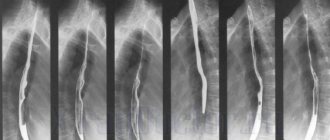
- A radiographic photograph, or radiograph, is some summary image of all shadows. This is a flat image of a three-dimensional object. Therefore, in order to obtain a reliable result of the object being examined, a couple of photographs are taken in different projections, sometimes more. Only a few images allow you to see the pathological focus from different sides.
- There is no way to evaluate the organ while it is functioning. An x-ray allows you to see only one moment of the functioning of the body.
In what cases is fluoroscopy indicated and contraindicated?
Indications for the use of this diagnostic method are suspicions of the presence of organ diseases:
- abdominal cavity;
- chest;
- of cardio-vascular system;
- genitourinary system.
Fluoroscopy is widely used as a way to monitor the following:
- vascular catheterization;
- vascular stenting (expansion of pathologically narrowed areas by introducing a thin metal tube - a stent) into the vessel;
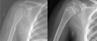
- repositioning of fractures (comparing bone fragments for the purpose of better fusion).
To diagnose the urinary tract, uterus and fallopian tubes, bronchial tree, esophagus, stomach, intestines, and blood vessels, a contrast agent is injected into the patient’s body, which accumulates X-rays well and makes it possible to accurately delimit anatomical structures, study the nature of filling, patency, and contraction of the hollow organ under study.
Contraindications to fluoroscopy are:
- pregnancy;
- reduced immunity;
- the presence of allergic reactions of the body to the contrast agent;
- childhood.
If the attending physician believes that refusing the procedure will cause more harm to the child than performing it, he has the right to prescribe such a diagnosis to his young patient as an exception.
Features of fluoroscopy in pediatrics include the use of personal protective equipment (lead aprons, ties, screens). First of all, the eyes, genitals, and thyroid gland need protection. During the examination of newborns, the entire body except the area being examined is covered with a lead screen, just like for adults.
Digital technologies in fluoroscopy
The main differences from film radiographic technologies are the ability to digitally process an X-ray image and immediately display it on a monitor screen or recording device with an image recorded, for example, on paper.
Digital technologies in fluoroscopy can be divided into:
- full frame method;
- scanning method.
Full frame method
This method is characterized by obtaining a projection of the entire area of the object under study onto an X-ray sensitive receiver (film or matrix) of a size close to the size of the area.
The main disadvantage of the method is scattered X-ray radiation. During primary irradiation of an entire area of an object (for example, a human body), some of the rays are absorbed by the body, and some are scattered to the sides, which additionally illuminates the areas that were initially absorbed by the X-ray beam. This reduces the resolution and creates areas where the projected points are illuminated. The result is an X-ray image with a decrease in the range of brightness, contrast and image resolution.
During a full-frame examination of a body area, the entire area is irradiated simultaneously. Attempts to reduce the amount of secondary scattered radiation by using a radiographic raster leads to partial absorption of X-rays, but also to an increase in the intensity of the source and an increase in the radiation dosage.
Scanning method
In this method we can distinguish:
- one-line scanning method;
- multi-line scanning method.
Single-line scanning method
The most promising is the scanning method of obtaining an x-ray image. That is, an X-ray image is obtained by a certain beam of X-rays moving at a constant speed. The image is recorded line by line (single line method) by a narrow linear X-ray sensitive matrix and transferred to a computer. At the same time, the irradiation dosage is reduced hundreds or more times, images are obtained with virtually no loss in the range of brightness, contrast and, most importantly, volumetric (spatial) resolution.
Multi-line scanning method
The multi-line scanning method is more efficient than the single-line scanning method. With the single-line scanning method, due to the minimum size of the X-ray beam (1-2 mm), the width of the single-line matrix of 100 μm, the presence of various types of vibrations, and equipment play, repeated irradiations are obtained. By using multi-line scanning technology, it was possible to reduce secondary scattered radiation by hundreds of times and reduce the intensity of the X-ray beam by the same amount. At the same time, all other indicators of the resulting X-ray image have been improved: brightness range, contrast and resolution. The priority of this method belongs to Russian scientists and is protected by a patent[2].