Ultrasound is still an effective and cost-effective way to find out about the condition of internal organs. Previously, examinations were carried out only for organs and soft structures, but now pathologies of the musculoskeletal system can also be detected. What does an ultrasound of the spine show?
For example, the condition of ligaments, nerve endings, intervertebral discs. Most often, a back examination is prescribed along with tomography and MRI. Therefore, below we will find out who can have an ultrasound, what it will show, how to prepare and what the technique is.
In what cases is an ultrasound of the spine performed?
Any pathology in the sacrolumbar region and spinal column has serious consequences for the entire body. If left untreated, problems with internal organs, blood vessels, blood circulation and brain function may develop.
The spine is responsible for posture, maintaining the correct state of the body, cartilage tissue is responsible for shock absorption, and the spinal cord ensures the functioning of nerve endings, the supply of cerebrospinal fluid and the functioning of internal organs.
An ultrasound of the spine may be recommended in the following cases :
- Pain in the back and entire spine.
- Difficulty performing simple movements.
- Shortness of breath, heaviness when inhaling.
- Migraines, constant headache.
- Dizziness.
- Scoliosis and poor posture.
- Problems with blood pressure.
- Problems with vision and hearing.
- Numbness of the limbs and loss of sensation.
- Memory problems.
- Disruption of internal organs.
If the patient experiences at least two of the above symptoms, constant, shooting pain in the back, then an urgent need to consult a doctor for diagnosis.
Features and advantages of the technique
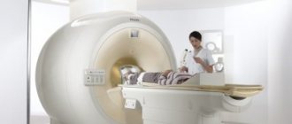
Diseases occurring in the vertebrae or soft tissues near the spine often require surgical intervention. Therefore, to make an accurate diagnosis and clarify all the data, the doctor resorts to various modern diagnostic methods. One of them is ultrasound examination. This is a non-invasive diagnostic test that operates on the principle of echolocation. It allows you to get a complete picture of the disease, determine the cause and stage of the pathological process.
The main features of ultrasound are:
- Safety. When performed, there is no ionizing cure, as when diagnosing back diseases using X-rays, CT or MRI.
- Multiple uses. No harm to the body, allows you to repeat the procedure the required number of times, without being tied to any time interval.
- Ease of implementation. It is especially important in infants, as it affects the result obtained. No special training or use of additional devices is required.
- No discomfort. During execution, the patient does not feel pain or discomfort.
- Versatility. Examination is prescribed even for pregnant women, during lactation or infants. All thanks to security and ease of use.
- Availability. Ultrasounds of the spine are performed not only in Moscow, but also in any other city in the country. This is one of the most inexpensive and simple diagnostic methods.
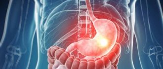
The development of the disease in the spine also affects the condition of the internal organs located nearby. An ultrasound examination allows us to timely detect not only the disorder itself, but also the cause, and prescribe comprehensive treatment.
Benefits of the survey
Not everyone knows what advantages an ultrasound examination provides and the accuracy of the data obtained :
- Affordable research price.
- Safety.
- Possibility of frequent use.
- Accuracy and information content of the data.
- Environmental friendliness of the survey.
- Possibility to see pathologies in intervertebral discs.
- Possibility of examination during pregnancy.
- Possibility of diagnostics for young children.
If we compare ultrasound with other examination methods, such as tomography, CT or X-ray, then ultrasound is absolutely safe, ultrasound waves do not contain harmful radiation, and the pictures are obtained in high resolution; it is possible to conduct a layer-by-layer examination of any soft structure.
But one of the main advantages is the ability to perform ultrasound frequently, which helps to monitor pathologies.
Ultrasound and MRI - what are they?
Both types of diagnostics have their advantages and disadvantages, but the choice of the preferred method is not only due to this. The intended diagnosis is of great importance, since in some cases the chosen method may not be very informative when studying certain areas and tissues of the spine. To answer the question of which type of examination is more effective, it is necessary to understand the essence of their work and know the advantages and disadvantages.
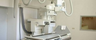
Ultrasound of the spine
Ultrasound diagnostics refers to non-invasive examination methods carried out using ultrasound radiation. The ultrasound system consists of an ultrasonic wave generator and a transducer. Linear and convex probes are commonly used to examine the spine. Devices with linear sensors have a short wavelength and allow you to evaluate tissue located at a depth of up to 11 cm from the point of contact with the skin. They are suitable for examining superficial structures such as small joints and small muscles.
Functional diagram of a traditional ultrasound system
To study large joints (for example, the hip) or organs located at a depth of 11 to 25 cm, it is better to use a convex probe. Not all clinics have such devices, so in some cases such diagnostics have to be carried out for a fee. Examination of the spine using a convex sensor is quite informative, but this type of diagnosis has a significant drawback: the resulting image is several centimeters larger than the size of the sensor itself, which requires a certain qualification of a specialist to make an accurate diagnosis.
Ultrasound of the spine
MRI of the spine
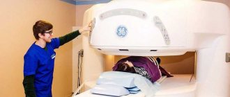
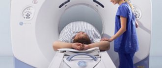
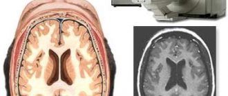
Magnetic resonance imaging is a diagnostic method based on the effect of resonant absorption. The method has many contraindications and requires proper preparation for the procedure. Before the examination, the patient must remove all metal jewelry, braces, and medicinal patches. If there are metal elements in the body (for example, metal plates), you must inform your doctor about this. MRI is prohibited for persons with any tattoos on the body: such patients will be offered alternative diagnostic options.
MRI machine diagram
The standard examination time is about 40 minutes. The tomograph is quite noisy, so you need to insert special headphones or earplugs into your ears. If necessary, contrast agents are injected into the vein 10-30 minutes before the scan.
Absolute contraindications
For pathologies of the lumbar and sacral spine, the doctor may suggest a magnetic resonance examination of the spine with verticalization. At the first stage, scanning takes place in the usual mode, after which the table, together with the patient and the tomograph, is raised to a vertical position. This allows you to create an axial load, as a result of which it will be easier to assess the degree of mobility and stability of the vertebrae and identify displaced intervertebral hernias.
MRI of the spine
Main types of ultrasound
Many people are interested in whether ultrasound is performed for osteochondrosis and what type of procedure is performed. In the presence of osteochondrosis, ultrasound is prescribed as part of a set of tests, and two-dimensional ultrasound examination is most often used. There are also 3D and 4D ultrasound, but for this the hospital must have special equipment, and more often it is used only in private clinics.
Video on the topic:
The type of examination is selected based on the approximate location and diagnosis of the patient.
Diagnostic cost
The price of the procedure depends on the popularity of the medical institution, the time it takes, and the location of the clinic. In Moscow, the cost of an ultrasound scan of the lumbar region ranges from 1,500 to 2,000 rubles. In St. Petersburg - from 1000 to 2600 rubles. In the regions - from 1000 to 1500 rubles. You can get tested for free at a municipal clinic with a doctor’s referral and a compulsory medical insurance policy.
Ultrasound examination is the only option for those for whom X-rays, MRI, and other diagnostic methods are contraindicated. This method allows you to assess the condition of ligaments and muscles and establish a diagnosis with maximum accuracy.
Have you had an ultrasound of the lumbar region? Share your experience, leave comments, tell your friends about the article on social networks. And be healthy.
What diseases can ultrasound diagnostics show?
Let us immediately note that it is impossible to find out about bone pathologies using ultrasound; this requires more accurate examination methods.
But ultrasound helps to identify such diseases and problems.:
| State | Localization |
| Degenerative and inflammatory processes in ligaments and soft tissues. | In all parts of the spine |
| Disc fission of the vertebral disc. | In the lumbar region |
| Scoliosis, osteochondrosis and osteoarthrosis. | In the lower back and cervical region |
| Injuries and damage to joints and muscle tissue. | In all parts of the spinal column |
| Herniated intervertebral discs. | In the spinal column |
| Congenital malformations in the spinal column. | All parts of the spine |
Indications and contraindications for the study
It makes sense to examine the lumbar region with an ultrasound if there are possible problems with soft tissues. Indications for the study are:
- development of oncological formations of various types;
- inflammation of soft tissues;
- myositis, or inflammation of skeletal muscles;
- development of cysts and hematomas.
Due to its harmlessness to humans, ultrasound has no contraindications. However, examination of the lower back is carried out with restrictions in certain categories of patients:
- those who are overweight, which will reduce the information content of the study;
- having scars of skin and soft tissues;
- in case of damage to the upper layer of skin in the area under study.
Preparation for the procedure
If we are talking about performing an ultrasound in the thoracic and cervical region, then no special preparation is needed.
If the lumbar spine is examined, then a day before the procedure it is necessary to cleanse the intestines of feces and gases.
This is done due to the fact that the procedure is performed in a supine position .
Experts advise avoiding foods that cause gas. You can also take anti-flatulence tablets.
Diagnostics in the lumbar region
When performing an ultrasound in the lumbar region, it is imperative to cleanse the intestines. To do this, three days before the procedure, it is better to give up dairy, legumes, fresh vegetables, and carbonated water . It is recommended to take Espumisan, activated carbon.
It is better to examine the lower back using ultrasound in the morning, on an empty stomach, after a five-hour fast. In the doctor’s office, the patient lies on his back, after which the abdominal area is smeared with a special gel, then diagnostics are carried out using pressing movements using a special sensor.
Ultrasound in this case helps to identify such diseases:
- Hernias.
- Deformation of bone and cartilage tissue.
- Violation of the integrity of the nerve roots.
- Tumors.
- Inflammation in the yellow ligaments.
Diagnosis of the cervical spine
When examining the cervical spine, the patient sits or lies on his back. The doctor uses a probe to examine the structures of the spine from the front and side of the neck. As a result, pathologies such as degenerative changes in soft tissues, stenosis, damage and injuries of the spinal column can be detected.
Diagnosis of the thoracic region
An examination of the thoracic region using ultrasound should be carried out in a supine position. Although ultrasound in this case is prescribed very rarely, since it is difficult to obtain information due to the surrounding structures in the sternum. Instead of an ultrasound examination in the thoracic region, an x-ray or tomography is often prescribed.
We recommend viewing:
The essence of the ultrasound examination technique
Ultrasound (sonography) is based on the ability of human tissue to reflect sound waves in the form of an echo. For the procedure, a special device is used - an ultrasonic wave generator. It sends signals inside the body, then receives and processes information through special sensors.
The data is displayed on a monitor connected to the device in black and white. The best option is to use 64 shades. Positive echoes are indicated in white, negative echoes are indicated in black. Depending on the operating mode of the device, the monitor displays a one-dimensional or two-dimensional image.
Ultrasound can be conventional, real-time, or based on the Doppler effect. Its essence is that during movement, ultrasonic waves are reflected from a moving organ at different frequencies. This type of ultrasound produces a three-dimensional image. It is often used to assess the condition of blood vessels.
Differences between ultrasound and other hardware diagnostic methods
In addition to ultrasound, other non-invasive methods of examining the body are used in medical practice:
- X-ray. The patient stands between the photographic film and the radiation source, which passes radiation through the human body. The doctor receives a black and white photograph, which shows cracks in the bones, blood vessels, and abnormalities of soft organs. Unlike ultrasonic waves, radiation has a negative impact on health, so x-rays are not indicated for everyone and are prescribed at certain intervals.
- CT. The method is based on computer processing of X-ray radiation. During the examination, a series of photographs is taken, resulting in a three-dimensional image. It is used to assess the condition of bones, teeth, and joints. The study reveals spinal pathologies such as scoliosis, hernia, tumor, and cyst. Due to radioactive radiation, the procedure is more dangerous than ultrasound and x-rays, but the pictures are better.
- Magnetic resonance imaging (MRI). The method is based on measuring the response of atomic nuclei, which are excited using electromagnetic waves. Using MRI, clear images of hard-to-reach organs, ligaments, and muscles are obtained; the speed of blood flow and cerebrospinal fluid can be measured. The method provides clearer images than ultrasound, but it cannot be done on people who have metal objects in their bodies (pacemakers, prostheses, piercings).
Article on the topic: Todd's palsy - long-term post-epileptic paresis of unknown nature
Advantages and disadvantages of the method
When performing an ultrasound, the body is not exposed to radiation, so the procedure is safe. It is prescribed to infants and elderly people. The study can be done several times in a row to monitor the success of therapy. Other benefits of the procedure:
- price (in clinics they do it for free);
- research speed;
- no invasive intervention;
- the opportunity to see in real time how internal organs work and assess their condition.
Ultrasound sonography of the spine has disadvantages:
- the image is limited by the sensor area;
- the resolution is lower than MRI and CT - the picture is worse;
- due to the heterogeneity of internal structures, there is a lot of interference along the path of ultrasound, distorting the results;
- The quality of the image depends on the angle of application of the sensor - errors in diagnosing the tumor process are possible.
Patient reviews
Polina : “After the injury, I had severe back pain, I endured it for a long time, but I decided to go to the hospital and see a therapist. I was prescribed an ultrasound, and I liked the procedure. Because I didn’t feel any discomfort or pain. And the data was enough to determine the course of treatment.”
Artyom : “I have osteochondrosis in the cervical region, so I often go for an ultrasound procedure to monitor the disease. The main advantage of the procedure is its low cost, even in private clinics, and it is also safe for health.”
Alya : “I’ve gone for the procedure before, only when there were problems with my kidneys. I can’t say anything bad: fast, inexpensive, reliable and safe. I don’t know how accurate the data can be obtained if the problems are with bones and not soft tissues. But the doctor recommended an ultrasound.”
How to prepare for the examination
No special preparation is required for an ultrasound of the back, although before examining the lumbar region it is necessary to cleanse the intestines - this will help increase the information content of the study. Before an ultrasound examination, you should not eat foods that cause gas formation:
- Rye bread;
- carbonated drinks;
- milk products;
- raw fruits and vegetables.
It is permissible to take medications that help in the fight against flatulence. The last meal should be at least 8 hours before the examination.
Ultrasound diagnostics of the lumbar spine
Diagnosis is mandatory in the following cases:
- Concerned about pain in the lumbar region;
- There is a suspicion of a herniated or displaced spinal disc;
- To identify congenital abnormalities;
- If there is a dysfunction of the lower extremities.
For maximum accuracy of the study, simple rules must be followed. The study is carried out on an empty stomach, so it is forbidden to eat in the morning. Two days before the upcoming test, you need to follow a diet that excludes foods that can provoke fermentation and increased gas formation. Immediately before the ultrasound, you need to cleanse the intestines, that is, do an enema or take a laxative.
Upon entering the office, the patient needs to remove clothes above the waist and lower his trousers a little. Next, the person lies down on the couch, first on his stomach. The area to be examined is treated with a special gel. During the examination, the specialist may ask you to change your body position, for example, sit down, stand up, arch your back, etc. The procedure usually lasts no more than 30 minutes.
In the absence of deviations in the structure of the lumbar spine, the vertebral discs have small nuclei with a homogeneous structure and have normal echogenicity.
What is the effectiveness of ultrasound diagnostics?
The main advantage of ultrasound diagnostics and its advantage over other practiced methods is safety. Therefore, according to indications, ultrasound can be performed even on newborn children.
The study can identify many pathological changes in the early stages, including:
- Changes in the structure of intervertebral discs (cracks, fibres, etc.).
- Anomalies in the formation and development of the spinal column.
- Damage to the vascular part of the spinal zone.
- The presence of protrusions and hernias (except for the thoracic and sacral regions).
- The appearance of narrowing of the spinal canals, etc.
A huge advantage is that ultrasound of the spine can be performed an unlimited number of times.
Ultrasound of the sacral region
Ultrasound examination of this area in medical practice began to be used relatively recently. This is due to the high bone density resulting in the reflection of UV rays.
Prescribed in the presence of pain localized in the sacrum and lower back, signs of osteochondrosis, lumbar lumbago, as well as pain in the buttock and hip area.
Helps identify the following problems:
- Instability of the sacral vertebrae.
- Displacement of spinal segments.
- Compression of vertebral bodies.
- Presence of injuries to the lumbosacral area.
Conducting this study is of particular importance at the treatment stage in order to monitor the dynamics of changes against the background of prescribed procedures and medications.