Signs of the disease
The doctor sees the vertebra itself and the spinal hernia on the x-ray if the patient has the following indirect signs of the disease:
- the gap of the intervertebral joints has a wedge-shaped appearance;
- the vertebrae of adjacent discs are displaced by a distance of more than 3–4 mm;
- a person suffers from constant spinal ailments;
- a person loses tendon reflexes.
Today, approximately 70% of workers in the Russian Federation complain of unpleasant back pain.
As a result, X-rays play a large role in diagnosing a hernia. In the presence of a hernial protrusion, the human spine becomes less elastic and loses various functions.
A spinal hernia occurs in the following situations:
- due to intense physical activity;
- after lifting heavy furniture or other objects;
- with increased pressure inside the spinal discs;
- in case of bleeding or tumor formation;
- due to various bacterial infections.
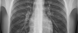
The x-ray image does not show the anatomical structure of the hernia. However, X-rays pass through soft tissue without any reflections.
What doctors will see
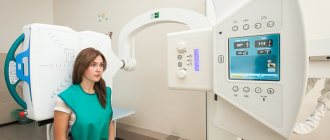
The two main projections of the procedure allow you to see the vertebral arch and spinous processes. Regardless of the degree of development of the hernial tumor, the image will definitely show the following nuances:
- Distance between disks.
- distinct contouring of each of the vertebrae: the slightest irregularities in the outlines can become a signal. For example, with a Schmorl's hernia, a peculiar concavity of the vertebra is present, and if additional processes are detected, osteochondrosis is diagnosed.
- Bone density level. Bones fixed in one place are particularly brightly colored in the image.
At first glance, all this is quite enough to make the correct diagnosis. In reality, it turns out completely differently. All these are indirect signs of a vertebral hernia; in fact, it will not be possible to consider a serious pathology. But at the same time, one should not underestimate the effectiveness of X-ray examination as part of the general identification of back problems.
Information content of the method
Is a hernia of an unhealthy spine visible on an x-ray? Such a study is considered insufficiently informative diagnostics.
After all, when studying the results of an X-ray examination, the doctor often does not see all the signs of a spinal hernia. The maximum that can be carefully examined in an X-ray image is the height of the vertebral discs, because it is not difficult for the doctor to calculate the distance between the vertebrae.
If the height of the spinal disc is reduced, this indicates the presence of degenerative or other changes in the back. However, during the formation of a hernia, prolapse of the spinal disc occurs, which in some cases is not associated with the destruction of cartilage tissue at all.
Thus, the main disadvantages of such a hernia diagnosis are the following:
- poor and unclear visualization. From an x-ray, the doctor cannot see cartilage, damaged ligaments or muscles.
- Causing harm to the body from ionizing radiation. Such radiation greatly affects the patient if he is diagnosed with stage 1 cancer.
At the same time, computed tomography (CT) is also considered a diagnostic test that imposes a significant radiation dose on a person. The doctor sends the patient for a CT scan for specific indications.
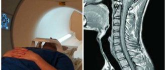
As a result, only MRI is the safest and most informative diagnostic method. She shows tissues filled with water.
When performing an MRI, the doctor identifies the following signs of a spinal disease:
- the vertebral hernia itself;
- vector of development of hernial protrusion;
- degree of damage to the spinal disc;
- damage to ligaments, muscle fibers, nerves in the back;
- various swelling and inflammation.
The doctor sends the patient for an MRI after he saw a wedge-shaped gap in the spinal disc from the X-ray image, which indicates the appearance of a large hernia.
So, when taking an x-ray, the doctor does not always see a vertebral hernia. However, a highly qualified and experienced neurologist identifies a hernial protrusion according to a number of specific criteria. In such a situation, the doctor determines the completeness of various tendon reflexes, sensitivity of the arms, legs, etc.
Indications for examination
When a patient comes in with back pain, a specialist, having collected the necessary medical history data, writes out a referral for an x-ray. Many people doubt the need for this procedure because they are not sure whether a spinal hernia can be seen on an X-ray. It is necessary to undergo an examination for one reason: only he will be able to determine with maximum accuracy the scale of the problem that has arisen: whether the muscles and ligaments are damaged, or whether discomfort and pain arise against the background of deformation of the osteochondral tissue. In addition, only x-rays will be able to show a change in the distance between the vertebrae, which is the first sign of neoplasms in the intervertebral space. Deformation of areas, bulging, unevenness, displacement, subluxation - all this is clearly visible during examination.
At the same time, today doctors are increasingly recommending that those who suspect a malignant tumor in the vertebral regions delay taking x-rays.
The same applies to suspicions of osteochondrosis and displacement. After all, the obvious problem will never be identified, and the person will receive a share of the radiation.
Contraindications
Not all patients with spinal hernia can undergo an X-ray examination. Radiation during such a diagnosis of the back is considered insignificant, however, there are conditions of patients in which the body is very sensitive to radiation exposure. In such cases, x-rays cannot be performed.
Thus, the doctor does not refer a patient with a hernia for a similar diagnosis in the following situations:
- in case of serious condition of the patient;
- if a person has a pneumothorax (air appears in the pleura);
- for any internal bleeding. In this case, the hernia on the x-ray will only worsen and the pain will become stronger;
- if the patient is pregnant (especially 1st trimester).
In the above cases, the neurologist sends the patient to the safest diagnostic methods: MRI, ultrasound (ultrasound), etc.
basic information
Between the vertebral bodies there are layers of cartilage - these are intervertebral discs. They consist of a ring-shaped element (annulus fibrosus), inside of which there is a jelly-like substance (nucleus pulposus), which has good elasticity. Intervertebral discs are the main shock absorbers of the spine. When walking, running, and even while standing, they help soften the vertical pressure (axial load) on the spinal column and dampen vibrations when the body moves.
Under unfavorable factors that worsen metabolism in the osteochondral components of the spinal system, degenerative and dystrophic processes in the structure-forming units of the shock-absorbing layer are activated, due to which the fibrous ring is destroyed and torn. The gelatinous substance, in turn, begins to put pressure on the damaged area, which leads to protrusion of the disc from a certain side. Since the protruding part is usually located in close proximity to the spinal nerve formations, irritation occurs of these nerves with which the pathological protrusion comes into contact. Thus, a person begins to experience painful symptoms in the affected area of the back or neck, often radiating to the leg or arm.
How to prepare for the procedure?
Can an X-ray of the spine show a hernia? Yes maybe. However, the patient needs to carefully prepare for such an examination. In such a situation, a person performs the following actions:
- cleanses the intestines with a regular enema. After all, intestinal gases attach to each other and do not transmit x-rays. As a result, the overall picture of the hernia is blurred.
- A few days before the x-ray, he follows a specific diet. For example, a person refuses various gas-forming foods (cabbage, carrots, milk, peas, etc.).
- After meals, he drinks tablets such as Festal, Mezim, and activated charcoal.
- He does not eat anything in the morning before the hernia x-ray.
Before performing an x-ray, the doctor asks the patient if she is pregnant, since such a procedure negatively affects the development and growth of the intrauterine fetus.
If an x-ray of the hernia is urgently needed, the doctor covers the pregnant woman’s stomach with a special leaded apron.
Preparing for an X-ray
Examination of the cervical or thoracic area does not require any preparation. The same cannot be said about a lumbar hernia. The main preparatory measures are aimed at eliminating gases in the intestines, since their fermentation can seriously distort the diagnostic result:
- A week before the x-ray, exclude all foods that provoke fermentation in the intestines (cabbage, rye bread, beer, legumes);
- Carrying out a cleansing enema immediately before the x-ray.
In addition, the information content of the image also depends on how correctly the patient behaved during the procedure: the laboratory assistant determines the correct position, which is important to follow to the end.
Principle of the method
When passing through different tissues of the body, X-rays are absorbed with different strengths. Thus, bones and spinal hernia on x-rays delay such radiation more strongly, and muscle structures and various organs - less. Therefore, on a computer monitor, the doctor sees an x-ray image, which shows the outlines of different parts of the body, first of all, bones.
X-rays were first used to diagnose various ailments in 1895. In the 19th century, 1 x-ray of the hand was taken. Over the past century, application methods have improved significantly, and the resolution of X-rays has also increased.
Doctors have not performed such fluoroscopy for a long time, during which the doctor saw a picture of the patient’s body on a special fluorescent screen. Today, more promising and accurate diagnostic methods are often used: computer tomography (CT), MRI.
However, today radiography is still considered indispensable in the diagnosis of spinal and other joint ailments, because it is an almost free procedure and has a high diagnostic value.
Will an x-ray show a herniated disc?
A hernia is not diagnosed on an X-ray of the spine, with the exception of a central Schmorl hernia. This study is carried out to identify complications that arise in the presence of disc herniation, as well as once again confirm with indirect signs changes in the spinal column that indicate a hernial protrusion.
What can you see on an x-ray?
The information content of images taken to diagnose a spinal hernia is extremely low.
A hernial protrusion is not visible on an x-ray, so doctors have to take into account a combination of indirect signs indicating a hernia.
For example, it is possible to declare a pathology with high accuracy if the gaps between the vertebrae are practically not detected.
With the help of this examination, degenerative processes in tissues are detected. Indeed, the result of such processes is an intervertebral hernia, but it does not always occur, so it cannot be said with one hundred percent certainty that pathological changes in the vertebrae necessarily indicate the presence of a hernia.
To make a more accurate diagnosis, additional diagnostics are used - functional radiography. This study also does not provide a direct indication of the presence of a hernia, but a great diagnostic success would be the detection of vertebral instability.
If the displacement of one vertebra relative to another is more than four millimeters, then there is a high probability that the patient has a hernial formation.
By the way, this technique does not allow you to see a hernia, but only indicates the predisposition of the spinal column to its occurrence.
Is a herniated disc visible on an x-ray if it is taken digitally? This question is often asked by patients to doctors, thinking that the “digital” prefix gives the study increased accuracy. Not at all, because the difference between these x-ray methods is in storing information, but digital is not any more informative.
Since the hernial protrusion cannot be seen, doctors prefer not to prescribe x-rays to patients - it is better not to irradiate the body with high doses of radiation, and to carry out diagnostics only when absolutely necessary. Much more informative is magnetic resonance and computed tomography, which helps to see back pathologies from several angles.
Contraindications
X-ray examination, like other diagnostic procedures, has contraindications. Doctors definitely do not prescribe spinal x-rays to the following patients:
- pregnant women;
- obese;
- those who had a barium examination in the last four hours;
- people who cannot remain still for some time.
Positive and negative aspects of diagnosis
An undoubted advantage of radiography is the diagnosis of complications associated with intervertebral hernia, as well as degenerative changes that could provoke it.
However, X-rays also have disadvantages, among which doctors note:
- inability to more clearly examine the intervertebral discs;
- X-ray does not see intervertebral protrusions;
- the body is exposed to radiation;
- low information content for pathologies such as osteoporosis, which is associated with a hernia.
X-ray examination is an ambiguous examination, which has both positive and negative sides. With regard to intervertebral hernia, images obtained by this method do not provide valuable information, since the hernia is not indicated on them. Is it possible to see other indirect signs - yes, but not the hernia itself, so doctors selectively prescribe x-rays.
https://youtu.be/SRlQmnVT0ec
Source: https://osnimke.ru/kosti-i-sustavy/gryzha-pozvonochnika-na-rentgene.html
What does radiography show?
Does an x-ray show a herniated spine? Yes it is. If a doctor sees a pathological deformation of the vertebral discs on an X-ray, he diagnoses the patient with a “vertebral hernia.”
To determine a hernia from an x-ray, the doctor performs the following steps:
- compares the right and left vertebral sides, checks the symmetry of the spinal column;
- measures the distance between the vertebrae, determines the places where the vertebral discs come together and the distance between them decreases;
- carefully examines the vertebral contours, studies their clarity;
- examines the bone structure of the spine for its color and uniformity;
- reveals visible vertebral growths.
Since a spinal hernia leads to the formation of pain, inflammation and swelling in neighboring tissues, the person takes the most comfortable position, in which the pain becomes less. Thus, the patient’s spine is curved, which is clearly visible in the picture.
When studying an X-ray of a spinal hernia, the neurologist compares 2 factors - large prolapse of the spinal disc and displacement of various vertebrae.
When performing radiography, those tissues in which a lot of calcium salts have accumulated are clearly visible. Calcium is clearly visible in the image.
Also, from a photograph of the spine, the doctor determines the bony base of the spine. Between the individual spinal discs there is a hollow space where the cartilaginous spinal discs are located.
The doctor makes a diagnosis of “intervertebral hernia” if the patient has the following signs of the disease:
- a decrease in the distance between the vertebral discs, during which the vertebrae themselves are destroyed;
- the formation of a growth on the vertebra at a specific location of the hernia;
- excessive growth of various bone tissues along the vertebral discs. Thus, bony processes appear on the front and side of the back;
- the appearance of osteophytes;
- pathological disorder of the location of the vertebrae;
- the structure of bone changes has changed. In such a situation, the patient experiences excessive growth of various bone tissues of the back;
- change in the curvature of the spine when examining the side view of x-rays;
- formation of vertebral osteoporosis or osteosclerosis.
Reliable methods for diagnosing spinal hernia
Since an x-ray cannot show the presence of hernia formations, doctors often diagnose the disease using other methods. If the neurologist is sufficiently experienced and competent, then he can make an assumption about the presence of a disease without resorting to radiography. He will pay attention to a decrease in tendon reflexes, a decrease in pain and tactile sensitivity. All of these signs give reason to assume the presence of a hernia in the spine.
Magnetic resonance imaging provides more accurate information about the condition of the spine and the presence of hernial bulges. An MRI shows the area where the nerve root was pinched and the fibrous cartilage fibers were torn.
This modern method also makes it possible to determine the location of swelling and inflammation. To date, MRI is the best method for determining hernial protrusion. The principle of its effect is that hydrogen is passed through a magnetic field, while registering radio waves. Tissues containing large amounts of water are studied.
A few words should be said about Schmorl's hernia. It has some differences from a regular intervertebral hernia. This is the place of its localization, the absence of compression of the spinal roots, and the absence of involvement of neurovascular bundles in the pathological reaction. However, the danger of such a pathology is still high. This is explained by the fact that Schmorl's hernia, not detected in time and not amenable to therapeutic intervention, often leads to serious complications.
Is it possible to see this type of hernial protrusion on an x-ray? The name of the disease itself is a radiological concept. You can install it in various ways:
- visual inspection and palpation;
- analysis of medical history;
- patient complaints.
Pros and cons of the study
The advantages of diagnosing a hernia using X-ray equipment are the following:
- X-ray is a quick way to study a spinal disorder;
- X-ray imaging of a spinal hernia is considered a simple, safe and accessible procedure for all patients, which shows even the smallest deviations in the back;
- when performing an x-ray, the doctor evaluates the bone structure of the vertebral discs, the density and thickness of the various cortical layers;
- In the X-ray image, the neurologist sees fluid accumulation and various deformations of the vertebral discs.
Moreover, x-rays of the lumbar spinal region are used in the diagnosis of various ailments of the spine, and it is the primary examination of the patient.
The only disadvantage of X-rays is that X-ray rays have a negative effect on the human body.
If you find an error, please select a piece of text and press Ctrl+Enter. We will definitely fix it, and you will get + to karma
Symptoms ↑
Intervertebral hernia causes unbearable pain, tingling, numbness of the legs, a feeling of “cottoniness”, “crawling goosebumps”. Often the patient is only bothered by pain in the leg, and he has no idea about the source and cause of this pain.
Read also…. Radiculitis of the cervical spine
The most typical symptoms of the disease:
- in the case of the formation of a hernia at the L4-L5 level, in addition to pain and stiffness in the lower back, weakness occurs in the area of the big toe, pain in the thigh, buttock, a feeling of “pins and needles” appears in the leg, with prolonged sitting there is numbness in the toes and other symptoms;
- in the case of a hernia in the L5-S1 area, the pain is characteristic of the ankle, knee, spreads to the inner thigh, numbness of the legs and other unpleasant sensations appear.
- In some patients, spinal hernias form in several places at the same time, so the symptoms are layered.
The symptoms described above occur during the typical course of the disease and may vary, it all depends on the individual characteristics of each patient. To make a correct diagnosis, additional diagnostic methods are used.
Dorsal hernia l5 S1
This is one of the most common and dangerous types of protrusion, and mostly occurs in the lumbosacral link l5-S1. This form can also occur in the neck, mainly involving the C5 C6 disc. The word “dorsal” means a hernia in which the displacement is concentrated directly towards the spinal canal (posterior location). That is, if the hernial protrusion is directed into the intervertebral lumen of the spinal canal, it will be called dorsal. Such a hernia, in turn, can be median (central) and paramedian (at an angle). As we said, this is the most typical form of the disease, but central protrusions are more severe because they prolapse into the spinal canal just along the midline.
Education at level I5 S1.
Since it is here that the central nervous system (spinal cord) and the adjacent abundance of nerve plexuses are localized, the appearance of this type of deformation is accompanied by extremely unpleasant symptoms and a more sophisticated set of neurological signs. And in the absence of adequate treatment - blocking the lumen of the canal and paralysis of the limbs. The reasons for its development are the same as for all other hernias.
Pregnancy and hernia
If there is a pregnancy, an experienced specialist recommends gentle conservative therapy that does not negatively affect the fetus and the well-being of the expectant mother. Pregnant women will require treatment according to a specific regimen, since its absence can lead to very bad consequences, including disability. The operation is postponed until the postpartum period. Typically, during pregnancy, wearing a bandage, light exercise, swimming, and the use of non-aggressive medications and folk remedies to relieve pain are recommended.
Paramedian hernia T6 T7
The disc, which forms a shock-absorbing “cushion” at the T6-T7 level, which corresponds to the thoracic area, is almost never damaged. This is due to the fact that it is in this part that the spinal structure is very securely fixed by a muscle corset. However, destruction of the T6 T7 disc with all the ensuing consequences cannot be 100% ruled out. It may be a tiny percentage, but it is there. Therefore, if you feel some painful discomfort between the shoulder blades and/or in the hypochondrium, a high-quality differential diagnosis is necessary to exclude pathologies with similar symptoms:
- pneumonia and pleurisy;
- abscess pneumonia;
- heart attack;
- angina pectoris;
- pericarditis, myocarditis;
- inflammation of the esophagus, pancreas, gastric mucosa, etc.
When it is confirmed that the disturbing signs are associated with paramedian protrusion, prolapse of the thoracic intervertebral plate, you need to start fighting it. After all, complications can be terrible - paresis and paralysis of all parts of the body that are located below the focal point.
