To diagnose gastric diseases, MRI is prescribed much less frequently than more traditional methods (FGDS, fluoroscopy and radiography).
This is due to the fact that MRI of hollow organs, such as the stomach, is less informative, and in addition, the procedure is quite expensive.
But some patients have difficult-to-diagnose situations in which MRI is used. To increase its information content, special methods for studying a hollow organ have been developed and used.
The advantage of MRI is the absence of radiation (unlike X-ray examination) and discomfort for the patient (characteristic of FGDS).
Introduction
The stomach
is a hollow muscular organ located between the esophagus and the duodenum. It is a pouch-like expansion of the digestive canal in which swallowed food accumulates and is digested. Gastric juice secreted by the glands of the gastric mucosa contains digestive enzymes, hydrochloric acid and other substances, digests proteins, partially fats, and has a bactericidal effect. In addition, mechanical grinding of food occurs in the stomach.
Magnetic resonance imaging of the stomach can be part of a comprehensive diagnostic system of abdominal organs or prescribed on an individual basis if there are signs of a pathological condition of the organ.
| Anatomical diagram of the structure and location of the stomach | MRI image of the stomach |
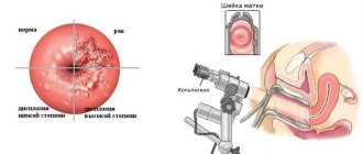
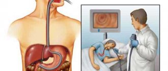
MRI of the stomach - an alternative to gastroscopy?
When it comes to stomach diseases, the “gold standard” for diagnosis is usually endoscopic examination - gastroscopy. What about other methods, in particular MRI? Is it possible to do an MRI of the stomach? Will it be possible to check this organ with its help? What does this diagnostic show?
We talked about MRI of the stomach with the executive director of Clinic Expert Orenburg, radiologist Yuri Andreevich Podlevskikh.
— Yuri Andreevich, how often do people come to your center with the question of whether they can do an MRI of the stomach?
Quite often: at least several times a week.
— Is it possible to see the stomach on an MRI? Do you follow this procedure?
We do not have an MRI of the stomach as a separate study. Currently, magnetic resonance imaging is not the “gold standard” for diagnosing diseases of this organ; it is not separately highlighted in the list of our services. We perform MRI of the abdominal organs. The stomach and some – I emphasize this word – we can see its pathological changes.
- Which? Will an MRI show, for example, signs of gastritis, erosion, stomach ulcers?
Gastritis - inflammation of the stomach lining - is not visible on MRI. Almost the same can be said about erosion and stomach ulcers. Some ulcers can be seen, but this is more an accident than a pattern.
Read materials on the topic:
How to and how not to treat gastritis?
How to protect your stomach from ulcers?
- In other words, if a person has pain in the upper abdomen, heartburn bothers him, you do an MRI of his abdominal organs and do not find pathological changes in the stomach, this does not mean that they are not there.
Absolutely right. The patient may have both gastritis and an ulcer.
— What about stomach cancer? Will an MRI show it or is it also uninformative for this disease?
With this pathology the situation is somewhat different. If you pose the question in the form “Is it possible to detect stomach cancer using MRI,” the answer will be both yes and no. And there is no contradiction here. I'll explain why. Cancer is a tumor that arises from the cells of the mucous membrane - the inner lining of an organ. If there is already cancer, but it is small, has not penetrated into the wall of the stomach, and does not protrude into its cavity, then the MRI will not see it. But if the tumor is already relatively large and protrudes into the lumen of the stomach, or has penetrated into the wall of the stomach and beyond (in the direction of neighboring organs), then MRI will be able to detect such a tumor.
What conclusions can there be? If the neoplasm is small and located on the gastric mucosa, then the best method for detecting it would be fibrogastroduodenoscopy (other names: gastroscopy, FGDS). Moreover, FGDS allows us to identify precancerous changes in the mucous membrane and take small fragments of it (biopsy) for further microscopic examination.
You can read more about gastroscopy in our article
On the other hand, if the tumor has grown beyond the stomach, then traditional FGDS will no longer be able to say anything about its extragastric location. An MRI will help here, which will tell you which organs and tissues the tumor has penetrated.
— That is, asking the question, which is better – MRI or FGDS of the stomach – is not entirely correct?
Yes exactly. Some pathological changes in the stomach are detected earlier and better with the help of FGDS; to clarify others, MRI may be needed.
— Yuri Andreevich, what if we compare MRI with X-ray methods of studying the stomach, in particular, fluoroscopy, radiography?
These methods also have strengths and weaknesses. If we are talking about an X-ray examination with barium (a type of contrast agent), then we can examine the inner surface of the stomach, ulcers, and some formations protruding into the stomach cavity. In this vein, fluoroscopy and radiography of the stomach are more sensitive than MRI. However, like MRI, x-ray examination will not be able to assess either the color of the mucous membrane or its less prominent, less noticeable changes. Of course, a biopsy cannot be done.
At the same time, fluoroscopy of the stomach, like gastroscopy, will not provide information about a tumor that has spread beyond the stomach and penetrated into neighboring areas - an MRI will be needed here.
Read material on the topic: X-ray of the stomach: an eternal classic or a step back?
A kind of “hybrid” diagnostic method, combining some properties of X-ray methods and MRI, can be called CT of the stomach. On the one hand, this method is based on X-ray radiation, and on the other, as in MRI, it allows one to obtain layer-by-layer images. However, CT scan of the stomach is also not “omnipotent”.
— To summarize, can we say that it is now impossible to completely replace gastroscopy with fluoroscopy/radiography, computed tomography or magnetic resonance imaging of the stomach?
Yes. Each of these methods has its own strengths. FGDS is best suited for identifying gastritis, erosions, small ulcers, precancerous changes and the early stage of a tumor located at the level of the mucous membrane. If the tumor is located in the wall of the stomach or has spread beyond its limits, an MRI or CT scan will provide additional information.
In other words, each method most accurately “hits” its target, and the totality of research results in a picture that is closest to objective reality.
In any case, I recommend that you first consult a clinician. For complaints from the stomach, upper abdomen, this can be a therapist, a general practitioner, or from narrow specialists (to whom they can be sent for consultation) - a gastroenterologist, a surgeon, an oncologist. Based on the survey and examination, the doctor will draw up a diagnostic plan, which will include those methods that will help make a diagnosis in each specific case.
You can make an appointment with specialists here
ATTENTION: the service is not available in all cities
Interviewed by Enver Aliyev
The editors recommend:
What to do if you suffer from heartburn?
Living in acid: what do we know about Helicobacter pylori
Colonoscopy of the intestines - is it scary?
When is CT indispensable?
Precancer: to be afraid or not to pay attention?
For reference:
Podlevskikh Yuri Andreevich
Graduate of the Orenburg State Medical Academy in 2012 with a degree in Pediatrics.
In 2013, he completed an internship in the specialty “Radiology”. In the same year, he underwent advanced training in magnetic resonance imaging.
Currently executive director of Clinic Expert Orenburg.
Receives at the address: Gagarin Ave., 17, room 1
Classification of tomographs
Magnetic resonance imaging is performed in special equipment called a tomograph
.
By type, tomographs are divided into open and closed
.
| Open MR tomograph | Closed-type MR tomograph |
The advantages of an open type tomograph are:
- It is suitable for patients suffering from claustrophobia and for children who may be scared in the confined space of a closed CT scanner;
- It has an air gap in which the patient is placed, usually larger than that of closed-type tomographs. This makes it possible to conduct examinations for overweight patients.
The advantages of a closed-type tomograph are:
- The examination time is shorter than that of the open one. The timing of the examination is important for those patients who, for one reason or another, cannot remain motionless for a long time.
- The quality of images from a closed tomograph is higher, because the quality of images depends on the magnitude of the created magnetic field strength, measured in Tesla units. Due to their design features, open-type tomographs cannot create tension comparable to closed-type tomographs.
According to the strength of the created magnetic field, tomographs are divided into
:
- Low-floor - up to 0.5 Tesla.
- Mid-field - from 0.5 to 0.9 Tesla.
- High-field - from 1.0 to 1.5 Tesla.
- Super high-field - 3 Tesla.
As a rule, a voltage of 1.5 Tesla is enough to obtain images of acceptable quality.
| Image quality depending on equipment power |
Indications
A local internist or gastroenterologist can refer you for an MRI of the stomach, or the patient can independently undergo an examination through the sphere of paid services. The need for the procedure arises when the following symptoms occur systematically:
- Aching, cutting, stabbing pain in the abdomen, appearing with a certain frequency.
- Flatulence.
- Feeling of heaviness and bloating.
- Nausea and vomiting for no apparent reason.
- Lack of appetite.
- Unexpected weight loss.
- Change in urine color.
- Traces of blood in the stool.
- Laboratory tests and/or ultrasound revealed signs of degenerative processes, neoplasms, and other pathologies.
- Helminthic infestation in acute form.
MRI scanning is also necessary in the work of oncologists and surgeons when:
- Determining the stage of the neoplasm.
- Assessing the extent of metastasis.
- Checking the effectiveness of the therapy.
- Preparing for surgery.
- Quality control of surgical intervention.
An MRI examination of the stomach allows you to establish an accurate diagnosis, select treatment, check the feasibility of surgical intervention and increases the chances of successfully combating the disease.
What diagnoses can MRI make?
From all that an MRI shows, several of the most common diagnoses can be identified.
- Stomach cancer.
At risk are men who abuse alcohol and tobacco. At the early stage of development of the disease, only some weakness, decreased appetite and ability to work, and an aversion to meat are noted. Then obvious clinical signs make themselves felt:
- frequent attacks of nausea and vomiting;
- the presence of blood in the vomit;
- sudden weight loss;
- fever.
With the help of MRI, the doctor will be able to determine not only the location of the tumor and its size, but also find out whether the pararectal lymph nodes, liver, and ovaries (if a woman is being examined) are affected.
2. Esophageal cancer.
Every fifth cancer patient dies from esophageal cancer. This is one of the most common and dangerous diseases, which already at the very beginning manifests itself with clearly defined symptoms - it becomes difficult and painful for the patient to swallow solid food, then discomfort occurs even when swallowing saliva, and frequent attacks of hiccups occur. As cancer progresses, it causes a severe cough with blood discharge. MRI allows you to visualize a tumor in the esophagus much more accurately than radiography or endosonography and determine whether the disease has spread to the lymph nodes and neighboring tissues.
3. Peptic ulcer.
The MRI image clearly shows all the ulcerative pits, their shape, depth and wall thickness. By supplementing the clinical picture with magnetic tomography, it is also possible to determine the nature of the ulcer - benign or malignant. To do this, measure the thickness of the stomach wall, determine the presence of irregularities, as well as a violation of the signal intensity that is reflected from the affected wall.
4. Hiatal hernia.
This means that part of the esophagus, upper stomach, or even intestinal loops has shifted through the esophageal opening of the diaphragm into the chest cavity. In this case, an MRI of the esophagus shows folds formed by its mucous layer, or areas of protrusion of the stomach into the posterior wall.
This is a fairly common pathology, affecting at least 5% of the population. But this is only an approximate figure, because in half of the cases the disease does not manifest itself at all, and the patient may not even be aware of the presence of a hernia. Often the pain is localized in the heart area and is mistaken for angina or even a heart attack. Patients who complain of girdle pain may be treated for pancreatitis. Hernia is difficult to diagnose, so an MRI is often the only way to find out the true diagnosis.
5. Esophagitis.
This is an inflammation of the lining of the esophagus caused by infection (most often candida or herpes) or mechanical irritation, such as due to frequent vomiting. With esophagitis, in addition to such common symptoms as heartburn, unpleasant taste and odor in the mouth, nausea, the patient complains that it is difficult and painful for him to swallow. He feels as if food is getting stuck in his esophagus and may have chest pain that radiates to his back.
Contraindications
Contraindications to MRI of the stomach are divided into absolute, when it is impossible to do an MRI, and relative, when it is possible to do it under certain conditions.
Absolute contraindications
:
- Installed pacemaker. Magnetic fields can interfere with the operation of electronic equipment, causing it to malfunction.
- The presence in the body of metal implants or other metal objects (for example, fragments) that react to the influence of a magnetic field. If the patient has implants in the body that do not respond to the magnetic field, then documents confirming this should be provided.
Relative contraindications
:
- Claustrophobia. This contraindication is relevant for closed-type tomographs and can be avoided by conducting examinations on open-type tomographs.
- First trimester of pregnancy.
- The patient cannot remain still during the entire examination. The images will be blurry and therefore not suitable for diagnosis if the patient moves during the examination. The solution may be a sedative or anesthesia.
- The patient's weight exceeds the permissible weight for which the tomograph table is designed. In this case, you need to look for tomographs with a large permissible weight. As a rule, open-type tomographs have an advantage in this regard over closed-type tomographs.
- The diameter of the circumference of the widest part of the body exceeds the length of the air gap of the tomograph. In this situation, you also need to look for a tomograph with the necessary parameters. As a rule, open-type tomographs have a larger air gap than closed-type tomographs.
- The patient has decompensated heart failure.
- The patient is in serious condition.
- Inappropriate behavior, including alcohol intoxication.
Indications and contraindications
As a rule, MRI is not a method of primary examination of a patient. Usually simpler and cheaper methods are used (FGDS and radiography). But in complex diagnostic cases, with an unclear clinical picture of the disease, with a lack of information obtained during the initial study, MRI or CT is indispensable for diagnosing and assessing the condition of not only the stomach, but also the organs adjacent to it. MRI is preferred due to the lack of radiation exposure to the patient.
MRI of the stomach can be prescribed for diagnosis when:
- peptic ulcer;
- chronic gastritis;
- malignant neoplasm of the stomach or suspicion of it;
- diaphragmatic hernias.
Using MRI, the results of treatment are monitored for any of the listed diseases, when for some reason an FGDS cannot be performed.
There are few contraindications for MRI:
- implanted pacemaker:
- presence of an electronic device in the middle ear;
- prostheses of large joints (excluding titanium prostheses);
- any metal objects in the body (bullets, shrapnel, hemostatic clips, braces, etc.);
- epilepsy or tendency to seizures;
- pregnancy;
- body weight over 200 kg.
Preparation
MRI of the stomach requires special preparation.
The stomach is a hollow organ and therefore, before performing an MRI, the patient must undergo special training to ensure the accuracy of the examination. To do this, you must follow the following rules: 2-3 days before the study, stop eating foods that cause increased gas formation: baked goods, legumes, vegetables, dairy products, cabbage, alcoholic and carbonated drinks. On the eve of the procedure, you can take the following medications: Espumisan, Smecta, White Coal, Enterosgel, etc. On the eve of the study, take dinner before 18.00 and perform a cleansing enema. Stop smoking 6 hours before the procedure.
How to prepare for an MRI examination?
This procedure does not require special preparation. Your doctor will recommend that you stop eating and drinking immediately before the test. To obtain the most accurate result, it is recommended to limit yourself in the consumption of tea, coffee and flour products a few days before the procedure. The tomograph creates a special magnetized field, as a result of which before the procedure it is necessary to remove jewelry and metal objects, glasses, piercings, watches, belts, remove your mobile phone, flash cards, coins, bank and transport cards from your pockets. Females are advised to limit themselves to minimal makeup, as some products contain metal particles.
How does the procedure work?
Carrying out an MRI of the stomach consists of three stages: preparatory, the procedure itself, and issuance of results.
Preparatory stage.
The patient needs to arrive some time before the appointment in order to:
- Complete the documents. If the procedure is paid, then a contract for the provision of paid medical services must be drawn up. If the procedure is for compulsory medical insurance or voluntary medical insurance, then the necessary package of documents is checked.
- Find out contraindications. Contraindications are described in the section of the same name in the article.
- If an MRI with contrast is required, contraindications for administering contrast are clarified. The doctor also needs to select the type and dose of contrast agent, which will take a certain amount of time.
- Next, before the examination, you should change into loose clothing without metal elements and remove all metal objects.
Procedure
:
- The patient is escorted to the tomograph.
- Placed on the table.
- The body is secured with soft straps to avoid micro-movement. Movements during MRI lead to blurring of the image and the examination needs to be redone.
- If necessary, fix the area being examined with a special coil.
- The examination is accompanied by specific noises produced by the tomograph mechanisms. To reduce the negative effect of noise, the patient is given headphones in which music is played, and the instructions of the doctor conducting the examination can be transmitted to the headphones.
- If the procedure is performed on a closed type tomograph, the patient is in a confined space.
Delivery of results
:
- Immediately after the examination, the patient is given an image on film and/or disk.
- For some time after the examination, the patient is given an examination report from a radiologist. Usually this time is about 30 minutes, but it can be longer depending on the doctor’s workload. It may be that the conclusion will be issued the next day. Some MRI centers send the report by email.
In some MRI centers, you can ask the radiologist any questions you have after the procedure.
Duration of the procedure
MRI of the stomach in a closed high-field tomograph with a magnetic field strength of 1.5 Tesla takes up to 30 minutes.
A similar examination using open-type equipment takes up to 60 minutes. If tomography using a contrast agent is necessary to make an accurate diagnosis, the duration of the procedure increases approximately 2 times. An initial scan is performed without contrast, then the solution is injected, and the manipulations are repeated.
Carrying out the procedure
Before starting the procedure, the doctor explains the entire examination process to the patient, talks about the rules and contraindications. Next, the patient lies down on a special table, which then smoothly moves into the tomograph. To obtain the most accurate results, you should remain calm and still. Often, during the examination, the stomach walls are straightened by slightly stretching with the help of an iron-containing solution, which is prescribed to the patient and relieves pain. Sometimes a specialist administers intravenous contrast, shows the most clearly needed areas, and this does not cause any discomfort.
What does it show
If the cause of the painful condition lies in the pathology of another organ, then the MRI image will demonstrate the correct location and size, the absence of signs of inflammation, deformations and neoplasms.
In the presence of pathologies, MRI allows us to establish the following conditions with an accuracy of 95%:
- Abnormal structure of the stomach from birth.
- Developmental defects of the organ.
- Inflammatory processes in colitis.
- Deformation of lymph nodes.
- Mechanical damage to blood vessels and internal bleeding.
- The presence of ulcers and erosions.
- Poor circulation in blood vessels (aneurysms, thrombosis).
- Tissue necrosis, peritonitis.
- Oncological diseases.
- Penetrating and intrinsic metastases.
- Cysts and benign neoplasms.
MRI allows you to distinguish fibrous compactions from an acute inflammatory process, determine the shape and size of the tumor, detect pathology and record relapse of the disease.
What's the difference with and without contrast?
The contrast consists of saline solution and a special substance, is injected into the body intravenously, and transported through the circulatory system to the object of study.
Thanks to the reaction of the components to the influence of the magnetic field, the area of localization of the pathology is “highlighted”, the size and shape of the defect is clearly indicated. Secondly, due to its properties, contrast affects pathological areas in such a way that they are shown on images more clearly (in contrast) relative to areas without pathologies.
This is what the doctor will see in the image taken with and without a contrast solution:
| On the left is an MRI performed with contrast; on the right without contrast solution | ||
There are contraindications for the use of contrast agents
:
- Hypersensitivity to components
- Allergic reactions
- Kidney failure
- Pregnancy
- Lactation.
Progress of magnetic resonance imaging
It's time to find out how an MRI of the intestines, stomach and esophagus is done:
- After collecting an anamnesis and consulting with a specialist about possible contraindications, the patient is placed on the machine’s couch.
- The couch automatically slides into the unit tunnel and the scanning procedure begins.
- During the examination, the patient is asked not to move.
- The duration of magnetic tomography of each gastrointestinal organ can be from 15 to 30 minutes.
Important recommendations before tomography
Knowing how to prepare for magnetic tomography of the gastrointestinal tract, you can prevent the occurrence of side effects, as well as prevent distortion of the results and blurred images:
- If a patient is indicated for hydro MRI of the intestine, then first you need to make sure that he is not allergic to the contrast agent.
- Before coming for an examination, you need to remove all metal objects and put on clothes without locks, hooks, buttons, or pins.
- Wear comfortable clothes so that they do not squeeze the skin and the person can lie comfortably in them.
- Be sure to inform the doctor about the presence of built-in metal objects, crowns, pacemakers, etc.
Restrictions, contraindications
If a person has no contraindications to the procedure, then it will not have any negative effect on the body. Tomography can be done as often as required by the doctor.
Is magnetic resonance imaging performed on pregnant and breastfeeding women? This research method is possible, but only without the use of a contrast agent, since contrast is toxic and can have a harmful effect on the fetus and infant. It is also prohibited to do an MRI in the following cases:
- if there are metal objects in the patient’s body;
- with renal failure;
- for claustrophobia (in this case, tomography is performed using an open-type apparatus);
- in case of severe pain (possible MRI, but subject to prior use of an anesthetic drug).
MRI of the stomach, intestines, and esophagus is a modern research method that allows, in one procedure, to examine in detail all organs of the gastrointestinal tract, detect existing problems and make an accurate diagnosis. Based on the MRI results, the specialist decides on the advisability of surgical treatment and other possible methods of therapy. Despite the fact that MRI of the gastrointestinal tract is used less frequently than gastroscopy, in some situations a specialist may prescribe it to confirm or refute a preliminary diagnosis.
CT or MRI, which is better?
It is not entirely correct to compare these methods with each other, since they are based on different principles of image acquisition. Each has its own strengths and weaknesses. The doctor decides whether to send the patient for a CT or MRI based on the purpose of the examination.
If we consider the difference between CT and MRI, then for comparison it is necessary to take the following factors:
Device capabilities
. MRI scans best soft tissue, ideal for assessing the condition of the muscles, ligaments, and tendons around the shoulder joint. CT most accurately demonstrates bone tissue. If a fracture, arthritis or bone tumor is suspected, it is better to entrust the diagnosis to a computed tomography scan. The device is capable of constructing a 3D projection of the joint based on the data obtained during scanning.
To establish an accurate diagnosis, you may need to use both methods.
Duration of the procedure
. For a computed tomography scan, it only takes 1-2 minutes. Scanning using magnetic waves will require the patient to remain motionless for 15 to 30 minutes.
Harmfulness
. A magnetic resonance imaging scanner has no restrictions on the number of examinations, since it emits impulses that are less harmful to the body than X-rays. A CT scan, on the other hand, is based on X-rays and cannot be performed on a single patient more than twice a year.
A visual demonstration of the capabilities of CT and MRI devices:
| CT | MRI of the stomach |
CT allows you to build 3D models:
| 3D model |
Purpose of procedures
Gastroscopy is prescribed when the doctor suspects the presence of stomach diseases (gastritis, peptic ulcer), as well as the presence of neoplasms and parenchymal bleeding. Gastroscopy makes it possible to examine the stomach and esophagus in detail, and check the structure of the mucous membranes. In addition, the procedure is prescribed to take scrapings from the walls of the stomach or to collect gastric contents for a biopsy. To check the organs of the digestive tract using a gastroscope, there is a more informative alternative to gastroscopy of the stomach as FGDS.
The purpose of FGDS has similar prerequisites as standard gastroscopy. However, the suspicion of a duodenal ulcer is added to the indications of the procedure, since an elastic “hose” is also inserted into the duodenum area.
CT scan is prescribed mainly for diagnosing neoplasms of various types. As a rule, CT, like MRI, is an accurate and expensive research method, which is most often used to confirm the diagnosis established during gastroscopy or FGDS, as well as in cases where standard endoscopic examinations turned out to be not entirely informative. Computed tomography is valuable in identifying anomalies in the structure of an organ, guarantees their detailed study and differentiation, and allows obtaining high-quality images that display the physical condition of the organs.
MRI of the stomach is the main way to diagnose cancer. The information value of the method is difficult to overestimate, since the MRI scanner distinguishes not only the presence of tumors, but also their nature. Magnetic resonance imaging is often prescribed to determine the stage of the oncological process, identify the exact localization of cancer metastases, to assess the condition of the organ and tissues after surgery and in the presence of persistent pain in the gasrum and duodenum of an uncertain nature. MRI is used in almost the same cases as CT, since both methods are highly informative. But if you have to choose what is better to use, CT or MRI of the stomach, then you should give preference to magnetic resonance imaging. MRI does not expose the patient to radiation, while the principle of CT is based on scanning with x-rays, and due to this fact, CT is not allowed to be performed frequently.
More information about MRI of the stomach can be found here
X-ray or MRI, which is better?
X-ray and MRI are fundamentally different examination technologies. If we compare them with each other, then according to the following criteria:
Image quality
. A tomograph scans the human body and builds an image in layers. The image has high resolution, detail, data on soft and bone tissues. An X-ray machine can highlight only one layer and demonstrate the condition of the tissues; the quality of the image will be worse.
| X-ray | MRI of the stomach |
Harmfulness
. X-ray radiation in large quantities is dangerous to health, while MRI can be done at least every day.
Availability of procedures
. An X-ray machine can be found in every ordinary clinic. Equipment for MRI requires large financial costs and the availability of specialists who know how to operate it, so large city and regional medical centers and commercial hospitals can afford a tomograph. You can get an X-ray quickly, but the appointment for an MRI is distributed over weeks in advance.
Price
. An X-ray examination can be done free of charge under the compulsory medical insurance policy in a regular clinic. Only patients from surgery and inpatient departments can get an MRI free of charge.
Ultrasound or MRI, which is better?
Both methods are safe, effective and informative. However, it is necessary to know their capabilities, advantages and disadvantages in order to determine what is more appropriate to apply in a particular case. To do this, let's compare the following aspects:
- Operating principle of the devices
. MRI uses magnetic pulses to visualize internal organs, and ultrasound diagnostics is based on the conversion of ultrasonic waves. People with metal implants, dentures, pins, cochlear devices, and pacemakers are not safe from exposure to electromagnetic fields. The ultrasound technique is suitable for them. - Research speed
. For MRI you need to spend 20-30-40 minutes and at least an hour for description. The ultrasound procedure takes 5-10 minutes, and the result is immediately available. - Device capabilities
. Ultrasound examination does not provide 100% accurate results. The processing of the received data is carried out by a person. The information content and reliability of the examination depends on the experience and skills of the specialist. An MRI machine processes signals, generates images, and interprets data using a computer processor. And only then does the radiologist begin decoding. The likelihood of errors and errors in this case is reduced. - Availability and cost of the procedure
. An ultrasound machine can be found in rural clinics, while an MRI scanner is the prerogative of large regional centers. It is easier to undergo ultrasound diagnostics through compulsory medical insurance. In the case of paid services, the cost of ultrasound is 2-3-4 times lower than MRI.
| Ultrasound | MRI of the stomach |
To kid
There are no age restrictions for MRI of the stomach.
To perform an MRI, a prerequisite is immobility throughout the entire examination. Children may find it difficult to lie still, so anesthesia may be considered.
Children may be afraid to be in the confined space of a closed-type CT scanner. In this case, an examination can be performed using an open-type tomograph. The parent will be able to stay next to the child and hold his hand.
Photo
| Gastrointestinal stromal tumor of the stomach |
| Gastric leiomyoma on MRI |
| Gastric leiomyoma |
| Abdominal cancer with metastases to the stomach |
| Enlarged stomach on MRI |
Price
On our website, many clinics list prices for MRI, including MRI of the stomach.
To see prices, you need to go to the list of clinics in your city. To do this, select your city on the “Addresses” page.
On the page for the list of city clinics, in the form for selecting a clinic based on parameters, in the “Research area” field, select “MRI of the stomach.”
The list will be filtered and the clinics will be sorted by ascending price, which will allow you to find out where to get an MRI of the stomach cheaper.
| List of clinics filtered by MRI of the brain using the example of the city of Moscow |
If prices are not indicated on the website, then you need to call all the clinics and find the best price for you yourself.
When talking with the operator, be sure to ask if there are any promotions or discounts. For example, some 24-hour clinics offer discounts on MRIs at night.
You can get an MRI with compulsory medical insurance or VHI insurance. To do this, you need to find out which clinic does MRI under insurance, using the information on the website or calling the clinics yourself if there is no information on the website. In the form for selecting a clinic by parameters, there is a field “MRI under insurance”. Next, you need to find out the list of documents that the clinic operator will tell you, collect them and you will be able to undergo an MRI with insurance, which will allow you to save a lot.
When is an MRI necessary?
When a patient comes to a gastroenterologist with complaints for the first time, he most often receives a referral for gastroendoscopy - with its help, you can easily visualize the gastrointestinal tract and find the cause of discomfort. But if during this study any neoplasms are identified, then it will no longer be possible to do without an MRI of the esophagus and stomach. After all, the endoscope scans the surface of the mucous membrane, and the organ can only be examined in a section using magnetic resonance imaging.
Of course, you can also see ulcers and inflammation on the MRI monitor, but most often tomography is needed precisely for:
- detailed examination of formations that were discovered during the initial examination with an endoscope or x-ray;
- clarifying the extent of the damage.
Reviews
On our website, people leave reviews for clinics about their MRI experience.
To read patient reviews, you need to go to the list of clinics in your city. To do this, select your city on the “Addresses” page.
The list in the block of each clinic will show a button to go to the list of reviews, if there are reviews, or a button to go to write a review, if there are no reviews yet.
| This is what review buttons look like, using the example of the city of Moscow |
You can also leave a review about your MRI experience so that other people can choose the most suitable clinic.
To leave a review, you need to click the “Your review” button and you will be taken to the review writing form. Or if there are already reviews, then you need to go to the end of the list of reviews and there will be a form for writing a review.
To open the review writing form, click the “Leave a review” button.
Fill out the form and send it for moderation.