Diaphanoscopy is a diagnostic method based on the effect of light on soft tissues. This procedure is also called transillumination and transillumination . Depending on their density, fabrics transmit light radiation differently. Air, as well as liquid substances, have good light transmission, and fabrics with greater density remain impenetrable.
In the case when there is liquid or liquid-like formations in one of the organs, for example, cysts, they can be seen in the lumen, and the organ itself will be highlighted in red. The saturation of red depends on the transparency of the liquid.
Pus is less permeable, and serous fluids are mostly clear. Cavities that contain air are even more permeable. Thanks to diaphanoscopy, a specialist can examine the sinuses, eyeballs, tissues near the eyes, scrotum, and mammary glands.
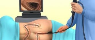
During a diaphanoscopy study, a specialist performs the procedure using a diaphanoscope. This device looks like a straight or curved tube that is powered by electricity. The diaphanoscope has many variations. In urology, this instrument can be replaced by another device called a cystoscope.
What is diaphanoscopy
Human tissue, depending on properties such as density, is determined by the ability to transmit light through itself. Using this property, it is possible to study some human organs.
This method is based on the use of a powerful light flux that does not heat up. This light flux is passed through the thickness of the organ being examined, for example, the scrotum. As a result of this method, tissue translucency occurs. The area that is translucent should have a bright red tint, which indicates the absence of pathologies and neoplasms. If the doctor who performs this kind of x-ray reveals darkened areas, then this suggests the presence of certain pathologies.
Through a diaphanoscopic examination, it is possible to determine the presence of a cyst in the organ under study, a tumor, inflammatory processes and other neoplasms, including foreign objects. The procedure is quite simple; if there is a dark spot on the translucent area, this indicates the presence of pathology.
Diaphanoscopy can be used to examine the following organs:
- eyeballs;
- scrotum;
- mammary gland;
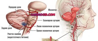
- intracranial space;
- eyelids and soft tissues of the orbit;
- frontal and maxillary paranasal sinuses.
In each of the above organs, characteristic diseases and pathologies are identified. The advantage of the diagnostic test procedure is the fact that the method is accessible, absolutely harmless, and also quickly feasible. After the study, results can be obtained almost immediately. During the study, the specialist does not take parts of the organ for examination, since the procedure does not involve endoscopic intervention. It also follows from this that the procedure is absolutely painless and therefore does not require the use of anesthesia.
Diaphanoscopy today is considered the best express method for the preliminary diagnosis of various diseases. What kind of diseases these are, we will find out further.
Indications for use
The procedure is indicated mainly for diseases associated with externally located organs:
- In a field such as ophthalmology, research is carried out to identify eye diseases and examine the tissues around the eyes. Diaphanoscopy absolutely painlessly helps to determine the presence of tumors in the visual organ, while ultrasound examination is not able to help in this matter.
- In otolaryngology, this method also provides therapeutic assistance. A doctor may refer a patient for upper respiratory tract diseases, in particular sinusitis and sinusitis, for diaphanoscopy. Indications also include cysts. The study helps determine the presence of tumors in the nasal tissues and is an auxiliary element in therapy, helping to monitor the patient's current condition. During transillumination, the beam penetrates the maxillary sinuses, as well as the frontal sinuses and soft nasal tissues.
- Thanks to diaphanoscopy, a mammologist can obtain certain information regarding the condition of the mammary glands and identify tumors under the skin. Tumors, cysts and tissue edema have different densities and transmit light differently. The diaphanoscope also helps to check the condition of the implants and turns out to be a very important element in monitoring the progress of therapy.
- In traumatology, this research method is used to see fragments and other foreign bodies in tissues.
- Urologists prescribe this diagnostic method in cases where diseases of the external genitalia occur. Diaphanoscopy of the scrotum is mainly used, as well as diagnosis of diseases of the penis.
Diaphanoscopy has no contraindications. Among its significant advantages are the absence of discomfort and a non-contact diagnostic method.
This procedure does not require any special preparation and is performed painlessly and simply. The only thing that a patient needs in order for a specialist to conduct a diaphanoscopic examination is compliance with basic hygiene rules .
What pathologies are detected by diaphanoscopy?
The basis of diaphanoscopy is the transillumination method, through which an organ is illuminated with a large light flux without heating it. This allows you to bring the light flux as close as possible to the organ being examined. Using this method, the following types of diseases can be identified:
- In the mammary glands, the diaphanoscopy technique makes it possible to detect the formation of tumors, inflammatory foci and cysts.
- In the paranasal sinuses, this method allows you to determine the presence of polyps, tumors, as well as inflammation and purulent exudate.
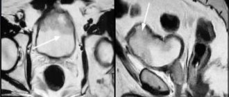
- In the eyeballs: retinal detachment, connective tissue formations, as well as areas of atrophy and intraocular tumors.
- In the scrotum, the diagnostic technique allows us to detect the presence of dropsy, cysts, tumors of the appendages, as well as a hernia.
- The intracranial space is characterized by the following diseases, which can be determined by diaphanoscopy: hydrocephalus, as well as intracerebral tumors and hemorrhages.
A preliminary diagnosis after light transillumination of the organ being examined allows for timely initiation of treatment. This method allows you to make an accurate diagnosis, but after it, if there are pathologies, you will need to undergo an additional ultrasound. Diaphanoscopy allows you to quickly and painlessly determine the presence of pathology, for which it is actually valued. Let's consider several methods for studying organs using diaphanoscopy.
Execution order
A diaphanoscope is a light source attached to a handle. Previously, ordinary small incandescent lamps were used, now preference is given to xenon light sources. The inspection is carried out in a darkened room.
- The device is placed in the patient's mouth, the patient wraps his lips around it, and the beam of light is directed upward. In this case, the maxillary sinuses are visible in the dark in the form of red symmetrical crescents in the upper part of the cheeks. In case of a pathological process (sinusitis, cyst, tumor), the resulting image will be asymmetrical, with different densities.
- To examine the frontal sinus, the light source is brought close to the skin in the area of the upper inner corner of the orbit. In this case, a cap is put on it, leaving only the upper part of the light bulb open. This way you can identify signs of frontal sinusitis and other diseases.
- Using a modern fiber optic diaphanoscope, it is possible to examine the ethmoid labyrinth. In this case, the nasal cavity is irrigated with an anesthetic and a flexible light guide is inserted into it. Normally, the inside of the orbit allows light to pass through, but with inflammation (ethmoiditis), the bony septum remains dark.
Diaphanoscopy can also be used to assess the effectiveness of treatment. In this case, it is carried out by one specialist at the beginning and after completion of therapy. In particular, the method is relevant for assessing the condition of the frontal sinus after frontotomy surgery.
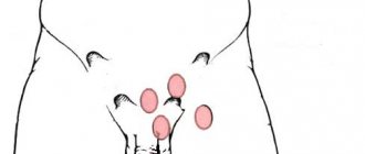
Diaphanoscopy of the testicles
In urology, the diaphanoscopy technique is widely used. With its help, a urologist can examine the scrotum and identify the presence of pathologies or confirm their absence. Diaphanoscopy of the scrotum is performed as follows:
- A special powerful flashlight is used for illumination. The procedure itself is carried out in a dark room.
- The patient needs to lower his pants and then sit on the couch.
- The doctor brings a flashlight to the back of the scrotum, and then evaluates the uniformity of transmission of light rays by this organ.
After a preliminary diagnosis through diaphanoscopy, an ultrasound examination is required. This method does not make it possible to make an accurate diagnosis, so if there are deviations, an ultrasound scan is performed.
- Hydrocele of the testicle is characterized by such a symptom as a painful enlargement of the scrotum, both of the entire organ as a whole and of a specific part. Dropsy is determined using diaphanoscopy, which, when examined, will turn bright red.
- A tumor of the epididymis manifests itself in the form of the formation of a dense lesion in the testicle. Most often, this tumor is located in the caudal region.
- Testicular tumor is one of the most serious and dangerous diseases. Diaphanoscopy allows you to determine such a disease by detecting a compaction in the form of a node. As a result, there is an increase in the size of the scrotum and its swelling.
- An inguinal hernia manifests itself as pain in the groin area of the abdomen. This is the most common pathology, manifesting mainly in young children. The diaphanoscopy procedure is prescribed if there is a suspicion of hydrocele.
Indications
The main indication for performing this diagnostic procedure is visual enlargement of the scrotum and surrounding soft tissues. Diaphanoscopy of the scrotum is recommended if the following pathological conditions are suspected:
- torsion of the spermatic cord, as a result of which the man develops secondary testicular edema;
- single or multiple hemorrhages in the scrotum;
- benign and malignant neoplasms in the area of the testicles and their appendages;
- foreign bodies and consequences of traumatic injury to the scrotum;
- spermatocele, which is a benign neoplasm resembling a cyst;
- hydrocele or hydrocele of the testicle.
In addition, the testicular diaphanoscopy technique is widely used for diagnostic purposes in young boys with suspected inguinal hernia. Despite its selective information content, this diagnostic technique is inferior to ultrasound.
Diaphanoscopy of the eye
Transillumination of the eyeball is carried out using a diaphanoscope. The technique can cause pain, so the use of painkillers is not excluded. For children at an early age, general anesthesia is used.
Once the patient is ready, the doctor turns off the light and turns on the diaphanoscope. The device is brought to the eye and moved along the surface of the conjunctiva. During the movement, the doctor changes the pressing force, which is necessary for the correct examination of the eye.
During the procedure, the specialist monitors the glow in the pupil area. The uniformity and brightness of the glow of the pupil indicates the absence of pathological abnormalities. Seals are detected by changing the intensity and brightness of the glow.
Diaphanoscopy of the eye can be performed in two ways: transscleral and transpupillary. The most common is transscleral diaphanoscopy, which is based on applying a light beam to the sclera to the equator and beyond it. The transpupillary technique involves applying the tip to the central part of the cornea. Pathology is detected in the presence of a shadow.
Diaphanoscope
The study is carried out using a device - a diaphanoscope. It is a powerful light bulb placed in a closed housing. Inside it there is also a lens system that collects light rays (a condenser) and a fan.
At the end of the diaphanoscope there is a tip - a conical-shaped light guide with a curved end. This design of the nozzle makes it possible to illuminate the eyeball from the opposite end of the equator.
The light guide is designed so that the rays are reflected several times on their way to the exit. By concentrating, they form a narrow beam of light. Often the kit includes tips with varying degrees of curvature. Light guides with a flexible end, made from a fiber bundle, are also produced.
The light of the diaphanoscope is cold, which eliminates the possibility of overheating the eye.
Diaphanoscopy of the paranasal sinuses
The principle of diaphanoscopy of the paranasal sinuses is based on the fact that light is applied to the inner corner of the orbit and passes through intact bones, as well as the soft tissues of the skull and paranasal sinuses. This creates a bright glow. The technique is carried out in a dark room.
To perform transillumination of the maxillary sinuses, the device is inserted into the patient’s mouth, after which it is necessary to wrap your lips around its cone. If there are no pathologies, then both halves of the face are due to a reddish glow, and two halves of the moon can be observed under the eye sockets.
If the patient has pathologies in the form of neoplasms, then there is no glow in the area of the maxillary sinus. In this case, the patient does not feel the presence of light with his eye. To illuminate the frontal sinus, the device is applied to the inner corner of the orbit. A healthy sinus is due to the appearance of a bright red glow.
The main advantage of a diaphanoscope over ultrasound and x-rays is absolute safety. The technique is allowed to be carried out even in the presence of damage to the skin in the areas under study.
Diagnosis of the testicle and scrotum
For ailments localized in the scrotum area, diaphanoscopy is mainly used. This method is especially effective when the doctor needs to find the cause of the enlargement of a given organ. Among the indications is testicular hydrocele. With this disease, fluid from the abdominal area collects in the testicular cavity. It is this phenomenon that affects the growth of the scrotum.
It is serous in nature and translucent. In contrast to swelling of the skin, tumors, inflammatory processes in the membrane of the testicle, as well as its appendages, with dropsy the permeability is much higher.
- The procedure is performed in the period after surgery if swelling occurs in the scrotum. To determine further strategy, diaphanoscopy can be of significant help.
- The technique also helps determine the condition of the sutures after surgery.
- When examining the testicle, the diaphanoscope is able to detect foreign elements in the soft tissues resulting from injuries. Also, a special device can accurately indicate the location of stones in the vas deferens.
Technically, the examination does not cause any difficulties for the patient. The procedure is performed in a dark room. A specialist armed with a diaphanoscope, directing a light beam at the external organ, monitors the permeability of light, illuminating the scrotum from all sides. The condition of the organ is determined by swelling and coloring.