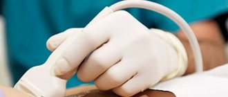
Ultrasound
Ultrasound waves are reflected differently by muscles, joints, and blood vessels. The computer converts the signal into a two-dimensional or three-dimensional image.
When used . For diagnosis in cardiology, oncology, obstetrics and gynecology. The device shows internal organs in real time. This is the safest method.
See: How harmful are X-rays and ultrasounds for health →
How it works
The mysterious machine through which luggage passes through at the airport is, overall, a fairly simple device. This is a short tunnel where suitcases and bags are transported using a conveyor belt. The inside of the tunnel is lined with lead, which can absorb most of the radiation created by the X-ray generator, the main element of the scanner.
“X-rays enter the tunnel through an opening about 1 cm wide and illuminate each piece of luggage. On the wall of the tunnel opposite from the source there is a detector that measures the amount of radiation that has passed through each point of the scanned object. Based on this data, the computer creates an image of the object,” explains Mikko Halonen, security specialist at Finavia, explaining how the device works.
Expert opinions
Ilya Gipp, Ph.D., Head of MRI-guided therapy:
— Many of these devices can be used for treatment. For example, a special installation is attached to an MRI machine. It focuses ultrasound waves inside the body, increasing the temperature in a targeted manner, and burns out tumors - for example, uterine fibroids.
Kirill Shalyaev, director of the largest Dutch manufacturer of medical equipment:
- What seemed impossible yesterday is reality today. Previously, during CT scans, a drug was administered to slow down the heart. The newest CT scanners make 4 rotations per second, so there is no need to slow down the heart.
| What radiation doses do we receive* | ||
| Action | Dose in mSv** | Over what period of time will we receive this radiation in nature? |
| X-ray of a hand | 0,001 | Less than 1 day |
| X-ray of a hand using the very first machine in 1896. | 1,5 | 5 months |
| Fluorography | 0,06 | 30 days |
| Mammography | 0,6 | 2 months |
| Mammography with MicroDose characteristic | 0,03 | 3 days |
| Whole body CT scan | 10 | 3 years |
| Live in a brick or concrete house for a year | 0,08 | 40 days |
| Annual norm from all natural radiation sources | 2,4 | 1 year |
| Dose received by liquidators of the Chernobyl accident | 200 | 60 years |
| Acute radiation sickness | 1000 | 300 years |
| Epicenter of a nuclear explosion, death on the spot | 50 000 | 15 thousand years |
| *According to Philips **Microsievert (mSv) is a unit of measurement of ionizing radiation. One sievert is the amount of energy absorbed by a kilogram of biological tissue. | ||
Entry of foreign particles into the esophagus
When a baby swallows foreign objects, they often become lodged in the esophagus. This is due to the presence of natural narrowings of the organ. If your baby swallows a pin, fish bone, or other sharp object, this can lead to a perforation of the esophagus wall. Urgent diagnosis and treatment is necessary.
Ball in the esophagus
Symptoms of an object entering the esophagus are unbearable chest pain, reminiscent of the sensation of a myocardial infarction. The patient may experience belching and increased salivation. Pain is also possible during the swallowing process.
Diagnosis of foreign objects in the esophagus
What does a foreign body look like on an x-ray? Foreign bodies of the esophagus are most easily identified on x-ray if their density differs from the density of the tissues of the organ. More dense particles appear as a light spot in the image, while less dense particles appear as a dark spot. If the density of a piece that has entered the esophagus is equal to the density of the tissue of the organ, it cannot be seen with conventional fluoroscopy. In this case, X-rays with contrast are used.
Oddly enough, the X-ray clearly shows pieces of glass or crystal that have entered the esophagus. These materials have high density and radiopacity.
Diagnostics for the baby
X-rays are taken in several projections to determine the size and location of the object. An aqueous solution of amidotrizoate is used as a contrast. When the wall of the esophagus is perforated, contrast leaks through the hole.
Preparation for the procedure
In most cases, X-rays are taken without special preparation. To undergo diagnostics using this method, you only need a doctor's referral. However, in some cases, preparation for x-rays is still required. This applies to intestinal x-rays. Before the procedure begins, he must be completely freed from food debris and feces. The preparation program for x-ray examination of the gastrointestinal tract includes:
- slag-free diet - fatty foods, sausages, spices, almost all cereals (except semolina and buckwheat), fresh vegetables and fruits are excluded from the diet;
- refusal to eat 18 hours and drink 10 hours before the procedure;
- taking laxatives 18 hours before the examination;
- cleansing enema 10 hours before the study.
Immediately before the x-ray, it is recommended to remove metal objects from the body in the area affected by the x-ray, as well as thick and synthetic clothing.
Pregnancy and X-ray
Pregnancy is a time of special care for yourself and your unborn child. Nutrition, avoidance of smoking and alcohol, and caution with medications become especially important factors during this period. X-rays and other medical radiation procedures of the abdominal area also deserve extra attention during pregnancy.
For starters, you most likely won't need abdominal x-rays during pregnancy. But sometimes, because of a certain medical condition, a doctor may deem a diagnostic X-ray of the abdomen or lower body necessary. If this happens, don't be upset. The risk to you and your unborn baby is very small, and the benefit of finding out your medical condition is much greater. In fact, the risk of not getting the X-rays you need can be much greater than the risk from radiation.
It is important to tell your doctor if you are pregnant or suspect you are pregnant. If you are pregnant, your doctor may decide it is best to cancel the x-ray, delay it, or choose a different test to reduce the amount of radiation. Or, depending on your medical needs, and understanding that the risk is very low, the doctor may decide that it is best to take an x-ray as planned. In any case, you should not be afraid to express any concerns you have to your doctor.
Why is it made?
An x-ray makes it possible to make a correct diagnosis in order to decide on further treatment. X-ray diagnostics must be carried out:
- for fractures and cracks of bones;
- to detect the location and extent of internal damage during trauma;
- during dental treatment and prosthetics;
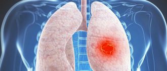
- for diagnosing cancer;
- when examining the condition of the lungs;
- while studying the cerebral cortex;
- for scanning the vascular system if ruptures are suspected.
Contraindications
Despite the widespread use of the method and its safety, X-rays are ionizing radiation that can have a negative effect on the retina, reproductive organs, bone marrow and epithelium. The procedure is contraindicated in the following cases:
- state of pregnancy at any stage;
- the child is under 14 years of age;
- in case of general serious condition of the subject;
- the patient has been diagnosed with an active form of tuberculosis;
- with pneumothorax or bleeding;
- for endocrine pathologies of the thyroid gland.
A doctor has the right to prescribe x-rays to children or pregnant women only in extreme cases when there is a threat to their life.
Examination technique
During the procedure, the patient lies on a couch or a special gynecological chair. The X-ray machine is located above it, and if echohysterosalpingoscopy is planned, the doctor uses a special vaginal sensor.
Before inserting a catheter into the genitals, the doctor disinfects the cervix, vagina and external genitalia with an antiseptic solution. The catheters and devices used in the process must be strictly sterile, and a special condom is placed on the ultrasound sensor.
Before inserting the catheter, the doctor examines the patient using a speculum. After this, a soft catheter is inserted into the cervix, through which a contrast agent is injected with a syringe. The substance enters directly into the uterine cavity and gradually passes into the fallopian tubes. At this time, the doctor takes a series of x-rays.
Echohysterosalpingoscopy of the uterine cavity follows a similar pattern: the doctor injects a saline solution into the uterine cavity, after which he carefully inserts the ultrasound device sensor.
The procedure usually does not cause severe pain or discomfort. The sensations during it are reminiscent of the pain syndrome in the first few days of menstruation. In this regard, most often, the use of anesthesia is not required. However, if the patient knows that the first days of her menstruation are very painful, and if there are no contraindications to the use of anesthesia, the doctor may administer local anesthesia before starting the diagnosis. General anesthesia is not used for this procedure.
If there is a possibility of spasms of the fallopian tubes or uterus, the doctor suggests taking antispasmodics, for example, the drug “No-shpa”. Most often, in such cases, an injection is given so that the active substance enters the blood faster.
What are the possible consequences of an x-ray?
There is scientific disagreement about whether small amounts of radiation used in diagnostic radiology can harm an unborn baby, but it is known that the baby is very sensitive to the effects of things such as radiation, certain medications, alcohol, and infections. This occurs in part because cells quickly divide and form new cells and tissues. If radiation or other agents could cause changes in these cells, the likelihood of birth defects or certain diseases such as leukemia may increase.
It should be noted, however, that most birth defects and childhood diseases occur even if the mother was not exposed to any harmful effects during pregnancy. Scientists believe that heredity and random errors during development are responsible for most of these problems.
Diagnostic results: what to do next
The obtained images of the uterus and fallopian tubes, or images of echohysterosalpingoscopy, are interpreted by a diagnostician. He draws up a conclusion on them, in which he displays the most objective and reliable information.
X-rays show the filling of the fallopian cavity and fallopian tubes with contrast material. If the drug passes freely through the tubes and is visualized in the abdominal cavity, then everything is in order with the patency of the fallopian tubes. If the image clearly shows that the fluid did not pass and stopped at a certain level, this confirms the presence of obstruction. The alternation of dark and light areas in the pipes in the image indicates that they have adhesions.
In addition, the peculiarities of the distribution of contrast throughout all examined cavities and organs makes it possible to see neoplasms, polyps, and foci of inflammation.
Also, the diagnostician draws conclusions about the size of the uterus and fallopian tubes, the features of their structure and location, and the structure of the inner wall of the uterus from photographs or the monitor of an ultrasound machine. For example, its uneven relief may indicate the presence of adhesions, inflammation, polyps or fibroids.
If the results of the examination give reason to suspect the presence of uterine cancer, it is necessary to order additional examinations, including taking tissue for a biopsy.
The diagnostic doctor's conclusion, along with the photographs, is transmitted to the attending physician, who referred the woman for examination.
After hysterosalpingography, the patient may experience mild vaginal bleeding for several days. Pain in the lower abdomen that appears during the examination usually goes away after 20-30 minutes. In the next 3-4 days, you should avoid sexual intercourse, visiting a bathhouse, sauna, or taking a bath.
Types of X-ray examinations in medicine
In modern diagnostics, there are several basic x-ray examination methods. They are used to identify pathologies of tissues and organs of different structure and structure:
- radiography of the gastrointestinal tract - to assess tone, peristalsis, size and structure, mucosal defects and foreign objects;
- radiography of the abdominal organs - to determine concentration and contractile functions, detect stones and neoplasms, foreign objects and perforations;
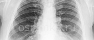
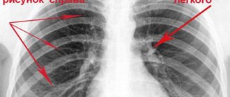
- X-ray of the chest and spine - to assess the structure of skeletal bones, identify tumors, degenerative lesions, infectious processes, etc.;
- radiography of peripheral parts of the skeleton - to identify congenital and acquired defects and injuries;
- radiography of soft tissues (breasts) - to identify tumor processes.
Different types of radiography are used to study different types of tissues and organs. Almost all studies have been well studied, but research is still being carried out on certain species, thanks to which the photographs become more informative.
Radiography of soft tissues
This type of radiography involves simple “transillumination” of soft tissues: muscles, joint capsules, tendons and ligaments, subcutaneous fatty tissue and skin. The essence of the diagnostic method is to assess the structure, density, and size of the elements under study. The photographs show all the pathological changes in the thickness of the tissue. To make a diagnosis, it is enough to take an x-ray in one projection, but in difficult situations 2-3 projections may be required.
Panoramic
This is one of the most high-tech x-ray examinations, which is used mainly in dentistry and orthodontics . The picture is a panoramic image of the maxillofacial region:
- teeth of the lower and upper jaw;
- soft tissues adjacent to them;
- maxillary sinuses.
Panoramic radiography is used to develop treatment tactics for defects of the maxillofacial apparatus, gum diseases, as well as for the correct installation of dental implants.
Intraoral (intraoral)
Intraoral or contact radiography is used in dentistry and periodontics. It allows you to obtain images of individual teeth in the upper and lower jaws and the adjacent soft tissues. To obtain a clear image, it is necessary to use lead plates that are placed on the inside of the jaw.
Sighting
Unlike simple radiography, targeted radiography allows you to get clearer images. It is used to diagnose pathologies of individual organs and areas of the body. In the resulting image, the study area is larger in size, so even small changes are visible. The scope of application of targeted radiography is practically unlimited, but most often it is used to identify pathologies of the chest organs.
Contrasting
The study, which uses contrast in addition to X-rays, is used to diagnose pathologies of hollow organs (intestines, blood vessels, kidneys and bladder, ureters, uterus) and fistulas. Such an X-ray examination helps to establish the structural features of organs, assess their functionality and determine the location of foreign bodies, tumors and lipid deposits, stones and stenosis.
Microfocus
Microfocus X-ray is an innovative method that allows multiple magnification of the object of study. In contrast to simple and targeted radiography, the area of study is limited to fractions of a millimeter. In this case, microscopic defects that are invisible during conventional studies come into focus. Microfocus radiography is used to diagnose pathologies of soft tissues, hollow organs, and bones.
X-ray according to Vogt
The eyes are examined using Vogt radiography. The method is used when small (microscopic) foreign bodies enter the eyeball. During the procedure, the laboratory technician takes two photographs in different projections, in which shadows are visible in the area where foreign bodies are located.
Important! Since the fundus of the eye reacts negatively to high doses of x-rays, the Vogt method involves the use of the softest radiation.
With functional tests
This type of x-ray is used to diagnose diseases of the musculoskeletal system. The procedure allows us to identify problems that occur when performing certain movements: defective fixation of bones, curvature of the spine and birth injuries in newborns. To identify them, during the examination the patient will have to tilt his head or take certain positions.
Isn't this dangerous?
When picking up their “irradiated” suitcase at the exit from the device, some passengers wonder: could irradiation somehow spoil the things in the luggage, or even “inject” radiation into them? Experts are sure: it can’t even theoretically. “During scanning, the object is exposed to radiation equal to 1 microsievert. You get the same amount for an hour of flight at an altitude of 10 km,” explains Joni Pekkanen. “And this is 10 times less than the background from an X-ray in a dental office,” adds Mikko Halonen.
The only thing for which radiation poses a potential danger is highly sensitive photographic or film film. It is recommended to pack it in hand luggage (or better yet, carry it in your hands) and notify airport employees about its presence in your luggage.