Scintigraphy of skeletal bones is a modern method for studying the skeletal framework and bone tissue using the latest radionuclide devices. This is an imaging, a skeletal scan. Thanks to the method, it is possible to detect diseases, bone deformations, and tumor growths in tissues in the early stages of development. The development of metastases is traced, if cancer is detected, splits and cracks in the skeletal base are visible.
In medicine, the method in question has been used recently, so most people are unfamiliar with it. The process of studying bone tissue takes place in a specially designed chamber equipped with radioisotope radiation and the necessary functions. Doctors say that skeletal bone scintigraphy is superior to standard radiography. The advantage is detailed and detailed visualization, allowing the study of the slightest deviations from the norm.
What is revealed during the examination
The radiation method, scintigraphic polypositional diagnostics allows you to see how intact the body tissue is and to examine the structure of the human skeletal base. The diagnostic method is also used in oncology, in order to see the presence of a tumor, bone formation, or spine. Thanks to the procedure, a malignant tumor can be seen at its inception stage, ahead of the appearance of external symptoms. Visualization of the structure of the body will reveal all pathologies, diseases, and explain the cause of ailments at the cellular level.
Scintigraphy shows metastases formed in the bones if a malignant tumor is present. Based on the examination, an effective and efficient set of therapeutic treatment is prescribed. The diagnostic method also monitors the benefit of therapy, changing the treatment regimen if necessary. The advantage of examining the supporting apparatus is the identification and assessment of pathological diseases before the appearance of the first visible symptoms.
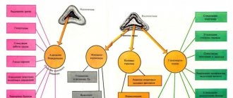
Radiopharmaceuticals
During the study, radiopharmaceuticals based on monophosphates, less often biphosphates, are used. Contains labeled technetium 99mTc. Compatible with developing bone tissue. They are easy to make. Preparation occurs during the procedure. A solution of sodium pyrophosphate with a labeled isotope is added to the radiopharmaceutical powder. The solvent is usually saline solution. In the Russian Federation, the following drugs are used on a regular basis:
- Technetril. Russian radiopharmaceutical combined with healthy cardiomyocytes. Used for single photon emission computed tomography (SPECT) of the heart muscle to diagnose coronary heart disease.
- Pirfotech. Russian radiopharmaceutical with a carrier pyrophosphate, tropic to the developing bone tissue. It is used in osteoscintigraphy to diagnose distant metastases.
- Technetium-99m-MDP. English radiopharmaceutical with the used carrier methylene diphosphonate.
The role of containers for vectors in radiopharmaceuticals used in bone scintigraphy are substances that can accumulate in bone tissue. Preparations containing zoledronic acid are considered advanced. They can detect areas of bone destruction and traces of malignant tumors. This technique is called subtraction. The principle is that based on the images with technetril, the results of scanning the interaction with iodine 123 are calculated.
Indications for diagnosis
A scan of the musculoskeletal structure of the body is prescribed by the attending physician after considering complaints and the results of related tests. The examination process is carried out if there are the following indications:
- Symptoms similar to the spread of metastases to internal organs with an active malignant tumor.
- To analyze the effectiveness of radiation, therapy based on hormones or chemical components. The method of treatment and the chosen scheme of therapeutic procedures are adjusted.
- Signs of inflammation, disrupted processes in the skeletal framework, which is not determined by a standard x-ray.
- Installed prostheses of bones, limbs, suspicion of an inflammatory process, mobility. If it is necessary to adjust the auxiliary prosthesis, scintigraphy of the supporting base is also prescribed.
- Injuries, bruises resulting in damage to bones and spine. The examination is done to exclude or confirm skeletal injury. Also, for osteoporosis, a scan of the bone base is prescribed.
- Slow or excessively fast metabolism and associated bone diseases.
If the above symptoms are observed, the doctor will prescribe a scan of the body’s supporting structures. After scintigraphy, an accurate diagnosis is established, followed by the choice of therapeutic treatment. Skeletal densitometry is performed. The difference with scintigraphy is the examination of bone structure. The procedure also affects a specific area of the body.
List of diagnosed diseases
In the process of studying the properties of radiopharmaceuticals in the body, a radiologist can accurately establish a diagnosis. Metastases of malignant tumors penetrate into bone tissue. When diagnosing diseases of internal organs, this is typical for:
- bladder cancer;
- spleen cancer;
- prostate cancer;
- with pulmonary embolism (pulmonary embolism, a condition when an artery and its branches are blocked by an embolus - a piece of blood clot);
- for pathologies of the lymph nodes;
- malignant formations in the excretory system;
- breast cancer;
- lung cancer;
- esophageal cancer.
Metastases are displayed on the scintigram as “hot foci” of an oval appearance. Scintigraphy is a universal method. It is used both to detect biliary, excretory, and endocrine diseases, and to summarize intermediate results of medical activities. With its help, foci of inflammation of the parathyroid glands are detected - small formations that are not detected by other treatment methods. The procedure is indispensable in identifying pathologies of the salivary glands.
Contraindications for scanning
Scintigraphy reveals a number of contraindications to the examination. The following are situations in which imaging should not be performed:
- Pregnancy, period of gestation. Radiation used in diagnosis will have a negative impact on the unborn child and contributes to the development of pathological processes.
- During the examination period, you are required to stop breastfeeding for three days. Hormonal processes can change the structure of the substance, causing harm to the child, the influence of negative radiation.
- It is necessary to inform the attending physician about the presence of active allergic reactions in order to exclude and avoid complications. Substances introduced into the blood before scintigraphy, the scanning process, can cause allergies of varying severity. It is important to notify the doctor and medical staff of the clinic about the likely readiness to provide emergency medical care.
- Men taking medications for potency health or erectile dysfunction should stop taking the medications during the examination. You need to stop using the drug three days before the scan to avoid complications.
- It is recommended not to have contact with children under 14 years of age for a day; this can cause harm to the fragile body.
- During scintigraphy, during the examination period, the consumption of alcoholic beverages and alcoholic substances should be excluded. Ethanol, penetrating into the blood, changes its composition, remaining in the components for a long time, forming an accumulation. Blood alcohol levels can cause complications and side effects from the skeletal framework scan procedure.
It is important to take into account contraindications for research and diagnostics. Ignoring these points leads to complications and side effects.
Possible complications
If the procedure is carried out with contraindications, complications are possible. Diagnostic scintigraphy is a safe type of bone examination that does not entail serious complications. But in some cases side effects are observed:
- Allergic reactions: swelling of the limbs, body parts. Severe itching, hives, redness and peeling of the skin.
- Dizziness, state of weakness, instability.
- Headache.
- Nausea, digestive tract disorders, discomfort in the stomach and intestines.
- Difficulty breathing, shortness of breath.
- Rapid heartbeat, tachycardia, arrhythmia.
- Deterioration of the general condition of the body, feeling of weakness, lethargy.
The type of complication and well-being depends on the characteristics of the body, the severity of the disease and the health of the patient. In the advanced stage of a serious disease, if contraindications are ignored, the complications are deplorable. It is important to cooperate with your doctor, follow his recommendations and consultations. Then the examination procedure will be painless and without consequences.
Treatment
The appearance of tumors in the spine is a secondary manifestation of cancer. In this case, treatment is carried out aimed at improving the patient’s well-being. Treatment of metastases does not solve the main problem, but treatment methods such as conservative, palliative treatment and surgery are still used. The doctor decides how to treat metastases depending on the case.
Conservative treatment includes chemotherapy, the use of hormonal drugs and bisphosphonates (prevent bone loss), and radiotherapy.
With palliative treatment, special medications are prescribed to relieve severe unbearable pain. Pain relief for metastases is carried out depending on the stage of the disease. At the initial stage of back pain, ibuprofen, ketoprofen, and nimesulide are prescribed. For moderate pain, the drugs promedol, tramadol and prosidol are prescribed. For persistent, intense pain, buprenorphine may be used, and for unbearable pain, fentanyl is prescribed.
Surgery is used to remove tumors that threaten the patient's life. Its use is not always permissible, for example, in the case of metastases in the cervical spine. In this case, endoscopy is used - a facilitated surgical intervention without much damage to soft and muscle tissue.
Preparing to Scan
To accurately determine the results of the examination, patients are warned by the doctor three days in advance about the importance of preparation. This will eliminate the possibility of complications and side effects. The procedure will be easy and effective if you prepare before the diagnosis:
- If you have recently had a computed tomography CT scan, PET scan, abdominal ultrasound, or PET scan, it is better to postpone scintigraphy until a later date. Otherwise, the survey results will be false and unreliable. A radionuclide substance used for previous examinations may adversely affect the procedure.
- A month before the scan, iodine should not be used for antiseptic purposes or wound disinfection. This substance may distort diagnostic results. For antiseptic purposes, it is better to use a similar disinfectant.
- For those who suffer from heart disease, it is better to avoid taking medications such as blockers before making a diagnosis. They accumulate in the body, negatively affecting the course of the examination and scanning.
- You should stop taking medications and drugs containing bromine: cough syrups, cold syrups, tablets. This component negatively affects the course of the examination and the accuracy of the obtained indicators.
- Young and middle-aged women should not be scanned during their menstrual cycle. It is better to do the examination on the tenth day after the start, when the cleansing processes in the body have ended. Hormonal changes that occur in the female body during a given period of time can have a negative impact on the course of the examination.
- Three days before the scintigraphy, you should give up alcoholic beverages and alcohol-containing substances. Ethanol, penetrating into the blood, changes its composition and remains inside for a long time. Therefore, alcohol, together with the irradiating effects of the examination procedure, can lead to serious complications.
- For viral and bacterial diseases. When you have a cold, it is harmful to carry out a scintigraphic scan of the bone structure of the body. This may have negative consequences.
The procedure for preparing for scanning does not require global decisions and actions. There is no need to be examined on an empty stomach or with an empty bladder. But preparation for scintigraphy is important. You will need to avoid actions and substances that can distort the accuracy of the results of examination and diagnosis.
Sequencing
For confidence and peace of mind, the patient must know how the examination is carried out in order to be prepared for it. Diagnosis takes place in specialized medical centers and clinics. First, the patient signs a written consent to undergo a bone scan. Then they proceed in the following sequence:
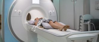
- A drug is administered intravenously - a radiotracer, which, spreading through the blood throughout the body, penetrates into the bone tissue. The body absorbs the contrast agent. The doctor conducting and monitoring the process must correctly calculate the amount of isotope. After three hours, the bone skeleton is saturated with liquid. While waiting for the process, it is recommended to drink plenty of clean water and bed rest. It is important to take a comfortable horizontal position and limit movements.
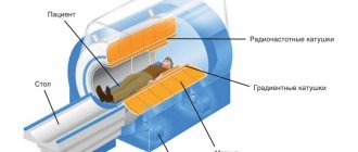
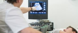
- Using a gamma camera, the musculoskeletal system is examined using an image. A radiopharmaceutical, a radiopharmaceutical, is used. The patient is placed on the treatment table. The examination process takes at least an hour, during which time the person must remain motionless. During hardware scanning, areas of damage are detected thanks to charged indicators that are concentrated in the damaged areas.
- After the procedure, it is recommended to drink a liter of clean, warm water. This will increase the rate of removal of the contrast agent from the body. A small number of them are dangerous for children under 14 years of age and pregnant women. After the procedure, radiation from a person occurs; contact with the child and pregnant woman should be limited.
The results of the examination should be given to the attending physician. Further interpretation of the results is carried out to establish an accurate diagnosis. It is strongly not recommended to read the indicators or diagnose the disease yourself, this is the job of a doctor! The type and regimen of therapeutic treatment is prescribed. Cooperation with the doctor and compliance with all recommendations contribute to effective treatment.
Features of scintigraphy of skeletal bones:
The study is carried out 3 hours after administration of the radiopharmaceutical. Takes from 10 to 30 minutes. The conclusion is issued on the day of the study.
When patients come for the study, they must have with them extracts from their medical records or an outpatient card, conclusions (if available) based on the results of X-ray studies, CT, MRI, as well as the results of previous scintigraphic studies.
Radiopharmaceuticals (RPs) used: diagnosis of skeletal bone diseases is carried out with labeled phosphate complexes, which strongly bind to hydroxyapatite crystals and immature collagen. 99mTs is used as a tag, which has a short half-life - only 6 hours. Gamma quanta leave the body and are registered by the detectors of the device, resulting in an image after computer processing.
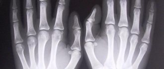
Normal scintigram of skeletal bones in anterior and posterior projection:
Advantages of the examination method
The described technique for diagnosing skeletal bones has a number of advantages and strengths. With its help, it is possible to identify pathological diseases at an early stage of development, even before the manifestation of clinical signs and symptoms. At the cellular level, the structure of fibers and the surface of tissues are considered. It is possible to detect a crack or split in the bone, deviations from normal. In oncology, this technique (hyperscan) is used to identify malignant or benign tumors and control metastases in the body.
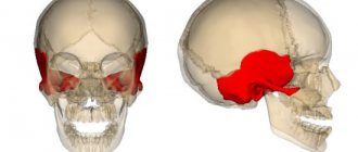
Cancer cell metastases
The scanning process lasts an hour, the patient remains in a horizontal position. There is no pain and no discomfort. The analysis results are ready immediately after the procedure. The increased level of information content makes scintigraphy in demand. During the diagnostic process, it is possible to identify deviations from the norm and the development of the disease before it appears on an x-ray. This contributes to timely therapy and cure. Bone scintigraphy for breast and thyroid cancer, in the brain, and reproductive organs will reveal the prevalence of metastases, the growth and size of the tumor. In some cases, this feature saves a person’s life and frees them from a grueling fight against a developed disease. The therapeutic treatment process can begin before metastases develop and spread.
Scintigraphy allows you to examine the entire body, the entire skeletal framework, and not a separate area. This provides a holistic picture of the body's health. In addition, it saves money and time, eliminating the need for several examinations of different parts of the body. Scanning the body does not entail serious health consequences and does not require grueling recovery time. Also, preparation for the procedure does not imply global, complex actions. The radiation level of the procedure is minimal and does not have a destructive effect on the body. The amount of radioactive influence does not depend on the time of diagnosis.
Treatment of metastases in the spine
The treatment plan is determined by the type and extent of the primary tumor, the general condition of the patient, the volume and manifestations of metastases in the spine. The main goals of therapy are to reduce pain, prevent or eliminate spinal cord compression, improve the quality of life and increase the life expectancy of patients. In the treatment of spinal metastases, chemotherapy, diphosphonates, radiation therapy, hormonal therapy and surgical interventions are used.
The decision on the need for chemotherapy and radiotherapy for spinal metastases is made taking into account the sensitivity of the primary tumor. For hormonally dependent neoplasia, hormonal therapy is carried out. Diphosphonates are prescribed to suppress bone resorption and eliminate hypercalcemia. Surgical interventions for spinal metastases are usually palliative. Indications for surgery are intense pain, progressive compression of the spinal cord, acute radicular syndrome with spinal instability and pathological fracture of the affected vertebra with compression of the spinal cord. The extent of intervention depends on the condition of the patient with metastases in the spine, the prognosis of the disease, the type of neoplasia and the extent of the lesion in the spinal column. All operations for metastases in the spine can be divided into two groups: decompressive and decompressive-stabilizing.
Decompressive operations (laminectomies) are relatively simple and easier to tolerate by patients. Their main disadvantage is the high probability of repeated deterioration of the patient's condition due to the progression of metastases and instability of the spinal column caused by laminectomy. Decompression and stabilization operations (using fixators, implants, auto- and allografts) allow early activation of patients, provide a long-term effect and significantly improve the quality of life of patients with spinal metastases. The main disadvantages of such interventions are their high traumatic nature and the impossibility of performing them in severe conditions and disseminated processes.
Prognosis for metastases in the spine
Metastases to the spine occur at stage IV of the oncological process, which is considered prognostically unfavorable. At the same time, bone metastases proceed quite favorably in comparison with secondary lesions of visceral organs. The average life expectancy for spinal metastases is 1 to 2 years. The rapid aggressive growth of primary neoplasia, multiple metastatic lesions of various organs, a short period of time between therapy for the primary tumor and the occurrence of metastases in the spine, the large size of the metastatic tumor, the absence of signs of sclerosis on radiographs of the vertebrae before and after therapy, and the patient’s serious condition are considered as unfavorable prognostic factors. . Favorable prognostic factors are the slow growth of the primary tumor, the single nature of metastases in the spine, the small size of secondary neoplasia, the presence of signs of sclerosis on radiographs before and after therapy, and the satisfactory condition of the patient.
Disadvantages of the procedure
The disadvantages of this examination technique include the lack of specifics. After diagnosis, the disease is suspected, but not confirmed. Scintigraphy results suggest additional tests and examinations to confirm the suspected diagnosis. You need to do a biopsy, computed tomography, MRI as prescribed by your doctor. Also, when identifying a tumor neoplasm, it is impossible to determine the nature and malignancy. For this purpose, additional examination methods are prescribed.
To remove a specific substance from the body and eliminate radiation exposure, it is recommended to prescribe foods that remove toxins from the body, and drink plenty of fluids. Preference should be given to clean drinking water. At the end of the procedure, you must take a shower, wash your clothes and wash your hair because it is radioactive. You should also not approach children or those whose bodies are weakened, because radioactive particles can cause harm. If pregnancy is planned, it is better to cancel the skeletal scan and undergo other diagnostics.
Decoding the results
At the end of the examination, the patient is given a picture with the results of the procedure performed. An experienced doctor should decipher the data obtained in order to establish a diagnosis and prescribe productive and effective therapy. It is not recommended to read the results yourself and assume a diagnosis. Self-medication can also be dangerous.
The image with the obtained data must be given to the attending physician and wait for a diagnosis to be made. The doctor examines the holistic clinical picture, taking into account additional tests and characteristics of the patient’s body. The calculated norm of healthy indicators is known, which is checked against the results obtained. Venous blood is collected for clinical examination. But it is important to remember that the concentration of a specific contrast agent affects the blood test.
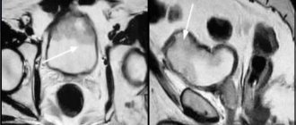
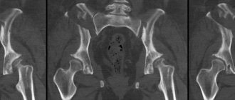
During the examination, dynamic, statistical indicators are obtained, which are compared with the norm. Based on examinations, the functioning of the skeletal support is studied. If pathological foci are diagnosed in posterior or anterior projections, then the concentration of radioactive contrast agents accumulates in them. In oncology, metastases most often spread to the pelvis and chest, knee joint and ribs, spine and bones of the brain, parietal part. In some cases, malignant cells are also found in the bone limbs. It is also possible to determine osteoporosis and pathology of the hip joint.
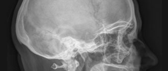
Cancer metastases on x-ray
Survey reliability
Patients doubt how much they can trust the scan and the results obtained. The scintigraphy procedure is imperfect, like all examination methods. In some cases, diagnostic results may turn out to be false and may not correspond to the actual condition. But among other imaging methods, the error statistics for scintigraphy is 1%. The reason for a false reading sometimes lies in the uneven introduction and distribution of a specific contrast agent. In this case, the indicators signal a pathological accumulation of fluid in the skeletal localization, the spread of metastases, or the development of pathologies. In fact, the reason is the unprofessional technique of distributing the excipient and the doctor’s insufficiently professional approach.
Also, signs of malignant, pathological tissue damage can be diagnosed in those places of the skeleton where injuries, bruises or fractures previously occurred. Therefore, before the examination, it is worth telling the doctor about all injuries to the musculoskeletal system of the body. This will help avoid unnecessary worries and false diagnoses. The doctor conducting the skeletal scanning process must filter the information received to focus on areas that indicate actual diseases and pathologies.
Scintigraphy is a reliable and accurate bone examination method. The negative impact on humans is minimal, and the results obtained about the health of the body are characterized by increased accuracy and reliability.
In isolated cases, unreliable images occur due to insufficient experience of the attending physician or equipment malfunction. False indicators may also be influenced by non-compliance with preparation for the examination and recommendations of medical personnel.
Where to do osteoscintigraphy
Scintigraphy of skeletal bones can be done in many large cities of Russia in specialized private and public clinics by appointment and consultation.
Medical institutions offer osteoscintigraphy services at the following prices:
JSC "Medicine" (2nd Tverskoy-Yamskoy lane, 10, Moscow). Cost – 11,490 rubles. Clinical Center for MMA named after I.M. Sechenov (Bolshaya Pirogovskaya str., 2, Moscow). The cost of “whole body” osteoscintigraphy is 5,000 rubles. Institute named after N.V. Sklifosovsky (Bolshaya Sukharevskaya Sq., 3, Moscow). Currently, for residents of the Western Administrative District of Moscow, if they have a specialist’s opinion on the need for radioisotope diagnostics, a scintigraphic study is carried out free of charge under the compulsory medical insurance policy. You must pre-register by phone or 8(985)741-25-46 on weekdays from 10 to 15 hours. The service is not provided for a fee. Research Institute of Oncology named after. N.N. Petrova (Leningradskaya str., building 68, St. Petersburg). Cost – from 4300 rubles, the price of high-resolution osteoscintigraphy and recording of study results on disk – 5900 rubles. St. Petersburg Research Institute of Phthisiopulmonology (32 Politekhnicheskaya str., St. Petersburg). Examination of the skeleton in the “whole body” mode – 3,700 rubles, recording the results on disk – 300 rubles, repeated scintigraphy of the skeleton bones will cost 3,000 rubles. City Hospital No. 31 (Dinamo Ave., 3, St. Petersburg). Bone scintigraphy with recording on disk – 4100-4500 rubles.
- Republican Hospital named after G.G. Kuvatov (Dostoevsky St., 132, Ufa). Scintigraphic examination of bones – 3500 rubles, recording on disk – 500 rubles.
- Republican Clinical Oncology Dispensary (29 Sibirsky Trakt St., Kazan). Scintigraphy of the skeleton in the “whole body” mode – RUB 2,204.
- Regional Children's Clinical Hospital No. 1 (Serafima Deryabina St., 32, Yekaterinburg). The cost of the study is 3000 rubles.
- Polyclinic of the Irkutsk Regional Clinical Hospital (IOCH) (Yubileiny microdistrict, 100, Irkutsk). Diagnostic bone scintigraphy – RUB 3,360.
- Regional Clinical Oncology Center (Voronezhskoye Highway, 164, Khabarovsk). The cost of radionuclide examination of the skeleton is 5560 rubles.
Although the cost of skeletal scanning is quite high, this radioisotope procedure is quite simple to perform and much more effective than x-ray examination. This type of diagnosis allows you to identify pathologies of the skeletal system at the very initial stages and begin timely treatment to maintain health and prolong the life of sick people.
Actions after scanning
After the examination procedure, it is recommended to thoroughly wash your hair with shampoo and take a shower. Particles of radioactive substances remain on clothes, so things should be washed and dried in fresh air. You cannot bring bandages, cotton wool, tampons or instruments that were used to carry out the examination procedure into the house. They need to be thrown away and not stored in the house; items can emit radioactive effects, causing harm to the body.
The medical diagnostic center has special bins and garbage containers for such objects. After the scan, you need to temporarily exclude communication and contact with small children and pregnant women. The dose of radiation to which the body is exposed can spread and have a negative, harmful effect. It is also recommended to drink plenty of clean, potable water at room temperature. An abundance of fluid will help remove specific substances from the body, speed up metabolism and metabolism. It is important to eat foods containing vitamins and beneficial microelements.
Fresh fruits and vegetables, dairy products will help the body remove radioactive substances and specific liquid administered for examination. Collaboration with your doctor and following recommendations and prescriptions will help you cope with the consequences of a skeletal foundation scan. The prescribed diagnosis, established through scintigraphy, will allow you to begin therapeutic treatment and speed up the recovery process. And proper preparation for the examination, the right diet and treatment plan will reduce the risk of complications, side effects and negative consequences.