0
Author of the article: Marina Dmitrievna
2017.08.13
3 104
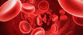
Red blood cells
The composition of blood is formed by its liquid part - plasma, and the formed elements contained in it: leukocytes, erythrocytes and platelets. When examining test results, both the number of formed cells and their size are of interest. And if deviations from the norms of this parameter are detected, further diagnosis will require a repeat study, followed by treatment if the diagnosis is confirmed.
Red blood cells - erythrocytes
Characteristics of anisocytosis, their types
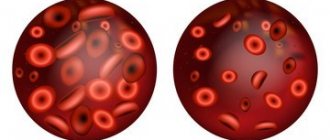
Red blood cells can have different diameters.
| Name | Diameter (µm) |
| Normocytes | 7 — 8 |
| Macrocytes | 8 — 12 |
| Megacytes | > 12 |
| Microcytes | < 7 |
A healthy person has red blood cells of all sizes. But a violation of their ratio is a pathology.
The proportion of normocytes should be 70% of the total volume. The volume of microcytes and macrocytes takes up no more than 15% separately.
Photo: https://pixabay.com/illustrations/heart-red-blood-cells-erythrocyte-2176218/
Classification of anisocytosis
Erythrocyte anisocytosis is divided into several categories.
Based on the size of the predominant cells, they are distinguished:
- Macrocytosis – increased number of macrocytes. When red blood cells are too large, there are fewer of them than needed and they carry less hemoglobin. This means that the blood is not sufficiently rich in oxygen. Low blood oxygen saturation causes a variety of symptoms and health problems.
- Microcytosis – the number of microcytes significantly exceeds the norm. Small red blood cells have a weak ability to carry oxygen, since they have a low concentration of hemoglobin.
- Mixed variant – increased number of macrocytes and microcytes.
Depending on the severity, the following types of anisocytosis are distinguished:
| Name | Designation in analyzes | Proportion of red blood cells with abnormal size (%) |
| Minor | + | 25 |
| Moderate | ++ | ≈ 50 |
| Expressed | +++ | ≈ 70 – 75 |
| Pronounced | ++++ | ≈ 100 |
What is macrocytosis, macrocytes
Macrocytes in a blood smear
Macrocytes are abnormally large cellular components of blood. The presence of such elements in the bloodstream is called macrocytosis. This condition is a consequence of a disruption in the process of hematopoiesis at the stage of cell generation. Incorrectly formed red blood cells are not able to perform their functions properly, that is, they cannot deliver hemoglobin through the capillaries, participate in the creation of immunity and metabolic processes.
In infants, macrocytosis is normal. And for pregnant women, it is a frequent companion throughout the entire period of bearing a baby, which is associated with the restructuring of processes in the bloodstream. Correcting nutrition and taking vitamins helps pregnant women cope with this disorder. For other groups of the population, this is a dangerous symptom.
In most cases, an unnatural increase in blood cells is accompanied by anemia - a low level of hemoglobin in the bloodstream. Anemia is a common symptom that reflects a number of diseases and is characterized by a lack of oxygen in the tissues of the body. An in-depth study of the causes and extent of macrocytosis allows us to determine the type of anemia and its treatment regimen.
Reasons for the development of anisocytosis
Etiology of macrocytosis
Vitamin B12 deficiency
Vitamin B12 deficiency is the cause of macrocytosis. A lack of vitamin B12 in the diet is rare and usually occurs only in older people on the “tea-and-toast diet” or in strict vegetarians.
However, deficiency can be caused by the following.
- Lack of intrinsic factor (an enzyme that ensures the absorption of B12) in patients who have undergone gastrectomy (removal of the stomach) or have pernicious anemia.
- Vitamin B12 malabsorption resulting from bacterial infection in the small intestine, tapeworm infestation, certain medications, ileal bypass, enteritis, intestinal malabsorption.
Folate deficiency
Folic acid deficiency can also lead to macrocytosis. Its disadvantage arises due to:
- dietary deficiency;
- increased need during pregnancy;
- congenital deficiency;
- malabsorption in the intestine;
- alcoholism.
Photo: https://www.pexels.com/
Drug-induced macrocytosis
Drug-induced macrocytosis is the most common cause in non-alcoholic patients.
The following categories of drugs are known to cause macrocytosis:
- antagonists (having the opposite effect) of folic acid (Methotrexate);
- purine antagonists (6-Mercaptopurine);
- pyrimidine antagonists (Cytosine arabinoside);
- alkylating agents (Cyclophosphamide);
- inhibitors (inhibiting the action) of tyrosine kinases (Sunitinib and Imatinib);
- Zidovudine;
- Trimethoprim;
- oral contraceptive pills;
- Phenytoin.
The tyrosine kinase inhibitors Sunitinib and Imatinib have been shown to induce macrocytosis in patients with various types of cancer, including renal cell carcinoma (RCC), gastrointestinal tumors, and breast cancer. In patients with RCC, the development of macrocytosis after treatment with Sunitinib may potentially serve as a positive prognostic factor for overall survival.
Persistent anemia of the following types can cause macrocytosis:
- myelodysplastic anemia;
- aplastic anemia;
- acquired sideroblastic anemia.
Photo: https://www.pexels.com/photo/colors-colours-health-medicine-143654/
Etiology of microcytosis
Microcytosis can occur with several different conditions, ranging from mild problems to more serious ones.
Thalassemia
This is a hereditary blood disorder that parents can pass on to their children due to gene mutations.
With this pathology, the body does not produce enough of a certain protein, usually contained in hemoglobin.
Without it, red blood cell formation does not occur properly.
Chronic conditions
Some chronic diseases can cause microcytosis:
- kidney disease;
- certain types of cancer (Hodgkin's disease, non-Hodgkin's lymphoma, and breast cancer);
- diabetes;
- heart failure;
- Crohn's disease;
- inflammatory bowel diseases;
- rheumatoid arthritis;
- lupus;
- infectious diseases (HIV, AIDS, tuberculosis).
Iron-deficiency anemia
The most common cause of microcytosis is a lack of iron in the blood. Iron deficiency anemia occurs due to:
- insufficient iron intake;
- inability to absorb iron due to certain pathologies (celiac disease or Helicobacter pylori infection);
- chronic blood loss due to frequent or heavy menstrual flow in women or gastrointestinal bleeding;
- pregnancy.
Lead poisoning
Children who are exposed to lead-based paint because they live in an old home or because of toys or other items can suffer lead poisoning.
Polluted water and heavy industrial pollution can also cause lead poisoning, although this is less common.
Sideroblastic anemia
Congenital sideroblastic anemia is an inherited blood disorder that affects the ability of the bone marrow to produce red blood cells. It also causes microcytosis, but is less common than other causes.
Symptoms indicating changes in red blood cells
There are various symptoms of anisocytosis. Some of them are mild, but they can gradually become severe.
A person with anisocytosis often experiences the following symptoms.
- Rapid breathing. This is a common symptom. Its occurrence is associated with a deficiency of hemoglobin, which leads to insufficient oxygen transport. Thus, patients often feel short of breath after minimal activity.
- Paleness . Since the necessary oxygen does not reach our skin, nails and eyes as it should, they become paler.
- Lethargy. With abnormal red blood cell sizes, oxygen distribution is inadequate. Consequently, general fatigue and lethargy are also common symptoms.
- Tachycardia. Palpitations occur not only after physical activity, but also in normal everyday life. The heart pumps quickly to compensate for the body's need for oxygen, and therefore the number of heartbeats increases.
Some other symptoms may also be present:
- headache;
- low body temperature;
- cold palms and feet;
- dizziness (feeling of falling).
Photo: https://pixabay.com/photos/sad-woman-upset-female-people-2385795/
How to determine symptoms?
With any provocateurs, microcytosis exhibits almost the same symptoms.
Obvious symptoms include:
- Low physical endurance, constant fatigue;
- Heavy breathing, both during physical activity and at rest;
- Heart rhythm disturbances;
- Acceleration of heart contractions;
- Paleness of the skin;
- Brittle hair and nails;
- Dry mucous membranes;
- Often “jams” appear in the corners of the lips;
- Difficulty swallowing (lump in throat).
If you notice any of the symptoms, immediately go to the hospital for examination.
Laboratory diagnostics. Criteria
The main way to diagnose anisocytosis is a blood test.
Red blood cell distribution width (RDW)
This parameter is used to measure the size and volume of red blood cells. RDW increases in accordance with changes in red blood cell size.
The normal RDW parameters are as follows:
| Adults | 13 % |
| Children under 6 months. | 16,8 % |
| Children from 6 months. | 13,2 % |
If results exceed the normal range, this indicates the presence of anisocytosis.
Doctors often compare the RDW result to the mean cell volume (MCV).
Results may show:
| results | Causes |
| Normal RDW and MCV | Anemia may be caused by a chronic illness or blood loss. |
| Normal RDW and low MCV | Anemia caused by a chronic condition or thalassemia. |
| Normal RDW and high MCV | Liver pathology or alcohol abuse. Use of antiviral and chemotherapeutic drugs. |
| High RDW and normal MCV | Deficiency of iron, B12 or folic acid. Possible chronic liver disease. |
| High RDW and low MCV | Iron deficiency, microcytosis. |
| High RDW and high MCV | Lack of B12 or folic acid, macrocytosis. Possible chronic liver disease. |
Photo: https://pixabay.com/photos/medic-hospital-laboratory-medical-563423/
Color index
Another indicator used in the diagnosis of various types of anisocytosis. They indicate the amount of hemoglobin in a red blood cell.
The normal value is in the range of 0.85 - 1.05.
Hypochromia in a general blood test, when the color index is less than 0.85, may indicate the presence of microcytosis. Hyperchromia (indicator more than 1.0) possibly indicates macrocytosis.
Preventive actions
Anisocytosis is not an independent pathology, but only signals the development of other diseases of the body, so due attention should be paid to the prevention of this condition. To avoid changes in blood composition, you should adhere to the following preventive measures:
- Correctly adjust your diet, saturate your diet with food containing a sufficient amount of iron;
- take blood tests regularly;
- promptly treat infectious diseases;
- to refuse from bad habits;
- pay due attention to sports. Physical activity has a beneficial effect on the body’s metabolic processes, which has a positive effect on blood composition;
- If you notice such signs as weakness, fatigue, apathy, you must inform your doctor about this.
By following a healthy lifestyle, you can prevent serious illnesses. Proper nutrition, hardening and exercise can strengthen the immune system and prevent the development of severe pathologies.
Treatment of anisocytosis
Treatment will depend on the cause of the anisocytosis.
It is important to determine the cause of the problem so that proper treatment can begin.
Anisocytosis is often associated with anemia, and anemia is usually caused by iron or vitamin deficiency.
The usual treatment for iron deficiency is taking iron supplements and changing your diet to increase your iron levels with iron-rich foods.
Iron-rich foods:
- dark green leafy vegetables;
- brown rice;
- beans;
- nuts and seeds;
- meat and fish;
- tofu;
- eggs;
- dried fruits.
Patients can also address vitamin deficiencies by taking supplements and making changes to their diet.
In severe cases of anisocytosis, your doctor may recommend a blood transfusion. This process will replace blood containing abnormal cells with blood containing healthy cells.
Photo: https://pixabay.com/photos/asparagus-steak-veal-steak-veal-2169305/
Classification by cell type
A common method for distinguishing between conditions is to determine which blood cells are involved within the disorder. The following types are called accordingly:
Change in red blood cells
Red blood cells ensure normal transport of oxygen to tissues, adequate supply and nutrition.
When changing sizes, anomalies are observed. Sometimes critical, posing an immediate danger to health and life.
Laboratory assessment of concentration, shape and size presents certain difficulties. Special analyzers are used.
Based on the results of the study, an index is derived that reflects the final state of formed cells; it is designated as RDW.
Platelet damage
Another clinical variant of the named pathological process is characterized by a disorder in the state of other structures. These elements ensure adequate coagulation.
Adhesion and then aggregation of platelet cells is a prerequisite for normal life. Otherwise, a person is constantly at risk of dying even from minor injuries.
Based on the results of the laboratory assessment, doctors receive an index designated as PDW.
Mixed form
A rarer form of the pathological process. Its typical course is severe; disorders require mandatory and immediate correction, since they are deadly. There is a change in the size of both platelets and platelets at the same time.
Anisocytosis and poikilocytosis
Anisocytosis and poikilocytosis refer to abnormalities in red blood cells. The key difference between the two is that in anisocytosis the cells are abnormal in size, while in poikilocytosis the red blood cells are abnormally shaped, meaning they do not have the standard biconcave shape.
Types of cell deformation.
| Name | Characteristic | Causes |
| Spherocytes | No flattened, lighter center | Hereditary spherocytosis, autoimmune hemolytic anemia, hemolytic transfusion reactions, erythrocyte fragmentation disorders. |
| Dental cells | The central part is elliptical or slot-shaped instead of round | Alcoholism, liver disease, hereditary stomatocytosis. |
| Codocytes | The cell looks like a target with a red dot in the center | Thalassemia, cholestatic liver disease, absence of spleen (splenectomy). Rarely, sickle cell anemia, iron deficiency anemia, and lead poisoning are possible. |
| Leptocytes | Thin flat cells with hemoglobin on the edge | Thalassemia and obstructive liver diseases. |
| Drepanocytes | Crescent elongated shape | Sickle cell anemia, thalassemia. |
| Elliptocytes | Slightly oval or cigar-shaped with blunt ends | Hereditary elliptocytosis, thalassemia, myelofibrosis, liver cirrhosis, iron deficiency or megaloblastic anemia. |
| Dacryocytes | Cells have one round and one pointed end | Thalassemia, myelofibrosis, leukemia, megaloblastic or hemolytic anemia. |
| Acanthocytes | Cells with abnormal spiny projections at the edge of the cell membrane | Abetalipoproteinemia, severe alcoholic liver disease, absent spleen, autoimmune hemolytic anemia, kidney disease, thalassemia, McLeod syndrome. |
| Echinocytes | Spiny projections distributed throughout the cell | Kidney disease, cancer, long-stored blood transfusion, pyruvate kinase deficiency. |
| Schizocytes | Red blood cell fragment | Sepsis, severe infection, burns, tissue damage. |
Photo: https://ppt-online.org/46925
Diet
When a patient's hemoglobin is low and iron deficiency anemia is confirmed as a disease, he will be advised to consume more of the following foods:
If a malignant tumor is detected in a patient, the issue with it will first have to be resolved, and only then microcytosis will be cured, since in the usual case the condition of the red blood cells will not change. In adolescents, causes of this phenomenon are also observed, but it is most often associated with growing up: decreased immunity, hormonal imbalances, increased growth. As you might guess, no treatment is required here. The boy or girl will have to adjust their diet and listen to the doctor’s advice. The problem should go away soon.
In a child, the causes of microcytosis are also often associated with a lack of iron in the body. Up to 90% of children suffer from iron deficiency anemia in one case or another. In this vein, parents may be alarmed by the pink color of their child's urine after he eats red beets. The fact is that the liver, with its enzymes, is obliged to discolor the natural dye of the vegetable. A child with iron deficiency anemia does not have enough of these enzymes in the body.
However, this symptom also occurs in unstable conditions, when cells experience temporary iron deficiency. However, you should consult a doctor. If the disease has been diagnosed in a child, ferrous salts and ferrous sulfate are most often prescribed. The preparations should contain: malic, citric, ascorbic acids, as well as amino acids. They promote the formation of easily soluble iron compounds in the gastrointestinal tract and their absorption.