Disruption of blood supply to brain tissue (stroke) is a pathology that occurs due to damage to blood vessels or their blockage by blood clots. Since the severity of the consequences and prognosis for this pathology depend on the time of initiation of treatment, patients with suspected stroke need immediate examination.
The standard in diagnosing lesions of brain tissue are various tomography methods, incl. and magnetic resonance. MRI for stroke allows you to determine the presence of pathological foci in the brain, clarify the area and severity of the lesion.
Is an MRI prescribed for suspected stroke: for what symptoms?
Magnetic resonance imaging is not necessary in all clinical cases of pathology. If symptoms are obvious, there are signs of a stroke in one of the cerebral hemispheres, and the patient’s medical history and visual examination data correspond, the diagnosis is established without special primary diagnosis.
However, in some cases, MRI is required. Indications for its implementation are:
- vague symptoms of stroke;
- discrepancy between the patient’s examination data and his medical history;
- suspicion of cerebellar damage or lacunar damage (limited to a small volume of brain tissue located in the deep parts of the hemispheres).
Magnetic resonance imaging is also performed to study the vascular bed of the brain after a stroke, assess the volume of edema or hematoma, and clarify the size of the area of necrosis.
An MRI may be prescribed for the following symptoms:
- Strong headache;
- dizziness, loss of consciousness;
- nausea, vomiting due to pain;
- speech dysfunction;
- numbness and cramps of the limbs, loss of facial sensitivity;
- loss of coordination, spasm or decreased muscle tone;
- noise in ears;
- general weakness, inhibited reaction.
What MRI should be done for a stroke?
The contrast and visibility of lesions on tomography directly depends on the method of examining the brain. The following MRI techniques are used to obtain images:
- standard (proton-weighted);
- diffusion-weighted;
- perfusion-weighted;
- MR angiography with contrast.
The diffusion-weighted tomography method is based on the study of metabolism (movement of matter) in areas of brain tissue. By clearly delineating areas of reduced metabolism, it allows you to identify areas of irreversible tissue damage and areas that are subject to restoration with intensive therapy (penumbra zones).
The perfusion-weighted technique involves studying the blood supply in various areas of the brain. Deviation from the norm in the direction of decreased blood supply is a diagnostic criterion for stroke.
Magnetic resonance angiography is a study aimed at visualizing cerebral vascular pathologies.
Indications for diagnosing pathology
MRI is recognized as the most informative method for determining the type of stroke and conducting differential diagnosis with other brain diseases. It is prescribed in the following cases:
- if the symptoms that arise are unclear;
- there is a discrepancy between the medical history and the patient’s examination data;
- it is necessary to determine a lacunar lesion or a deep lesion;
- to assess the size of the necrosis zone;
- if a cerebellar infarction is suspected;
- it is necessary to determine the consequences of the hematoma, including in the long term;
- in order to study the venous bed, the possibility of tumor metastasis, the presence of malformations or vasculitis.
The negative aspects of MRI include the relatively high cost of the examination, the duration of the procedure is more than an hour, which is especially problematic in the acute period, and poor detection of subarachnoid hemorrhage.
How does the procedure work?
Diagnosis of stroke using MRI is performed in the following sequence:
- After the patient arrives at the hospital, the doctor differentiates the symptoms of a stroke from similar signs of other pathologies (for example, hypoglycemia).
- All metal structures are removed from the patient. The presence of non-removable steel objects and devices in the patient’s body may be a contraindication to the procedure.
- If there are no contraindications to MRI, the patient is placed on the tomograph table, his head is fixed during the procedure, and he is pushed into the tunnel. To perform MR angiography, the patient is first injected with contrast agents – gadolinium preparations.
- Using high and ultra-high fields, we study and obtain images of the brain in various projections.
In case of stroke, it is recommended to perform ultra-high-field MRI, because this allows you to reduce the time of the procedure, obtain a less distorted picture of the lesion, and track even small positive dynamics during rehabilitation.
After the procedure, a diagnostician analyzes the images obtained and describes pathological changes for further treatment of the patient in intensive care units.
Field of pathology research
The area of study for various tomography techniques is: the structure, metabolism and blood supply of brain tissue or the lumen of blood vessels supplying nutrients to the probable affected area.
What is a stroke?
Stroke is a condition associated with a sudden interruption of blood supply to the brain. The ischemic form of the pathology is most often detected.
There are several forms of the disease depending on the cause of its appearance and symptoms.
1. Hemorrhagic stroke
associated with damage to the blood vessels of the brain, resulting in hemorrhage. With pathology, areas of the soft tissue of the brain are compressed, and the resulting voids are filled with blood. There are several forms of stroke:
- subdural - a hematoma forms above the membranes of the organ;
- spidural - blood clots are located under the membranes of the brain;
- parenchymal – gray matter is affected;
- intraventricular;
- mixed.
2. Subarachnoid stroke
, in which the hemorrhage is localized in the subarachnoid space. The pathology affects the brain and spinal cord.
3. Ischemic stroke
associated with blockage of an organ’s vessels by an atherosclerotic plaque or thrombus. Due to insufficient supply of oxygen and nutrients to certain parts of the brain, tissue death occurs.
4. Lacunar stroke.
If there is a problem, damage to the small perforating artery is observed.
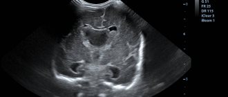
MRI results: what pathological lesions look like on the images
Depending on the nature of the lesion and the volume of the area of tissue necrosis, a stroke looks different on MRI images:
- The ischemic type occurs when an embolism of a cerebral vessel and cessation of blood supply in one of its areas. The images identify areas of pathological changes (foci of ischemic damage) and the site of thromboembolism of a large vessel.
- Hemorrhagic - occurs when a weakened vessel wall ruptures and hemorrhages into the brain. The hemorrhagic type of lesion is more dangerous than the ischemic one, because the released blood volume forms a hematoma, which compresses the surrounding tissue. MRI is not sensitive to impulses from blood movement, so in the early stages hemorrhage cannot be recorded.
- Subarachnoid hemorrhage. Blood entering the space between the membranes of the brain may be associated with a ruptured aneurysm of a large vessel or a traumatic brain injury. MRI does not determine the movement of blood in subarachnoid hemorrhage, but does reveal massive cerebral edema, which causes neurological pathologies.
Features of data in acute cerebral circulatory disorders
In the most acute stage of stroke (up to 6 hours after vessel thromboembolism), there are no changes on standard MRI. Diagnosis of the lesion at later stages sharply worsens the prognosis, because the use of the most effective methods of therapy with minimal loss of functionality of brain tissue becomes impossible.
To visualize stroke in the most acute stage (no later than 2-4 hours after the onset of hypoxia), the diffusion-weighted MRI technique is used. Early diagnosis of pathology allows the use of thrombolysis and restoration of blood supply to ischemic areas of the brain.
In the acute period (up to 3 days), foci of ischemic lesions on MRI are distinguished by light spots that are clearly demarcated from adjacent healthy tissue. In the longitudinal relaxation mode (T1), a weakening of the signal is observed, and in transverse relaxation (T2), its amplification. Also, the acute stage of the pathology is accompanied by thickening of the cerebral convolutions and loss of a clear boundary between the white and gray matter.
Edema is visualized around the area of necrosis during ischemic stroke on MRI. In the subacute period (from 3 to 14 days), the area of edema decreases. The pattern observed on MRI images in the subacute state persists until the stage of residual effects.
Hemorrhagic and subarachnoid bleeding is recorded by MRI based on indirect signs (the presence of swelling and vascular abnormalities), therefore other methods are used to diagnose these pathologies. In the acute stage of hemorrhage, swelling around large lesions is visualized on MRI as an oval area with reduced density.
Within 3 days, MR angiography reveals the focus and extent of vascular disorders.
What does an MRI look like in the early and late recovery phases?
During recovery, ischemic or hemorrhagic stroke appears on MRI as dilatation of the ventricles and sulci of the brain. Repeated tomography during rehabilitation makes it possible to assess the dynamics of narrowing of the lumen of blood vessels, the size of edema, the timing of formation and the volume of foci of necrosis.
The consequences of the pathology are determined on a tomogram for an indefinitely long time after the end of the subacute period. In the chronic stage, stroke on MRI images is manifested by foci of glial transformation and areas of local tissue atrophy.
In the late phase of recovery, the development of alternative neural networks is observed, which cause the disappearance of speech, motor and other disorders.
Types and stages of the disease
A stroke is an acute disturbance of blood circulation in the brain, which comes in four types:
- Ischemic stroke (80% of cases). It develops when a cerebral artery is blocked by a blood clot, which prevents the supply of blood rich in nutrients and oxygen to the overlying brain tissue. Signs characteristic of a stroke develop quickly: within one minute. Irreversible changes in the brain occur within 5 minutes. Emergency medical assistance is required.
- Microstroke, or TIA – transient ischemic attack (2-3% of cases). A micro-stroke can also be caused by a blocked artery. But the body quickly dissolves the clot, restoring blood supply to the brain. The symptoms of a TIA go away as quickly as they begin – within a few minutes. But in the absence of medical care, with a high degree of probability, a microstroke can develop into an ischemic one within 1-2 days.
- Hemorrhagic stroke with cerebral hemorrhage (15% of cases). The artery ruptures and blood escapes into the medulla. The prognosis is less favorable compared to the ischemic form.
- Hemorrhagic stroke with subarachnoid hemorrhage (2-3% of cases). Blood from a burst artery does not flow into the brain tissue, but into the space between the brain and the skull.
There are several stages of stroke:
- The most acute period (three days from the moment of the attack). In a matter of seconds, characteristic symptoms develop: unexpected weakness, numbness or paralysis of half the body, dizziness, headache and disorientation in space, nausea and vomiting, spots before the eyes. The patient experiences speech and hearing problems, spasms or relaxation of muscle tissue. If symptoms resolve on their own within 24 hours, a transient ischemic attack is diagnosed.
- Acute period (time period between the 3rd and 21st day from the onset of the attack). Signs of the disease gradually disappear. If by the end of this period the patient has recovered completely, a TIA is also diagnosed.
- Early recovery period (starts from 21 days from the onset of the disease to 6 months). During this time, the severity of residual symptoms slowly decreases. If active rehabilitation is carried out, previously lost functions are partially restored.
- Late recovery period (time from 0.5 to 2 years from the moment of the attack). Functions continue to be restored to the maximum possible extent. If necrosis of certain parts of the brain has not occurred, functionality can be restored to 100%.
Now let’s find out how MRI can be useful for a person with signs of a stroke.
Is it necessary to undergo an examination after a stroke?
Carrying out an MRI of the brain after strokes or hemorrhages is a mandatory procedure both during the entire recovery period and during subsequent monitoring of the patient’s condition for preventive care.
In the later stages of rehabilitation, MRI most accurately identifies lesions and allows you to monitor their changes. Brain tomography is recommended to be done at least once a year.
Methods of treatment and prevention of stroke
Regular measurement of blood pressure is considered one of the important preventive measures. Modern electronic tonometers allow patients to carry out such measurements independently at home. If there is a significant systematic increase in blood pressure and deterioration in health, you should immediately seek help from your doctor. In addition, innovations in the field of vascular surgery are actively used for the treatment and prevention of strokes. Surgical intervention is resorted to in the presence of a life-threatening narrowing of the artery and in case of a cerebral aneurysm.
Author: Telegina Natalya Dmitrievna
Therapist with 25 years of experience
Magnetic resonance imaging: pros and cons
The advantages of MRI are:
- the ability to obtain sections in various projections, as well as images of areas of the brain adjacent to the bone;
- high contrast of images;
- no radiation exposure;
- the ability to record changes in tissue density.
The disadvantages of the method include:
- a ban on MRI in the presence of implanted devices and objects made of ferromagnetic materials;
- low sensitivity to hemorrhage compared to CT;
- Duration of research and stay in a closed space (up to 1-1.5 hours).
CT photo for hemorrhagic stroke:
Photo1. Massive subarachnoid and intraventricular hemorrhages. Arrows mark places where blood accumulates. The cause of hemorrhage in this case is arteriovenous malformation. Photo source: Dale Birenbaum, MD, Laura W. Bancroft, MD, and Gary J. Felsberg, MD, Imaging in Acute Stroke. West J Emerg Med. Feb 2011; 12 (1): 67–76. https://www.ncbi.nlm.nih.gov/pmc/articles/PMC3088377/
Photo 2. Large intracerebral hematoma on the right. The image shows that the brain structures are shifted to the left. The right cerebral ventricle is compressed, the left is dilated. Photo source: Dale Birenbaum, MD, Laura W. Bancroft, MD, and Gary J. Felsberg, MD, Imaging in Acute Stroke. West J Emerg Med. Feb 2011; 12 (1): 67–76. https://www.ncbi.nlm.nih.gov/pmc/articles/PMC3088377/
Photo 3 . The same patient as in the previous photo. CT angiography was performed. Photo source: Dale Birenbaum, MD, Laura W. Bancroft, MD, and Gary J. Felsberg, MD, Imaging in Acute Stroke. West J Emerg Med. Feb 2011; 12 (1): 67–76. https://www.ncbi.nlm.nih.gov/pmc/articles/PMC3088377/
Other diagnostic methods
Alternative diagnostic methods for stroke include angiography and CT scan of the brain. Their common disadvantage is the radiation exposure that the patient receives during the procedure. In addition, some contrast agents used for angiography are toxic.
Computed tomography is used for early diagnosis of hemorrhages in brain tissue. If the clinical picture of the pathology is inaccurate (for example, with the rapid development of ischemic stroke against the background of atrial fibrillation), CT is prescribed to exclude the hemorrhagic nature of the lesion.
Home tests
No one is immune from stroke, even young people.
In order to contact a diagnostic and treatment center within the first minutes of the disease, it is necessary to identify the neurological symptoms of the disease as quickly as possible. Therefore, even an ordinary person without medical education needs to know the signs of cerebrovascular accident.
To pre-diagnose a stroke, it is necessary to conduct a test. It consists of only 4 points:
- You need to ask the person to smile. A crooked smile is a consequence of paralysis of part of the brain.
- Repeating words and sentences. Action is difficult, words are not pronounced clearly.
- Raise both arms up at the same time. When hit, it is impossible to perform an action.
- Show tongue. If it has shifted to the side, this is a sign of illness.
If at least one of the symptoms is present, you should urgently call an ambulance.
Useful video on the topic:
Contraindications to computed tomography
Computed tomography is contraindicated during pregnancy, since the examination uses x-rays, which can harm the fetus. It is difficult to conduct a study in an obese patient: as a rule, CT scanners are not designed for loads of more than 150 kg.
CT angiography is contraindicated in case of iodine allergy: intravenous administration of contrast can provoke a severe allergic reaction. Relative contraindications:
- diabetes;
- renal dysfunction;
- bronchial asthma;
- diseases of the thyroid gland, in which the level of thyroid hormones in the blood increases ( hyperthyroidism ).