Among the various methods for examining the kidneys, ultrasound is the most commonly used. Diagnosis using ultrasound of the renal vessels is called Doppler or Doppler sonography. This method makes it possible to detect changes in the blood supply to the organ.
The effectiveness of the diagnostic method is based on the Doppler principle, named after the Austrian physicist who discovered an interesting pattern.
The fact is that ultrasonic waves passed through the human body are reflected from blood cells and make it possible to visualize the vascular system. Important! Ultrasound of the renal arteries and renal vessels has important diagnostic value in assessing the state of blood flow.
If the blood supply to the kidneys is impaired, their key function - the ability to excrete urine - will suffer first.
When do they do it?
An examination of the kidneys does not always involve attention to their blood supply, that is, to the vessels and arteries.
However, there are indications that make it necessary to assess the blood flow of the excretory system:
- Renal colic. In this acute condition, along with examination of the veins and arteries, urine analysis, intravenous urography and chromocystoscopy .
- Difficulty urinating - may be associated with insufficient blood supply to the kidneys, and here it is important to assess the condition of the blood vessels in order to establish the cause of the negative process before it leads to inflammation.
- Swelling of the face and limbs may be a consequence of impaired excretory function.
- High blood pressure – problems with the kidneys can cause arterial hypertension. That is, high blood pressure is not necessarily associated with hypertension; it can be caused by a number of chronic diseases or certain types of tumors.
- Changes in urine analysis, for example: the appearance of red blood cells, protein, changes in density and an increased number of white blood cells.
- Late toxicosis in pregnant women. In this case, ultrasound of the renal vessels and arteries is an important procedure in assessing the need for urgent delivery .
- Tissue bruises in the kidney area (kidney bruise) – after an ultrasound, the doctor will assess how damaged the organ is and how things are going with its blood supply.
- Chronic or acute kidney diseases - the examination will reveal the completeness of urine outflow and help determine whether the blood supply to the kidneys is impaired.
- Diabetes mellitus, vasculitis or other systemic diseases, which, if uncontrolled, can lead to oxygen starvation or inflammatory processes in the kidneys.
- Suspicion of tumors, which can also negatively affect the blood supply to organs due to the fact that the vessels are pressed or deformed.
In addition, ultrasound of the renal arteries and vessels is used as a preparatory measure before kidney surgery .
What does an ultrasound show?
The blood supply to the kidneys is carried out due to the coordinated work of blood vessels (arteries and veins). The arteries arise from the aorta and then divide into smaller branches. The processed blood leaves the kidneys through the venous pathways.
If these processes are disrupted, the function of the organ will suffer. And since the kidneys are responsible for electrolyte and water metabolism, this threatens serious systemic disorders. Therefore, it is important to undergo timely diagnosis if any changes are suspected.
During the examination, most attention is paid to the condition of the arteries.
There are three main types of diagnostics of the renal vascular system:
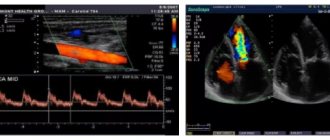
- CDC of renal veins and arteries. The essence of color Doppler mapping is that the doctor receives a color image from which he evaluates blood flow. The brighter the color, the better the blood supply.
- Doppler ultrasound is a study of the renal vessels using an ultrasound machine with a Doppler effect, obtaining blood flow graphs. This method allows you to find out the features of blood flow in graphical form.
- Ultrasound duplex scanning (ultrasound duplex scanning) - an image of the vessel is displayed on the screen, so you can study its characteristics.
The principle of Dopplerography of renal vessels is that ultrasound waves that are sent through the sensor are able to be reflected from red blood cells. By scanning arteries and veins, a converted image is projected onto the screen, from which you can judge:
- diameter of the lumen of blood vessels (normal or there are narrowings and dilations);
- the presence of pathological changes (thrombi, atherosclerotic plaques, protrusions of the vascular wall);
- characteristics of blood flow (volume, speed, signs of lack of blood flow);
- effectiveness of previously prescribed treatment.
Additionally, read the article about ultrasound of the kidneys, indications and procedure.
Types of vascular diagnostics
Several popular methods are used to assess renal blood flow:
- Color Doppler mapping (CDC) allows you to combine a black-and-white image of the kidney with Doppler blood flow assessment.
- Duplex ultrasound scanning (USDS) makes it possible not only to assess the speed of blood flow, but also to find out the anatomical features of the vessels.
- Doppler ultrasound (Dopplerography, ultrasound of renal arteries and vessels) is intended to study the patency of the vascular bed using blood flow graphs.
Indications
Ultrasound scanning of renal vessels is prescribed to identify the causes of the following pathological processes:
- pain in the back, in the lumbar region;
- the occurrence of renal colic;
- difficulty urinating or discomfort;
- swelling of the lower extremities;
- changes in blood pressure;
- diseases of the heart and vascular network;
- with the development of toxicosis in a pregnant woman in the later stages;
- disorders in the endocrine system;
- any diseases of the genitourinary system;
- if nephrosclerosis is suspected;
- with hematuria;
- if you suspect hypertension of a renal nature;
- in case of deviation from the norm according to the results of a urine test;
- falling on your back or other injury to the lumbar region;
- to identify the presence of neoplasms in an organ;
- before surgery in the kidney area.
In addition to identifying the disease, Doppler ultrasound of the renal vascular system can be performed to assess the quality of treatment throughout the course.
Ultrasound of the kidneys in children excludes congenital anomalies of the renal vessels and also helps to determine vesicoureteral reflux.
There are no contraindications to ultrasound of the renal arteries.
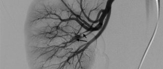
The only drawback of ultrasound examination of the renal vessels is the inability to view the fine network; in such cases, it is necessary to carry out additional research methods (angiography, CT, MRI).
Preparation
The information content of ultrasound of the renal vessels depends on a number of factors, including the competence of the doctor and the high-quality preparation of the patient for the study.
Important! For vascular ultrasound to be informative, it is necessary to reduce the amount of intestinal gases.
Before the procedure, the patient must take a number of measures:
- For several days before the examination, you should refrain from foods that increase gas formation (cabbage, grapes, yeast baked goods, legumes, nuts, carbonated drinks).
- Use enterosorbents or simethicone-containing drugs, such as: “Simethicone”, “Espumizan”, “Enterosgel” or “Polysorb”. You can check the dosage and frequency of administration with your doctor or diagnostician.
- A prerequisite for examining the vessels of the kidneys is an empty stomach. You should not eat, drink, or take medications before the procedure.
If the ultrasound is performed in the afternoon, then the last meal should be 6 hours before.
Exceptions are made for seriously ill patients - they may not eat for 3 hours.
In addition, the doctor may make an exception for patients who need constant medication and a strict diet.
These include, for example, diabetics and people suffering from coronary heart disease.
There is one more feature of ultrasound with Doppler - this procedure should not be performed after gastroscopy or colonoscopy. These research methods cause air to accumulate in the intestines, and examination of the vascular system will be difficult.
Service cost
Moscow clinics offer ultrasound of kidney vessels for 1000–5100 rubles, St. Petersburg clinics for 800–4000 rubles, clinics in other regions for 1300–2300 rubles.
The average price varies between 1500–2000 rubles.
You can contact specialists from both private and public clinics for this service. Consult your doctor, he will recommend a good clinic with affordable prices.
Are you familiar with ultrasound of renal vessels? Have you or your loved ones had this examination done? What was revealed? Share your experience. Repost on social networks. All the best.
How do they do it?
Reference! The technique of studying the blood flow of the kidneys using ultrasound is simple and comfortable, it does not require effort on the part of the patient.
How is this done?
A conductive gel is applied to the lumbar region of the patient (who is sitting or lying on his side), and the doctor uses a sensor to read information from the computer screen.
After the procedure, the patient is given a report, which is commented on by the attending physician. The difference between CDK, ultrasound dopplerography and ultrasound doppler scanning lies in the features of the equipment; for a patient, examination by any of the three methods will be the same.
Progress of the procedure
Ultrasound of the renal arteries is a painless and simple procedure that does not require any effort from the patient during diagnosis.
- Ultrasound examination of blood vessels is performed with the patient sitting or lying on his side.
- A contact gel is applied to the lumbar area, facilitating the movement of the sensor across the body and absorbing external waves.
- From a sensor installed on the patient’s body in the projection of the kidneys, the doctor receives an image of the organ on the screen of the device, where it is possible to select and measure the visualized fragments, examine the organ in volume and determine the pathology.
- At the end of the procedure, the doctor gives the patient a conclusion on which a diagnosis will be made.
After an ultrasound scan of the renal arteries, the patient can immediately begin his usual activities; no recovery period is required. The procedure itself takes no more than 20-30 minutes of pure time.
Decoding
When deciphering the data obtained from an ultrasound of the vascular system, the doctor compares the resulting parameters with the norms. Any deviations will indicate pathological processes in the kidneys.
Table 1. Interpretation of vascular ultrasound results
| Systolic blood flow velocity (cm/sec) | Diastolic blood flow velocity (cm/sec) | Normal dimensions of the artery, mm | |
| Main trunk | 73+/-26 | 37+/-1 | 3,3 – 5,6 |
| Interlobar arteries | 32+/-3 | 13+/-4 | 1,4 – 1,6 |
| Segmental arteries | 45+/-8 | 22+/-4 | 1,9 – 2,3 |
| Arc arteries | 23+/-3 | 10+/-2 | 0,9 – 1,2 |
In addition, the shape and size of the kidneys themselves are standardized - the organs must have the shape of a bean and be no more than 15 cm in size.
Research results
It is very important how correctly the ultrasound report is interpreted. What should be revealed as a result of dopleography:
- anatomical location of arteries;
- places of origin of additional branches;
- state of blood flow in the vessel;
- elasticity of the vessel wall;
- deviations in the structure of the blood supply;
- condition of the vascular wall for ruptures, aneurysms, thinning, thickening;
- when the arteries are blocked or narrowed, it is determined whether the cause is an external factor (tumors, abscesses in fatty tissue, hematoma) or an internal one (atherosclerotic plaques, air embolus, thrombus).
Duplex scanning of the renal arteries is not difficult. And this must be done if there is evidence.
It should be remembered that the quality of the results depends on the experience of the specialist and on the quality of the diagnostic equipment (the higher it is, the more sensitive the sensors and the higher the accuracy of the data obtained).
One of the most effective and safe methods for diagnosing kidney disease is ultrasound. It allows you to determine the size, contours and internal structure of the urinary organs, as well as see changes in the structure of the parenchyma and pyelocaliceal system.
Additionally, the patient may be prescribed a duplex scan of the renal vessels. With this non-invasive test, your doctor can evaluate the blood flow in your renal arteries.
Advantages and disadvantages of the procedure
Doppler ultrasound, a method for determining blood flow in medium and large human vessels, is done not only to identify vascular pathological changes, but also to assess the effectiveness of treatment, as well as to detect indications for organ surgery.
There are many advantages associated with Doppler ultrasound:
- ultrasound examination of the kidneys is not accompanied by pain, since the procedure is minimally invasive (performed without the use of a needle or injection);
- ionizing radiation is not used for visualization of blood vessels;
- Doppler ultrasound has no contraindications or age restrictions;
- Unlike X-rays, ultrasound images provide a clear picture of the condition of the soft tissues;
- the survey is carried out in real time;
- the procedure is not expensive and is widely available.
The main disadvantage of the method is the inability to qualitatively study small blood vessels
Ultrasound waves are not able to replace the study of the functional state of blood vessels and the characteristics of blood flow using contrast X-ray examination - angiography. To clarify the diagnosis after ultrasound, a more thorough examination using a magnetic resonance imaging scanner or computer may be required.
Features and differences
Given that the duplex testing format provides a more accurate and at the same time detailed information picture on the health of the renal arteries, the price of the procedure will be higher than that of a standard ultrasound. But the result will please you with the opportunity to accurately determine whether the vessels are included in the list of possible affected organs of the body.
The basis of the Doppler effect relies on providing data on the difference between the frequencies sent to the organs and the response echo signals of the ultrasonic pulse. The waves directed at the area under study are reflected from the red blood cells and allow one to obtain a picture of the condition of the problematic arteries. All collected information is transferred to a computer monitor and then deciphered by a diagnostician.
To make the technique easier to use, experts have sorted it into two broad categories: visualization and spectral. The last option is necessary to assess the current state of blood flow, which is expressed as a curved line. And the visualization component gives a general idea in black and white or even color, depending on the features of a particular device.
Experts insist that color resolution is more informative, as it reduces the risk of errors to almost zero. Color mapping mode uses two colors: red and blue. The first is responsible for indicators of blood flow directed to a highly sensitive sensor. And the blue color demonstrates the flow in the opposite direction.
Also, the specialist will definitely pay attention to the brightness of the colors, since the richer the tone, the higher the speed of blood flow.
The main advantage of vascular duplex over traditional ultrasound is the ability to examine not only the suspected diseased organ itself. Ultrasound standards do not allow high-quality identification of arterial pathologies, which significantly reduces the productivity of treatment.
With a thorough examination, it is possible to detect anomalies at an early stage of development, which helps to almost completely nullify its negative consequences for the organ. The technique makes it possible to detect defects in the renal arteries, even if the patient does not have any characteristic complaints.
Technique for studying ultrasound of the renal arteries
- Content:
- The principle of studying the renal arteries using ultrasound
- What does Doppler ultrasound of the kidneys and renal arteries show?
- What is the difference between ultrasound and ultrasonography of renal vessels?
Dopplerography of renal vessels Today, Doppler ultrasound of renal vessels, as an important addition to ultrasound examination, is used in almost all clinics. And this is not surprising, because this diagnostic method reveals abnormal, pathological and structural changes in the renal system, various complications when examining patients with kidney disease.
The principle of studying the renal arteries using ultrasound
Having a high resolution, the USDG method of the renal arteries has significantly improved the quality of the examination, allowing one to look inside the vessel to determine existing disorders in real time.
Improving the quality of diagnostics has become possible due to the ability of ultrasonic waves to reflect from red blood cells (blood particles) that are in constant motion. In turn, the effect of reflected pulses - the Doppler effect - depends directly on the speed of blood flow in the vessel.
According to the Doppler principle, the blood flow velocity observed during ultrasound scanning of the renal arteries consists of two components:
- absolute blood flow velocity
- directional angle of the ultrasonic beam emitted by a special ultrasound sensor
The reflected wave energy in the form of sound pulses is captured by the same sensor and displayed on the instrument screen. During ultrasound scanning of renal blood flow, specialists can visually observe the movement of blood in the vessels under study (in the area of interest) using the resulting graphic images.
The cartogram of the reproduced echo signals, performed in the gray-scale echography mode using spectral and color Dopplerography, contains information about the quantitative and qualitative parameters of blood flow (velocity, intensity of their changes).
What does Doppler ultrasound of the kidneys and renal arteries show?
The process of ultrasound examination of the kidneys itself takes place according to a protocol with established rules for conducting the research procedure. A qualified specialist, adhering to standard standards for ultrasound examination of kidney ultrasound with Dopplerography, performs the following actions:
- Determines the location, size of the kidneys, their mobility
- Visualizes the contours and structure of surrounding tissues
- Detects abnormal and pathological changes in the organ being diagnosed
- Evaluates the structure of the renal sinus:
- presence of compactions (stones, low-grade chronic inflammation)
diffuse changes that may indicate some pathological process that has passed from the acute stage to the chronic stage
In this case, Doppler ultrasound of the renal arteries is simply necessary to establish the true cause of such metamorphoses, as well as to promptly begin the necessary therapeutic treatment to avoid negative pathological reactions in the kidneys.
- cysts (can be either benign or malignant)
- Study the condition of blood vessels in the kidneys
Doppler ultrasound, a method for studying renal blood flow, is an integral part of conventional ultrasound, with the only difference being that it is carried out using Dopplerography, which visualizes blood flow in the arteries with the ability to measure its speed and direction.
Combination of examination modes, what is the difference between ultrasound and Doppler ultrasound of renal vessels
Ultrasound as an independent research method in practical medicine is used quite rarely. This is due to the fact that during a typical ultrasound examination of the kidneys, only the condition of the organ being diagnosed can be determined.
Today it is unthinkable to imagine using one method without the other. Ultrasound of the kidneys with Doppler ultrasound complement each other. We can say that Doppler-encoded ultrasound is just an improved version of conventional ultrasound.
kidney diagnostics Duplex scanning, which combines both methods of examination, allows, in addition to a general examination of the condition of the kidneys, to diagnose blood vessels. Dopplerography complements the ultrasound research method, having greater information content and the ability to examine and evaluate:
- renal circulation
- vascular architectonics (general structure)
- renal perfusion
- blood flow speed, resistance value in the renal vessels
Ultrasound scanning of the renal vessels makes it possible to detect a damaged area of the renal artery, i.e. identify stenosis, aneurysm. This test is almost universally used in clinics when examining patients with kidney disease. This has already become a mandatory way to diagnose chronic pyelonephritis.
Two complementary methods of ultrasound examination are carried out everywhere, thanks to their non-invasiveness (painlessness), harmlessness, ease of scanning and reasonable cost of examination, accessible to the majority.
In addition, special preparation for ultrasound examination of the renal arteries is not needed, unless a simultaneous examination of the ureters and bladder is carried out, which is necessary as additional confirmation of the normal functioning of the kidneys or to refute it in case of detected disorders.
Materials used in the article:
https://1uzi.ru/uzi-brushnoy-polosti/podgotovka-k-uzi-pochechnykh-arteriy-s-cd/
https://infomrt.ru/149-uzdg-pochechnyh-arteriy.html
https://klinika-zdorovya.ru/simptomy/bolit-golova-v-viskakh/
Post Views: 85
Prerogative aspects of Doppler ultrasound
The advantages of using this research method are as follows:
- pathological changes in the kidneys do not have age criteria; ultrasound scanning of the renal vessels is allowed to be performed at any age;
- the method is non-invasive, that is, there is no direct intervention in the patient’s body;
- it is possible to assess the state of kidney health immediately after the study;
- Doppler sonography is not a rare or unavailable diagnostic method;
- Ultrasound scanning does not require much time.
At the present stage of development of accessible medicine, Doppler sonography is the prerogative diagnostic technique for renal diseases. A timely procedure diagnoses the disease at the beginning of its development.
Technique for performing ultrasound of renal vessels
The study is prescribed in the morning, on an empty stomach. Last meal 8-12 hours before the procedure. If for some reason it is postponed to the daytime, a light breakfast is allowed, which consists of liquid porridge, tea, 6 hours before the ultrasound.
During the procedure, the patient lies down on the couch, the doctor applies a sensor lubricated with gel to the skin of the abdomen and turns it at different angles, receiving an image of the kidneys and blood vessels in different projections, which is displayed on the monitor.
Then, using Doppler ultrasound, blood flow is studied. A special computer program processes the information received from the sensor and displays it on the screen in the form of color pictures and graphs. The higher the blood speed, the brighter the image. Therefore, it is possible not only to judge the speed of blood flow, but also to determine obstacles to blood flow, their location, and size.
The patient may be asked to turn to one side, stand up, and take a breath to get a good look at the structure of the kidney tissue and blood vessels. The examination is painless and lasts 20-30 minutes. Upon completion, a conclusion is issued describing the results obtained.
Norms and pathologists
During the diagnosis, vascular pathologies can be identified. They are determined by comparing the norm with the obtained data. You need to go with the research protocol to your attending physician, he will be able to decipher the data and prescribe treatment as necessary.
Conclusion. The combination of several methods in the study of renal vessels is considered the most effective and efficient method, which makes it possible to identify pathologies at the very beginning of its development. Regular preventive examinations for risk groups are mandatory.