- Home /
- Branches /
- CT scan /
- CT coronary angiography (heart vessels)
MSCT coronary angiography is a minimally invasive method for visualizing the arteries of the heart (coronary arteries).
During the examination, the contrast agent is injected into a peripheral (most often ulnar) vein, and not into an artery, which makes the examination safe for the patient. The method is similar in principle to standard X-ray contrast coronary angiography, but has important additional properties: it allows you to determine the condition of the coronary artery wall and the structure of the plaque that causes its narrowing (stenosis), namely whether it is calcified or not. CT shuntography is performed in patients with coronary artery bypass grafting to assess the condition of the shunts (occlusion, stenosis), being the method of choice.
In addition, the study allows you to assess the condition of the heart chambers, the thickness of the heart muscle (myocardium), and evaluate a number of functional indicators.
Before the procedure, the calcium index (the severity of calcification of the vessel walls) is assessed to determine the feasibility of a study with intravenous contrast enhancement.
What does the examination reveal?
The procedure is considered quite informative; it allows one to identify the structural features of the local circulatory system quickly and with great accuracy.
However, the technique is used within the framework of static tissue imaging. That is, you can get a picture of the current state of affairs, anatomical structure and all deviations except functional ones.
They are not shown in real time, which makes coronary angiography, although effective, a limited technique that requires additional research. To verify the diagnosis or obtain auxiliary information.
What a check of the heart vessels can show in the end:
- Anatomical congenital changes. Many heart defects, including fatal ones, are not always visible during classical diagnostic procedures. This is a big problem. Coronary angiography provides the opportunity to confirm abnormalities in the structure of the heart and blood vessels the first time.
- Traumatic lesions of the chest. They occur in different forms and variations: from fractures to serious bruises. According to statistics, in approximately 30-60% of cases of such damage, cardiac structures also suffer. This is deadly, since hemopericardium or other changes may develop.
- Coronary angiography provides more information about the condition of tissues, but is used only as an auxiliary technique. And then, in case of injuries, such an invasive examination is not always possible due to the general position of the patient. Indicators of blood pressure, myocardial contractility and other vital levels. The activity is contraindicated for persons in serious condition. This is a load on the body.
- Heart failure in the chronic phase. It represents a drop in the pumping function of a muscular organ with a gradual decline in activity. It progresses constantly, but not in all cases it is possible to detect the causes of this phenomenon. Including highly sensitive methods.
Coronary angiography in such a situation acts as an auxiliary method. It is quite possible that the cause of the developing deficiency is atherosclerosis. Changes in the lumen of the arteries, its narrowing.
- Angina pectoris. Classic disease. A characteristic feature of this is insufficient blood circulation in the muscle layer of the organ. This is the result of atherosclerosis or vascular spasm, which no longer nourishes the tissues sufficiently.
It is important to identify the pathological process in the early stages, since the outcome without therapy is a heart attack, and then it is not far from death.
- Atherosclerosis. The disease is of a generalized type. Its essence lies in the deposition of excess cholesterol on the walls of large arteries. Including hearts.
Next, the plaque becomes fixed and grows, blocking the lumen of the blood vessels. They are unable to conduct blood in sufficient quantities, and the tissues begin to die.
In addition, pressure increases, which can lead to disruption of the anatomical integrity of the artery, rupture and massive bleeding.
Another variant of atherosclerosis is narrowing (stenosis) of the lumen as a result of spasm. This is a classic situation for smokers, alcohol drinkers, and even for individuals who exceed the individual norm for physical activity.
The diagnostic method provides a lot of information. However, it is specific and usually the procedure is used in combination with others. To get the clearest and most comprehensive picture. Whether it's ultrasound, ECHO-CG or MRI. ECG. There are many options.
Preventive examination
We have discussed above the cases in which a computed tomography scan of the coronary vessels of the heart is mandatory. For example, to make an accurate diagnosis, assess the patient’s condition, exclude the fact of pathogenic changes, and plan specific treatment.
However, there are a number of cases when this procedure is not mandatory, but is recommended in the form of effective prevention of serious diseases. After all, the fact is that this examination can detect pathology at an early stage, when it can be completely cured without harm to the body.
In this case, CT coronary angiography is indicated for patients who belong to the so-called risk groups:
- According to the age. Men over 40 years old, women over 45 years old.
- By way of life. Obesity, weight problems, poor diet, constant emotional stress, nervous strain, hard (physically, emotionally, mentally) work, smoking, addiction to alcohol.
- According to diseases in the medical history. Previous stroke, persistently high blood pressure, diabetes, high levels of “bad” cholesterol, vascular disease, hereditary predisposition to heart pathologies.
Types of coronary angiography
The examination method is classified according to such criteria as the essence of the procedures and the technique used (a comprehensive basis for typification).
Based on this, the following forms are called:
- General. Classic. It involves inserting a catheter through the femoral artery, injecting a contrast agent, and then assessing the condition of the entire heart using a special detector. This method is used most often. It acts as a kind of overview to identify the general condition of cardiac structures.
- Selective coronary angiography. It is used to determine pathologies of individual vessels. The same invasive research. However, the catheter insertion point is not necessarily located in the femoral artery (according to Seldinger). A puncture of the forearm is possible, it all depends on the diagnostic purposes and the expected findings.
- CT coronary angiography. Does not imply invasiveness. It is carried out without contrast enhancement or with a minimal amount of it, since blood itself is a good coloring agent. In fact, this is a different technique, although it is called coronary angiography. And it does not replace, but only complements the previous two methods. Another name is MSCT coronary angiography (using a multislice computed tomograph).
The question of choosing a diagnostic method is complex and depends on the specific clinical case. Everything is in the hands of a cardiologist.
Coronary angiography is almost never prescribed first, so the specialist’s decision is based on the results of other objective studies.
What is coronary angiography?
Coronary angiography is an invasive (internal) type of examination of the condition of the vessels of the heart muscle, which can be used to diagnose coronary artery disease. At the time of the manipulation, a contrast agent is injected into the patient’s inguinal or radial artery, which can be detected by X-rays. A general view of the vessels being examined is displayed on a monitor of special computer equipment when X-rays are examined.
There are 2 types of CT coronography: general and selective.
| Type of study | a brief description of |
| Selective | Coronary angiography of this type is used to study one or more vessels. The catheter is installed in a position that ensures immediate filling of the corresponding vessel with a contrast agent. A unique feature of this method is the maximum speed of manipulation. |
| General | All vascular elements of the heart are examined. A special substance, contrast, is injected into the coronary vessels, allowing the condition of the arteries to be assessed, then a detailed video recording is made. |
Indications for diagnosis
There are many reasons for prescribing the procedure. Among them:
- Severe pain in the chest of unknown origin. Occurs as a result of many possible disorders. The measures allow you to clarify the structure of the arteries at the local level and determine the degree of changes in hemodynamics (blood flow). If deviations are detected, additional procedures must be carried out.
- Preparation for surgery. Cardiac surgery is unthinkable without a thorough plan. Coronary angiography solves this problem. It allows you to clarify the scale of the future surgical field and work out treatment methods.
- Coronary angiography of the heart is also done after surgery to evaluate the effectiveness of treatment and the degree of success of the therapy. Including much later than the surgical procedure in order to study long-term results.
- Shortness of breath for no apparent reason. Paroxysms of severe pain (attacks that last more than a few seconds or minutes), decreased tolerance to physical activity, and other similar moments accompany a process such as coronary insufficiency. Diagnostics are prescribed to exclude or confirm this disorder. Read about other causes of shortness of breath and lack of air in this article.
- Lack of effect from the drugs used to treat cardiovascular pathologies. The cause must be sought in the coronary arteries, especially if other diagnostic paths have not given any concrete result.
- Suspicions of heart failure. This is not a technique of primary importance; it is not carried out at the start of diagnosis. But it allows you to identify a disorder if the circulatory system is involved.
Symptoms of heart failure according to stages are described in this article.
- Chest injuries. Research is not always necessary, only when there is a suspicion of certain damage to the arteries.
- Suspected atherosclerosis. Narrowing of the lumen of blood vessels as a result of spasm or cholesterol deposition on their walls. Read more about the causes of high blood cholesterol and what to do about it here.
Coronary angiography may be required to diagnose congenital anatomical and developmental defects. As a confirmation method.
The list is incomplete.
The main advantages of the procedure
Now we know what it is - CT coronary angiography. Let us highlight its important advantages, which patients note in their reviews:
- Almost complete absence of invasiveness - procedures associated with any medical penetration through the protective barriers of the body - mucous membranes, skin.
- Possibility of conducting a comprehensive detailed examination of the coronary vessels without hospitalization.
- A relatively quick procedure.
- Possibility of identifying hidden pathologies. Such as, for example, vascular stenosis.
- The ability to determine the type of atherosclerotic plaques at an early stage of pathology development - soft or calcified formations.
- Assessing the correct placement of shunts and walls, monitoring them after some time after the operation.
It is noted that CT coronary angiography of the heart is periodically prescribed to specialists whose work involves constant nervous strain and stress on the heart, without consequences for their health. In addition, this activity is not prohibited even in acute myocardial infarction.
Contraindications
The list of grounds for refusing an event is not so wide. But these recommendations must be followed strictly so as not to provoke life-threatening complications.
- Diabetes mellitus in the sub- and decompensation phase. The glucose level is unstable, the condition of the blood vessels also does not allow diagnosis. We must first bring the situation back to normal and restore the functioning of the body. Without this, it is dangerous to prescribe coronary angiography. Bleeding, ruptured arteries and death from the consequences are possible.
- Elderly age. It is not an absolute contraindication. But the event creates a certain load on the body, which can threaten human health and even life. It is necessary to carefully weigh all the pros and cons, only then begin to act. The issue is resolved by a cardiology specialist.
- Cardiovascular disorders in the acute phase. For example, a severe attack of angina pectoris, heart attack and others like that, up to inflammatory processes (myocarditis and others). It is necessary to first bring the condition back to normal, only then carry out diagnostics.
- Bleeding.
- Increase in body temperature. Infections are an absolute contraindication for coronary angiography at this time. As soon as the person returns to normal, an examination can be scheduled.
- Lung diseases. Respiratory failure. Diagnosis is postponed until better times.
For the most part, these are relative contraindications. That is, the procedure can be carried out only after eliminating the factor that prevents this. Monitoring the dynamics of a person’s condition is carried out by a specialized doctor, as well as a cardiologist.
When the study is contraindicated
Absolute contraindications:
- pregnancy;
- severe calcification of the coronary arteries;
- arrhythmias;
- tachycardia, in which the pulse is higher than standard;
- allergy to iodine;
- renal failure;
- severe diabetes mellitus;
- diseases of the thyroid gland (thyrotoxicosis).
Relative contraindications:
- taking metformin-containing medications;
- taking nephrotoxic drugs: NSAIDs, aminoglycosides, dipyridamole;
- the patient's inability to hold his breath;
- inappropriate behavior;
- Exceeding the patient weight limits for that particular CT machine.
If you have allergies or intolerance to medications, you should inform your specialist in advance!
Preparing for the study
Since coronary examination is invasive, albeit to a minimal extent, the preceding steps are identical to those for any surgical procedure.
About a week or a few days before diagnosis, patients undergo the following procedures:
- General blood and urine tests.
- Coagulogram. Coagulability study.
- Electrocardiography. To exclude cardiac disorders, at least those that could become an obstacle.
- Ultrasound, ultrasound examination, duplex scanning.
- Chest X-ray.
- Specific measures to detect HIV, syphilis.
Seven days before the examination you must stop drinking alcohol.
Approximately 12 hours before the procedure, you are prohibited from eating or drinking.
Next you need to follow the instructions:
- You should not take medications on the day of diagnosis, unless given permission by a specialist.
- The clinic needs to take all the medications that the person usually takes.
- It is important to empty your bladder before testing.
- When entering the office, you must remove all metal jewelry.
- During coronary angiography, the patient should follow the instructions of the medical personnel. They may ask you to take a deep breath and change your body position.
Preparing for diagnosis does not present any difficulties. After the procedure, you need to check with your doctor what to do next to minimize the risks of complications.
Relative contraindications to the procedure
Now let's talk about relative contraindications to CT coronary angiography (we discussed the indications above). The following are situations in which the procedure is undesirable, but is possible after normalization of the patient’s condition and permission from his treating doctor:
- Uncontrolled ventricular arrhythmia. The specialist allows CT scanning only if the disease is classified as controlled.
- History of intoxication through cardiac glycosides.
- Uncontrolled hypokalemia.
- High body temperature.
- Uncontrolled hypertension.
- Infectious endocarditis.
- Pathological processes associated with poor blood clotting.
- Some diseases of internal organs.
- Decompensated heart failure.
How is coronary angiography done?
The procedure is performed in the cath lab. Diagnosis begins with treatment of the catheter insertion site. Hair in the groin or forearm area is shaved, depending on the point of impact. Next, the patient is placed on the couch.
Anesthetics are administered, and sedatives can be used if necessary. To reduce the degree of psychological discomfort. Especially for impressionable people.
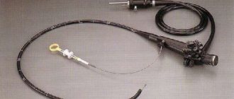
An incision is made in the skin in the area of a large vessel. The probe is a small, thin and long tube. Under video control, the catheter moves along the aorta until it reaches the heart.
A contrast agent is administered. At the same time, special devices recording the frequency of contractions are placed on the chest to eliminate problems during the event.
After the drug is administered, the patient is placed under the detector. The X-ray itself is performed in different projections.
Attention:
Coronary angiography with contrast can provoke allergies; during the procedure you should inform your doctor if any sensations arise.
As a rule, patients do not feel pain or discomfort throughout the procedure. A doctor or nursing staff (or nurses) can give instructions on behavior. This is necessary for more efficient advancement of the catheter through the vessels.
The entire diagnosis lasts about 1 hour, perhaps a little more. Upon completion, the person remains in the clinic for some time. At least for 2-4 hours.
Depending on the institution, hospitalization for up to a day is possible. To monitor the period after the event.
The results are analyzed by three specialists: a cardiac surgeon, an x-ray surgeon and a cardiologist, and a written conclusion is usually issued the next day.
The images themselves can be received in hand as a printout or electronically on a CD or flash drive on the day of the diagnosis.
Selective coronary angiography
What is special about this method? Essentially, this is a modernized version of the above. During the examination, the doctor does not examine all the vessels, but concentrates on several or even one. This selective approach is reflected in the name.
The procedure requires installation of a special catheter for the patient - it is through this that the contrast agent is supplied. A special wish for the event is a good film for the X-ray machine. Its poor quality may affect the resulting image. If it is fuzzy and vague, then the doctor will not be able to make an accurate diagnosis based on it.
A significant advantage of the selective method is that it does not require the introduction of large volumes of contrast agent. Another feature is the fast completion of the procedure. Moreover, in a short period of time, a specialist can obtain images of vessels of various locations.
However, selective coronary angiography also has a significant disadvantage - the procedure cannot be performed using the same probes. They need to be replaced, and this action can lead to atrial fibrillation. Another disadvantage of the method is the fact that the selective type of coronary angiography is carried out only with the help of complex equipment - such that it is capable of taking a large number of frames per minute. It is important to pay attention to the quality of the examination probes.
Risks and possible problems
Possible consequences of coronary angiography include:
- Massive bleeding due to accidental iatrogenic arterial injury. It occurs infrequently, but poses a mortal danger to the patient.
- Heart attack. An episode of angina. It is quite capable of ending in a heart attack.
- Rupture of blood vessels, heart.
- Allergic reactions to the injected contrast agent. This is a risk because it is not known how the body will respond to the use of the medication.
- Possible thrombosis. Including after a certain time, from several hours to a couple of days. Therefore, it is recommended to see a doctor during the critical period.
- Arrhythmias.
Coronary angiography is a relatively safe technique, but observation is necessary to exclude negative consequences. Even if their probability is not great.
Possible complications
Since coronography is carried out with the direct introduction of certain drugs and devices into the human body, the consequences of the procedure can be quite dangerous to health. During coronography, in rare cases, thrombocytopenia may occur - a critical decrease in platelets in the blood, usually associated with the introduction of heparin into the body.
Among the most common threats are:
- contamination or infection;
- respiratory failure;
- heparin-induced thrombocytopenia (decrease in the number of platelets in the blood);
- kidney damage;
- allergic reaction;
- decreased blood pressure;
- heart attack and stroke;
- arterial dissection;
- hematomas at the puncture site;
- local vascular damage.
See this article for more details about complications.
Recommendations after the procedure
There are some tips:
- You need to stay in the clinic for a couple of hours or up to a day. Depending on what the treating specialist or diagnostician decides.
- All manifestations must be reported to medical personnel. This is a security issue.
- It is important to drink fluids after the event. This will prevent the formation of blood clots.
- Intense physical activity is contraindicated for 2-3 days, as is overheating and hypothermia.
- You should not consume alcohol for at least the same period.
Technique for CT coronary angiography
Since the procedure requires the use of a contrast agent, the patient must undergo an allergic reaction test and diagnosis for diseases, the presence of which is a contraindication to the use of contrast, before undergoing it.
Before the procedure, he will be asked to remove any metal objects from himself, as they may negatively affect its quality. Before scanning begins, the patient lies down on the tomograph table, which smoothly moves into the scanner.
The scan begins after contrast is administered and is accompanied by clicking sounds, which are normal and should not frighten the patient. On average, the procedure lasts from 15 to 20 minutes, during which you need to remain motionless and follow the diagnostician’s commands - for example, hold your breath.
In order to make an appointment with our specialists, fill out and send us the form online or call: +7 (495) 788-33-88.
CT coronary angiography or angiography - what is the difference?
With CT coronary angiography, the vessels of the heart are visualized using intravenous contrast. The only danger for the patient is a possible reaction to iodine-containing contrast.
In turn, traditional angiography is an invasive procedure. The procedure begins with local anesthesia of the puncture site, followed by puncture of the femoral artery with a needle and a catheter, which is advanced towards the heart. Passing through the iliac artery, abdominal and thoracic aorta, contrast is alternately injected through the catheter into the right and left coronary arteries, then X-rays of the heart area are taken. In addition to the risk of a reaction to contrast, bleeding, arrhythmias, thrombosis of the coronary arteries, dissection of the inner wall of the vessel, and a heart attack may occur after the procedure.
In this case, angiography has a higher spatial resolution, although the resolution of MSCT in most cases is quite sufficient to accurately determine the vascular lesion. The main advantage of MSCT is the ability to visualize not only the lumen, but also the vessel wall.
As for the received radiation dose, with MSCT it is on average 1.5 times greater than with traditional angiography.
Risks, complications and consequences
Coronary angiography, like any invasion, can have side effects caused by the body’s improper response to the intervention and the patient’s stress. Rarely, but the following events occur:
- bleeding at the insertion gate;
- arrhythmia;
- allergy;
- detachment of the inner layer of the artery;
- development of myocardial infarction.
Pre-procedural testing is intended to prevent these conditions, but sometimes they happen. The doctors involved in the examination cope with the situation, the procedure is stopped at the first unfavorable signs , the patient is removed from the dangerous state and transferred to a hospital for observation.
Is this examination dangerous?
Everyone who is about to undergo this procedure is interested in what the consequences and complications may be. This type of diagnosis is considered a safe manipulation; the process is controlled using an X-ray or computed tomograph. Nevertheless, the risk of complications is still present; the most dangerous consequence is damage to blood vessels by the catheter.
The likelihood of such a complication is very low; it can occur from sudden movements or carelessness of the doctor. If the patient has not undergone proper preparation, an allergic reaction or bleeding may occur (with poor clotting). All these risks can be reduced if you carefully prepare and relax before the procedure.
If you are overcome by fear and anxiety in the X-ray operating room, you can warn the doctor about this and he will prescribe sedatives.
How coronary angiography and stenting are performed: indications and contraindications
Coronary angiography is a reliable way to diagnose the heart and blood vessels; it allows you to study in detail the anatomical features and the presence of changes in the arteries. Which, with timely diagnosis, contributes to effective treatment and prevention of the development of serious complications.
Coronary angiography is an x-ray of the heart vessels into which a contrast agent has been previously injected. Thanks to this, the lumen and inner walls of the arteries are clearly visible. This allows timely diagnosis of blood clots, tissue tears, etc. To create contrast, the substance urografin is used.
The procedure received this name because coronary means the vessels that bring blood to the myocardium, and graphy is the generalized meaning of all x-ray studies.
You can also find the following designations:
- coronary angiography;
- coronary angiography;
- angiogram.
Types of coronary angiography
Angiography is divided into several types depending on the volume of vessels that need to be examined. Also in modern medicine, computer tomographs are increasingly used instead of X-rays. As a result, coronary angiography is distinguished depending on the research apparatus.
General coronary angiography
This is a classic X-ray contrast diagnostic method, with which all the vessels of the heart are examined. Contrast is injected into all arteries, a picture is taken, and the image is displayed either on film or on a disk. Allows you to assess the condition and functionality of all vessels in the complex.
Selective coronary angiography
The main difference from general coronary angiography is the study of only certain vessels. The catheter is positioned in such a way as to quickly deliver contrast to the desired artery. Next, they take pictures at a speed of 2 to 6 per second on cinema or wide-format film, since these are the ones that produce the highest quality pictures.
In selective examinations, a small amount of contrast is used and the procedure itself is carried out at a fast pace. This allows you to do the study several times in different projections. The main disadvantages of the method are the risk of atrial fibrillation, as well as the need to change catheters, which are only enough for 6 injections of the drug.
MSCT – coronary angiography (CT coronary angiography, computer coronary angiography)
This procedure is called multislice computed tomography. During this procedure, all blood vessels and valves in the heart are examined using a 32-slice tomograph. The arteries are also filled with contrast material. And then the patient is placed under a tomograph and a three-dimensional image is obtained.
Advantages of MSCT compared to traditional coronary angiography:
- a quick and minimally invasive procedure that does not require the patient to be hospitalized;
- low risk of complications after manipulation;
- allows you to determine the type of atherosclerotic plaques, the condition of shunts and stents;
- It is possible to assess the condition of the heart from all sides thanks to a three-dimensional image.
Virtual coronary angiography
This procedure is the simplest and safest of all types of angiography. Contrast is injected into the cubital vein and a series of images are taken on a CT scanner. The entire procedure takes a couple of minutes and does not require anesthesia or hospitalization of the patient.
Virtual coronary angiography allows you to study the level of patency of stents after bypass surgery without the risk of developing myocardial infarction. But it cannot replace the traditional type of angiography, since only the proximal parts of the vessels and shunts are visualized.
Indications for coronary angiography
Angiography is prescribed mainly for the development of coronary heart disease. Using the procedure, the doctor determines the optimal type of treatment in each specific case. Doctors recommend mandatory testing before open heart surgery for patients over the age of 35 years.
Angiography is prescribed to identify the following criteria:
- anomalies of the arteries, their condition and the level of blood supply to the myocardium;
- degree of damage due to atherosclerosis;
- myocardial bridge and the degree of vasospasm.
Suspicion of coronary heart disease in the absence of its clinical symptoms
Indications for angiography:
- angina pectoris 3 and 4 classes as a result of taking medications;
- angina pectoris, in which a high risk of myocardial infarction has been determined using stress tests;
- repeated attacks of rapid heartbeat;
- a history of resuscitation due to sudden cardiac arrest;
- negative results of stress tests in people with emotionally stressful work;
- diagnosed ischemic disease in the presence of concomitant problems in the body that do not allow other studies to be carried out;
- angina pectoris grade 3-4 with stable manifestations, which decreases to level 1-2 when taking medications.
Atypical chest pain
Indications for angiography for pain in the chest:
- signs of ischemic disease according to the results of special tests;
- several hospitalizations due to severe chest pain;
- Controversial laboratory and functional test results.
Unstable angina and suspicion of acute myocardial infarction
Indications for the procedure are as follows:
- unstable angina, in which medications do not help or the result of treatment is short-lived;
- Prinzmetal's symptoms;
- unstable angina, which was detected during treatment in the hospital;
- angina pectoris of an unstable nature along with serious risks for stress tests;
- a drop in blood pressure combined with congestion in the lungs.
Recurrent angina after coronary artery bypass grafting or stenting
Indications for the procedure after surgery (shunt, stents):
- symptoms of a blood clot in the heart arteries;
- angina pectoris that occurred within 7-9 months after stenting or angioplasty;
- chest pain within a year after coronary artery bypass surgery;
- the presence of criteria for the risk of developing a heart attack based on stress tests and tests in the laboratory after the operation (regardless of the period after its implementation);
- symptoms of recurrent coronary stenosis within 30 days after angioplasty;
- deterioration of functional test scores in the absence of signs of disease development;
- angina pectoris a year or more after heart surgery with a low risk of heart attack.
Suspicion of acute myocardial infarction
Indications for angiography at risk of acute infarction:
- Less than 12 hours have passed since the onset of the acute phase of the disease;
- symptoms of shock in the first 1.5 days after the development of the disease;
- low blood pressure, in which medications do not improve the condition;
- failure of thrombolytic therapy.
Conditions for which coronary angiography is recommended
Factors in which it is advisable to conduct research:
- sudden chest pain and shortness of breath during treatment for myocardial infarction;
- before heart surgery;
- before surgery on other organs in a patient who has had a heart attack in the past;
- heart failure;
- arrhythmia of a malignant nature (without improvement during treatment);
- uncertain pathogenesis of infarction;
- before organ transplantation (liver, lungs, heart, kidneys);
- diagnosis of infective endocarditis;
- angina pectoris that cannot be treated;
- sudden cardiac arrest with unknown pathogenesis;
- chronic heart failure in combination with angina pectoris or pathology of left ventricular contraction;
- disorders in the aorta in combination with problems associated with the condition of the coronary arteries;
- fresh sternum injury;
- Kawasaki disease;
- cardiomyopathy.
Preparation and carrying out the procedure
Before coronary angiography, the patient must perform a number of preparatory procedures to obtain a more accurate result.
These include:
- laboratory tests of urine and blood, ultrasound diagnostics of the heart, electrocardiography;
- refusal to eat the evening before and on the day of angiography;
- hair removal in the groin area (in case of contrast injection into the femoral artery);
- lack of stress and physical activity a couple of days before the test;
- prohibition of blood thinning medications for 7 days before angiography;
- drinking about three liters of water per day in the last days before the procedure;
- emptying the bladder;
- removing jewelry and contact lenses;
- informing about the use of medications and the presence of chronic illnesses.
The procedure process (duration ranges from 20 to 60 minutes):
- The patient signs consent for coronary angiography.
- It is placed on a couch and secured to prevent the catheter from dislodging.
- Local anesthesia is given and a contrast agent is injected.
- A cardiac monitor is connected to monitor heart rate and blood pressure.
- Pictures are taken (from 2 to 10 minutes, depending on the type of procedure) in different projections with data recording.
- Apply a bandage to the contrast injection sites to prevent wound infection.
The video shows how coronary angiography is performed. Filmed by Moe Serdtse.
Decoding the results
The procedure involves examining the coronary arteries for their location, wall thickness, and narrowing of the lumen. Based on these data, various pathologies of the heart and blood vessels are diagnosed, and a decision is made on the type of treatment.
Angiography interpretation options:
- Occlusion – characterized by complete blockage of the arteries. High risk of heart attack.
- Stenosis – a narrowing of the lumen in the arteries is visible, which leads to poor circulation. This pathology leads to coronary artery disease.
- Anomalies in the location of blood vessels are often a congenital defect. The detection of local narrowing in the arteries on an x-ray indicates a blockage, which is a symptom of atherosclerosis.
- Narrowing of the artery 3 mm from its origin is a sign of thrombosis, arteritis or atherosclerosis.
- Calcium deposits on the walls indicate diabetes mellitus, endocarditis or hypercalcemia.
In addition to the results of coronary angiography, the doctor takes into account other laboratory and functional tests to make a final diagnosis for the patient.
Possible complications
Coronorography is an invasive procedure, and therefore the risk of complications or death is very small (about 1%). But to prevent possible risks, it is recommended to check the kidneys and the reaction to iodine (the main contrast agent), and also strictly follow the doctor’s instructions. If all measures are followed, the risk of problems is virtually eliminated.
The following possible complications during the study are identified:
- ventricular fibrillation;
- blockage of the radial artery;
- stroke;
- extensive myocardial infarction;
- renal failure;
- infectious infection and, as a result, inflammation;
- anaphylactic shock.
Contraindications
Coronary angiography has no absolute contraindications, so it can be performed on almost all patients. But there are a number of conditions in which it is recommended to postpone the procedure or use other diagnostic methods.
These include:
- anemia;
- infections;
- problems with blood clotting;
- stroke;
- chronic diseases of internal organs.
Whether or not to refer a patient for angiography is decided by the cardiologist on an individual basis. You may need to consult another specialist depending on the person’s illness.
What is the price?
The cost of the procedure varies in regions.
| Region | Price | Firm |
| Moscow | from 9876 rub. | "SM-Clinic" |
| Chelyabinsk | from 8355 rub. | Road Clinical Hospital |
| Krasnodar | from 7900 rub. | "Clinician" |
Photo gallery
Coronary angiography of the femoral artery Introduction of anesthesia Carrying out the procedure
"Coronary angiography of cerebral vessels"
From the video you will learn about the possibilities of angiography for diagnosing brain pathologies. The lecture of Ph.D., an employee of the 3rd department of the National Scientific Research Center for Pedagogical Sciences named after. ak. N. N. Burdenko, Dmitry Nikolaevich Okishev.
Source: https://hromosoma.com/diagnostic/koronarnaya-angiografiya-28769/
Features of the study
Veins, arteries and blood vessels absorb X-rays, so it is impossible to assess their condition using standard photographs. Angiography was created for such studies: it is possible to examine the vessels in detail only thanks to a special X-ray contrast agent.
Angiographic examination in a specially equipped sterile room, which contains:
- apparatus for examining blood vessels;
- apparatus for video recording and multi-filming under X-ray conditions;
- high-speed fluorographic camera.
The method is widely used to identify a wide variety of pathologies associated with blood vessels, kidneys or heart. Angiography of the heart vessels helps to identify:
- stenosis;
- aneurysm;
- cysts;
- benign or malignant tumors.
Thanks to angiography, specialists are able to visualize vessels of different sizes, from large aortas to small capillaries. In some cases, research is required before surgery.
Most often, angiography of the heart vessels is a routine examination. But if the patient is in the hospital and suddenly signs of rapidly increasing angina appear, angiography is performed on an emergency basis.
How to do radiography
The duration of the procedure is several minutes. The patient enters a special chamber and faces the screen. Your arms need to be bent at the elbows and raised up so that they do not obscure the chest. If contrast is needed, the patient first drinks a barium suspension. Then the data in the images is quickly recorded, and the subject is asked to turn at different angles to the screen and hold his breath on command.
Radiography
There is no unpleasant or painful sensation during the examination. At the end, the resulting images are processed, developed and dried, after which the radiologist describes the identified changes.