You are here: Blood test -
Red blood cells -
Red blood cells in a child's blood
- Norm and reasons for deviation
- Symptoms
- Diagnostics
- Treatment
- Prevention and prognosis
Red blood cells in a child’s blood are the most important component of the basic biological material of the human body. In each age category, the normal values of the parameter will be individual. There may be cases of decrease or increase in indicators.
In most situations, deviations from the norm indicate the occurrence of some pathological process. The possibility of influence of completely harmless reasons cannot be excluded.
The clinical picture indicating an increase or decrease in level will be slightly different. There are common signs, for example, headaches and dizziness, weakness and fatigue, and irregular heart rate.
Red blood cells in children are counted during a general clinical blood test, which has no contraindications. To find the causative factor, a comprehensive examination of the body is required.
The value can be brought back to normal using conservative therapeutic methods, including medication, diet therapy and folk remedies. In any case, treatment is carried out under the strict supervision of the attending physician.
Indicators of the norm and reasons for deviation
The norm of red blood cells in the blood of children differs depending on the age category. The concentration of red blood cells is measured in these units - 1 million cells per 1 cubic meter. milliliter of blood or x10^12 per liter of liquid.
The following table shows normal red blood cell levels by age:
| Age | Norm (x10^12/l) |
| Cord blood | 3,9–5,5 |
| 1–3 days | 4–7,2 |
| 1 Week | 4–6,6 |
| 2 weeks | 3,6–6,2 |
| 1 month | 3–5,4 |
| 2 months | 2,7–4,9 |
| 3–11 months | 3,1–4,5 |
| 1 year | 3,6–4,9 |
| From 3 to 12 years | 3,5–4,7 |
| 13–18 years old | 3,5–5,6 |
It is worth noting that gender is not a criterion that clinicians pay attention to. This factor is taken into account only in adults.
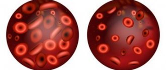
If a child's red blood cell count is elevated, the disorder is called erythrocytosis. Pathology increases the chances of blood clots forming and blocking blood vessels of vital internal organs. Hemoglobin will increase.
Reduced red blood cells in a child’s blood – erythropenia or erythrocytopenia. In such situations, there is an increased risk of developing internal bleeding or extensive internal hemorrhage.
The consequences are formed not only due to the deviation of red blood cell values from the norm - the situation in which platelets are low or high plays an important role. Often, a violation of one blood cell leads to problems with other components (for example, monocytes, reticulocytes, etc.).
When the volume of red blood cells in children is increased, this can be caused by the following pathologies:
- congenital heart defects;
- heart failure;
- diseases of the hematopoietic system, including thalassemia and erythremia;
- disruption of the production of blood cells in the bone marrow;
- dysfunction of the adrenal cortex;
- diseases of the respiratory system, such as bronchitis, asthma, COPD or pneumonia;
- diabetes;
- oncology - kidney or liver cancer is the most dangerous for a child;
- hypertonic disease;
- any illness whose symptoms include fever, profuse diarrhea or frequent vomiting;
- hypernephroma;
- extensive burns;
- Itsenko-Cushing's disease;
- polycythemia;
- surgical excision of the spleen.
When the erythrocyte distribution index is reduced, the content of iron-containing protein simultaneously decreases: hemoglobin and erythrocytes are closely related.
The problem may be caused by:
- acute blood loss;
- chronic bleeding, for example, from the nose or in the gastrointestinal tract;
- deficiency of iron, vitamins or folic acid;
- chronic renal failure;
- severe intoxication with chemicals;
- long-term chemotherapy or radiation therapy;
- leukemia and lymphoma;
- glomerulonephritis;
- myelodysplastic syndrome;
- multiple myelomas;
- hemoglobinopathies;
- autoimmune diseases;
- neoplasms in the bone marrow;
- aplastic type of anemia.
The level of red blood cells in the blood can change due to reasons not related to disease. Less harmless provocateurs:
- ingestion of large volumes of contaminated water;
- poor nutrition;
- exposure to stress;
- professional sports;
- living in an area with thin air;
- mental and physical fatigue;
- active (in adolescents) and passive (in newborns) smoking;
- prolonged refusal to eat;
- irrational use of drugs;
- previous surgical interventions.
It must be taken into account that a decrease or increase in red blood cells in a child’s blood may be a disorder inherited from the parents.
Red blood cells
Red blood cells are low in infants
A blood test for hematocrit is another important indicator by which doctors determine the child’s health status and the presence of disease processes.
The study helps to identify deviations from the norm in the ratio of red blood cells to the amount of plasma, either upward or downward.
Minor fluctuations are considered acceptable, but if the balance is significantly disturbed, the doctor, based on the indicators, diagnoses a specific disease.
In addition to standard tests, the level of red blood cells in the child’s blood is checked, which makes it possible to diagnose diseases.
How is a hematocrit test done?
To calculate the hematocrit number, blood taken from a child is sent into a special test tube with divisions marked on its wall, inserted into a centrifuge and processed for an hour and a half. The centrifugation process allows you to separate red blood cells from plasma: heavy elements, including red blood cells, sink to the bottom of the vessel, and the plasma remains at the top.
Visually, the ratio of red blood cells to plasma is determined using a scale on a test tube. Hematology analyzers are used to accurately calculate hematocrit. The resulting Ht indicator expresses the number of red blood cells without assessing their quality, but such information is enough for doctors to accurately make a diagnosis. The Ht value is expressed as a percentage and written as a fraction - liter/liter.
Age norms of hematocrit in children
A general blood test shows the level of hemoglobin, hematocrit and other blood characteristics (we recommend reading: what kind of hemoglobin should a newborn child have?).
When assessing the numerical value of red blood cells to determine whether the hematocrit is low or high, doctors rely on their standard value, characteristic of a healthy newborn.
In addition, doctors have derived the norm for each age period of childhood, from birth to adulthood.
Hematocrit blood test results vary depending on the child's age
For clarity, we have combined all the numerical indicators of the norm into a separate table, which looks like this:
| Child's age | Standard indicators in % |
| Newborns (1–28 days) | 44–62 |
| From 1 month of age to 3 months | 32-44 |
| from 6 months to 1 year | 36-44 |
| 2–6 years | 36–40 |
| 6–12 years | 37–44 |
| 12–18 years old, boys | 37–48 |
| 12–18 years old, girls | 36–44 |
What does deviation from the norm look like?
Experts consider deviations from the norm in the form of a decrease or increase as a sign of a painful pathology or physiological characteristics of the child. High Ht indicates an increased number of formed elements, which make the blood viscous and thick.
Having blood in this state provokes the formation of blood clots. However, exceeding the norm by 10-12% in infants and children under one year old is considered a physiological factor that does not cause concern. The picture looks alarming if the hematocrit is increased in a baby after one year.
In babies under one year of age, deviations from the normal Ht are most common, and this is not a sign of disease
The hematocrit is reduced - this means that the volume of blood cells has decreased, and they are responsible for supplying nutrients and oxygen to the organs. There is a violation of the acid-base balance, leading to oxygen starvation of the cells, which negatively affects the general condition of the baby. Weakness appears, the heartbeat quickens, the skin becomes pale, and there is shortness of breath.
Why does hematocrit increase?
A marked increase in Ht is directly related to hyperproduction in the bone marrow (an increase in the number or size of red blood cells). The reason for the increased concentration of cells lies in the area of pathological changes occurring in the body of a small patient. However, the process does not always occur due to pathological disorders in the body.
The hematocrit will be higher than normal if the patient did not drink before taking the test or his body was dehydrated as a result of vomiting or high fever.
Main factors of increase
We will necessarily increase Ht in case of disease processes that affect the growth of the number and size of red blood cells. The main factors for high hematocrit in children can be:
- True and redistributive erythrocytosis.
- Excessive reproduction of red blood cells or erythremia (Vaquez disease, polycythemia). It manifests itself as uncomfortable tingling in the fingers and toes, heaviness in the head, sudden redness of the skin, and cardialgia. Similar symptoms accompany diseases that cause an increase in the size of red blood cells.
- Compensatory reaction of the cardiovascular system to certain factors: pulmonary insufficiency, being at high altitude, kidney pathology, “blue” heart defects.
An increase in hematocrit may be associated with serious diseases, including heart disease
Relative reasons
Incoming processes can provoke an increase in the Ht level. Doctors include these:
- frequent bouts of vomiting;
- profuse diarrhea, causing blood thickening;
- intestinal obstruction, in which fluid moves into it;
- increased sweating (hyperhidrosis);
- peritonitis;
- hereditary and acquired kidney diseases;
- pathology formed in the respiratory organs;
- leukemia (more details in the article: causes of leukemia in children);
- burns and injuries;
- erythremia;
- asymptomatic oxygen starvation;
- long-term use of certain medications (for example, glucocorticosteroids).
What affects the decrease in Ht?
A decrease in hematocrit is recorded when its level is clearly low, that is, it drops to 25%. Having discovered that the hematocrit is reduced, the doctor prescribes an additional examination for the small patient to determine the cause.
Anemia is the most common cause of low hematocrit in a child
As a rule, a decrease in Ht is a consequence of the accumulation of excess fluid, due to which the blood thins and the percentage level of red blood cells and plasma changes (overhydration), and hemoglobin decreases (see also: how to treat high hemoglobin in a child?). The cause may be hyperproteinemia (excessive concentration of proteins). Such changes are caused by:
- Severe blood loss.
- Violation of the formation of red blood cells in the bone marrow towards a decrease in their number or size.
- Hereditary or acquired hemolytic anemia, typhoid fever, malaria, hemolytic poisons, in which rapid destruction of red blood cells occurs.
- Intravenous administration of large amounts of fluid for impaired renal function, leading to an increase in blood volume.
- Various types of anemia.
- Overhydration - blood thinning due to disturbances in the excretory system, insufficient blood circulation, water intoxication.
- Increased protein levels in the blood (hyperproteinemia). Occurs with frequent vomiting, acute infections, diarrhea, paraproteinemic hemoblastoses, Hodgkin lymphoma, myeloma.
Anemia is a common cause of decreased hematocrit. The child needs medical supervision and the establishment of a proper diet. The doctor may suggest taking medications that increase Ht.
The baby's menu is made up of foods rich in iron: liver, apples, nuts, eggs, meat. It is useful for a child to take hematogen, which is sold in all pharmacies. Parents should ensure compliance with all settings recommended by the pediatrician.
Symptoms
Regardless of which direction the erythrocyte norm fluctuates in children, characteristic external manifestations will be expressed. In some cases, pathology may go unnoticed due to the progression of the underlying disease. The problem is that children cannot verbally describe what exactly is bothering them, which is why parents should closely monitor the child’s behavior.
If red blood cells are low, this is expressed by the following symptoms:
- lethargy and weakness;
- constant drowsiness;
- increased heart rate;
- cold skin;
- secretion of sticky sweat;
- decrease in blood pressure;
- excessive pallor of the skin;
- headache;
- lethargy;
- dizziness;
- increased tearfulness and frequent mood swings;
- lack of appetite;
- growth retardation.
When red blood cells increase in children, the clinical picture will include:
- redness of the skin, especially on the face;
- skin itching of varying severity;
- increase in blood pressure values;
- bleeding from the nasal cavity;
- blurred vision;
- headache;
- decreased physical activity;
- muscle and joint pain;
- attacks of dizziness;
- ringing and noise in the ears;
- shortness of breath.
A set of procedures for determining blood density
Blood during bowel movements in a child under one year old from the anus after stool
To determine the degree of blood thickness, blood is taken from infants for analysis. It determines the level of red blood cells, their sedimentation rate, hemoglobin, hematocrit and platelets. All these indicators are important for diagnosis. Blood is drawn through a vein if related tests have been prescribed. In other cases, a puncture is made from a finger. If it is not possible to take blood from it, then the side of the heel is pierced.
The manipulation is carried out according to certain instructions, which are strictly followed:
- Before starting the procedure, the laboratory assistant washes his hands with soap and prepares the workplace.
- Wear sterile disposable gloves.
- Use a cotton swab to lubricate the puncture area.
- He holds his finger with one hand and with the other he makes a puncture with a scarifier.
- A glass capillary with a bulb makes a fence.
- Use a new cotton pad moistened with an antiseptic to blot the injection site.
- Press the wound with dry cotton wool.
Newborns clench their hands into fists, so drawing blood from a finger can be problematic. In this case, the big toe or the corner of the heel is pierced. The same method is used if the hospital does not have children's needles available.
Red blood cells
The procedure for collecting blood from a vein is carried out according to the same principle. The largest and most accessible veins in a newborn are located on the head. Usually all tests are taken from there. This is why a catheter is installed on the head. Also after birth, an analysis is taken from the umbilical cord. It shows the level of bilirubin, which indicates the presence of physiological jaundice.
Important! There is no need to worry that your newborn is in pain. After birth, children sleep constantly, they feel little, all procedures take place in their sleep.
To be on the safe side, doctors prescribe additional tests that will help recreate the overall picture of the disease:
- determine venous and peripheral hematocrit;
- examine the level of glucose and calcium in the blood;
- control bilirubin levels;
- determine the acid-base state of the blood.
A set of tests will help make an accurate diagnosis and prescribe the correct treatment.
Diagnostics
High or low levels of red blood cells are detected during a complete blood count. This laboratory test may require either capillary or venous blood.
Parents should ensure that the child is hungry before taking blood, since the most reliable results will be obtained only when the biological material is taken on an empty stomach. If this is not done, a repeat analysis will be required, which is extremely undesirable for children, especially younger ones.
The results are interpreted by a hematologist, who pays attention not only to the number of red blood cells, but also to their sedimentation, or rather the speed at which this occurs. After this, the doctor transmits the information received to the pediatrician - this specialist draws up a further individual diagnostic program for each patient. This is necessary to find the root cause.
Diagnostic measures common to all are:
- a clinician's examination of the medical history to look for an acute or chronic underlying disease;
- familiarization with family history - to establish the influence of genetic predisposition;
- collection and analysis of life history - to confirm or refute the influence of harmless causes;
- a thorough physical examination and assessment of the condition of the skin;
- a detailed survey of the patient’s parents - to determine the first time of occurrence of characteristic signs and compile a complete symptomatic picture.
Additional diagnostic tools include:
- specific laboratory tests;
- instrumental procedures;
- consultations with pediatric specialists from other areas of medicine.
Normal red blood cell concentration and anemia
Clinical blood test
A blood test is a laboratory test that provides information about your health status. Depending on what needs to be examined, the blood is collected in different tubes and analyzed in the laboratory. Tests will indicate the presence of infection, anemia or leukemia.
This test provides information about the number of red and white blood cells and platelets in children. This test provides the doctor with evidence of anemia. Using a blood smear in newborns, the doctor will evaluate the size, shape and possible pathological changes in children's red blood cells under a microscope. Bleeding disorders such as hemophilia are also detected through the test.
Blood is considered connective tissue because it consists of a fluid in which living cells are suspended. Indications of deviations from the normal value allow us to draw conclusions about diseases:
- Salts in tissues such as iron, sodium, potassium or calcium.
- Sugar level.
- Lipids such as cholesterol and triglycerides.
- Proteins and enzymes, eg albumin, gamma GT.
- Metabolites: uric acid or creatinine.
- Blood clotting factors.
- Tumor markers.
Chronic diseases increase the number of enzymes in studies. Using the number of red blood cells in a child, the therapist can determine whether the little patient is suffering from an infection in which bacteria have entered the blood, and what pathogenic microorganisms are involved.
Important information: What do low platelets in the blood indicate during pregnancy (increasing foods)
Treatment
If the red blood cells in children are low or high, the pathological predisposing factor should be treated. In addition to conservative methods, surgical intervention may be necessary. More often, therapy has an integrated approach.
You can normalize red blood cells in a child’s blood using the following measures:
- taking medications - limited to vitamin-mineral complexes;
- blood or red blood cell transfusion;
- adherence to a therapeutic diet - when the concentration is reduced, it is necessary to introduce foods fortified with iron into the menu, and when the concentration is increased, ingredients that thin the blood;
- use of traditional medicine recipes.
The correction will be individual and carried out under the strict supervision of the attending physician.
Functions of red blood cells
Erythrocytes (red blood cells), with the help of hemoglobin protein, perform the function of distributing oxygen throughout the body and removing carbon dioxide from tissues. Hemoglobin levels affect the health of any person. Its lack causes great damage, especially for the growing body of children. The brain and kidneys suffer the most from lack of oxygen.
What are the responsibilities of red blood cells?
Prevention and prognosis
To ensure that red blood cells are always within normal limits, parents need to monitor compliance with the following simple preventive recommendations:
- nutritious and healthy nutrition;
- prevention of mental and physical fatigue;
- absence of stressful situations;
- constant strengthening of the immune system;
- taking only those medications prescribed by the clinician;
- eating quality foods and drinking purified water;
- protection from bad habits, radiation and the penetration of toxic substances into the body;
- regular visits to the pediatrician.
High or low levels of red blood cells in themselves are a threat to life. If other components of the blood deviate from the norm and there is complete lack of treatment for the provoking disease, dangerous complications can arise.
Danger of condition
Large fontanel in a child - causes and consequences
A baby with thick blood is at risk of developing chronic diseases. If the symptoms are not severe, the condition most often goes away after several days of treatment with a saline dropper. If medical assistance is not provided in a timely manner, then some consequences arise. In 40% of cases, the symptoms of polycythemia do not indicate the presence of the disease, although it may be sepsis or heart problems. Complications and consequences:
- Oxygen starvation of the brain, impact on mental and mental development.
- Frequent convulsive syndromes.
- Partial necrosis of tissues of internal organs.
- Stagnation of blood in the area of the heart with weak output.
- Thrombosis of the renal veins, problems with urine excretion.
- Increased insulin levels, hypoglycemic coma.
Complications arise if the mother refuses treatment. Usually doctors immediately determine the presence of a problem and begin therapy. Serious complications occur in only 20% of cases. In the rest, children recover within a week and begin the normal life of a healthy newborn.
Newborn baby's heel and blood patch
What affects the number of red blood cells
Erythropoietin
In the human kidneys there are special cells that recognize the lack of oxygen in the body. They produce erythropoietin, a substance that stimulates the appearance of red blood cells in the red bone marrow. (In a person from newborn to 4 - 5 years, hematopoiesis is observed in all bones of the body, and then remains only in flat and large tubular bones.) When maturing, the erythrocyte enters the blood and performs its functions for 110-120 days and is then utilized in the spleen , as well as in the liver and red bone marrow.
Symptoms
In the early form, hemorrhages often begin in the prenatal period. The child is born with intracranial, pulmonary and skin hemorrhages. Internal bleeding into the liver, spleen, adrenal glands, as well as into the abdominal organs, and vomiting with blood are typical.
The disease is characterized by cutaneous hemorrhagic effusions
The classic reaction is characterized by the presence of blood in the stool and vomit around the 7th day of life. Poor coagulation is visible in the bleeding of the navel, in the long-term non-healing in case of circumcision of the foreskin in boys, in nosebleeds, cephalohematoma on the head and bruises on the skin. The wounds do not heal for a long time after injections. In severe cases, anemia and hemorrhagic damage to internal organs are detected.
The late form develops as a result of liver diseases and dysfunctions of the gastrointestinal tract along with breastfeeding. Leading symptoms:
- hematemesis (bloody vomiting);
- in half of the cases there are intracranial hemorrhages and cephalohematomas;
- extensive bruising on the skin and mucous membranes;
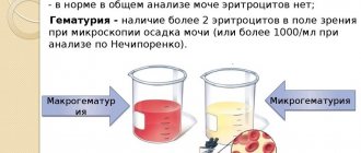
- hematuria (blood in the urine);
- melena - a disease accompanied by black stools and indicating gastrointestinal bleeding; the development of melena is possible with gastroesophageal reflux;
- the umbilical wound is bleeding.
Melena is often accompanied by hyperbilirubinemia, since red blood cells in large quantities disintegrate and die in the intestines. Ulcers appear on the mucous membrane of the stomach and duodenum. This condition is explained by the stress of birth, during which glucocorticoids are released in large quantities.
In severe cases of the disease, hypovolemic shock is possible - a condition characterized by a rapid decrease in circulating blood volume due to fluid loss due to persistent vomiting and diarrhea. The child's blood pressure and temperature drop, he is weak, and his skin is pale. The condition requires urgent resuscitation.
Change in leukocyte formula
When studying a newborn's blood test, important information is conveyed not only by the total WAB indicator, but also by changes in the level of all types of leukocyte cells. Each type of white cell performs specific tasks in building a natural defense system. Deviation from the norm of a certain group of leukocytes clarifies the developing disease and its nature.
Leukoformula in newborns (normal)
| Baby's age | WBC 10^9 units/l | Leukocyte formula (quantitative indicator of cells in the field of view) | |||||
| neutrophils | eosinophils | basophils | monocytes | lymphocytes | |||
| stab | segmented | ||||||
| 1 day | 20 | 5-12 | 50-70 | 1-4 | 0-1 | 4-10 | 16-32 |
| 5 days | 12 | 1-5 | 35-55 | 1-4 | 0-1 | 6-14 | 30-50 |
| 10 days | 11 | 1-4 | 27-47 | 1-5 | 0-1 | 6-14 | 40-60 |
| 1 month | 10 | 1-5 | 17-30 | 1-5 | 0-1 | 5-12 | 45-60 |
| 1 year | 9 | 1-5 | 20-35 | 1-4 | 0-1 | 4-10 | 45-65 |
Reasons for high white blood cell counts:
- Neutrophilic leukocytosis: bacterial lesions, inflammatory processes in the body, cancer, vaccinations, blood loss, intoxication, a consequence of the use of immunomodulators.
- Eosinophilia: allergies, rheumatism, malaria, mononucleosis, parasitic infection.
- Basophilia (rarely observed): nephrosis, colitis, anemic condition, allergies, chickenpox.
- Lymphocytic leukocytosis: poisoning, severe viral infections (influenza, tuberculosis, whooping cough, syphilis).
- Monocytosis: parasitic, viral infections, rheumatism, leukemia, lymphogranulomatosis.
- Reasons for low performance:
- Low basophils: severe allergic reactions, long-term stress, infectious lesions, disorders of the adrenal glands, hyperthyroidism, acute pneumonia, after chemotherapy and hormone therapy, disorders of the endocrine system and bone marrow.
- Eosinopenia: psychoemotional disorders, appendicitis, sepsis, starvation.
- Neutropenia: anemia that occurs when iron levels are low, severe infections (rubella, hepatitis), neoplasms, dysfunction of the spleen and pancreas.
- Monocytopenia: leukemia, sepsis.
- Lymphocytopenia: bone marrow pathologies, anemia, infectious lesions, postoperative period.
Forms of the disease
There are primary and secondary hemorrhages. Primary is said to be when the fetus initially had a deficiency of the vitamin, and its supply through mother’s milk is minimal. By the 5th day, the deficiency can be compensated for by its intestinal production.
The secondary form is diagnosed if liver lesions are present, when the synthesis of polypeptide precursors of plasma factors (PPPP) is impaired.
The disease is classified according to the time of onset:
- early - bleeding makes itself felt on 1, maximum 2 days after birth;
- classic - appears on days 3-5;
- late - occurs at any time during the first 8 weeks of life.
About vitamin K and its role in the body
The very name of the vitamin, designated by the letter K, comes from the phrase “coagulation factor,” which directly indicates its most important function. It has been proven that blood clotting requires at least 10 active proteins, 5 of which are synthesized with the participation of vitamin K. The liver needs it to produce prothrombin, a substance that allows blood to thicken. Vitamin K is necessary not only for the circulatory system, it also helps retain calcium in bone tissue.
Without vitamin K, normal functioning of the circulatory system is impossible.
In adults, a deficiency of this vitamin is rare, since it is produced in sufficient quantities by intestinal bacteria, and it is also present in many vegetables without breaking down after heat treatment. But in babies, deficiency is possible for a number of reasons, then hemorrhagic syndrome develops in newborns.