If a patient has to frequently consult an otolaryngologist with throat diseases, then a laryngoscopy may be prescribed by the doctor to obtain objective data on the condition of the larynx. What it is? The question is quite logical. It’s better to clarify some details in advance instead of getting nervous and stressing yourself out. In this article we will analyze in detail what this procedure is, what are the indications for its implementation and whether there are any contraindications.
Indications for the procedure
The doctor makes the decision to perform laryngoscopy if it is necessary to identify:
- the cause of a sore throat or ear;
- the cause of difficulty swallowing;
- presence of a foreign body in the throat;
- the reason for the appearance of blood in sputum;
- the reason for the voice change;
- the reason for the lack of voice;
- the presence of laryngeal pathologies.
In addition, this manipulation is prescribed for foreign body removal, biopsy and removal of polyps on the vocal cords.
Indications and contraindications for the study
Laryngoscopy examination is prescribed for the following symptoms:
- muffledness, hoarseness, lack of voice;
- pain in the oropharynx, larynx, ears;
- expectoration of mucus mixed with blood;
- narrowing of the larynx, difficulty inhaling, swallowing;
- lump in the throat - a feeling of the presence of a foreign body in the respiratory tract.
The procedure is also prescribed if it is necessary to remove a foreign body stuck in the larynx, obtain a sample of biological material for histological analysis, remove polyps, or differential diagnosis of cancer.
Hypopharyngoscopy has no absolute contraindications. Endoscopic examination is not performed on persons:
- with an unstable psyche, diagnosed with neurotic disorders;
- with diseases of the cardiovascular system, especially those who have suffered a stroke;
- with stable hypertension. After normalization of blood pressure, the restriction is lifted;
- with injuries of the cervical spine. This is due to the fact that the patient will have to tilt his head back, and the doctor can rotate it in different directions during the examination. In case of serious injuries, this is fraught with complications;
- with blood clotting disorders. During manipulation, minor damage to the laryngeal mucosa may occur, but if there are problems with hemocoagulation, massive bleeding develops.
Pregnancy is a relative contraindication. The study is safe for the expectant mother and fetus, but the patient’s anxious state can be harmful.
Preparation for laryngoscopy
Carrying out indirect laryngoscopy does not require serious preparation from the patient. It is enough to refrain from eating and drinking a few hours before the procedure. This is necessary in order to avoid vomiting. Well, the patient will have to remove his dentures.
Before performing direct laryngoscopy, the otolaryngologist collects a complete medical history of the patient's condition. It is important for the doctor to know about all the medications the patient has taken recently. He checks for drug allergies and asks questions about blood clotting. Be sure to find out the presence of cardiovascular pathologies, rhythm disturbances or problems with blood pressure. For women, the doctor checks the possibility of pregnancy.
Next, patients undergo all necessary measures related to general anesthesia. Sedatives and agents to suppress mucus secretion are administered. Immediately before the procedure, the patient removes dentures, contact lenses and jewelry.
What is indirect laryngoscopy?
Most often, during an appointment with a patient, the doctor determines that indirect laryngoscopy is necessary. What it is? Let's try to explain. This is the simplest and most painless type of examination of the larynx. The procedure uses a small hand mirror, the diameter of which does not exceed 16-30 mm, and a special frontal reflector. This procedure is optimal for examining older children, but it is also quite informative when examining adult patients.
Varieties of the method
The indirect method was the first to be developed and introduced into clinical practice. In this case, the larynx is examined using a small mirror, which is inserted into the oropharynx. In this case, the mirror can reflect a directed beam of light, allowing you to examine the structures of the larynx. The method is most often used for a quick examination of the respiratory tract, and therefore is widely used in clinics and during medical examinations of the population.
A direct type of laryngoscopy is performed using a special laryngoscope, which can be flexible or rigid. Recently, endoscopic varieties of the procedure have been increasingly used, allowing one to obtain high-quality and even enlarged images of the structures being examined. The procedure is performed in many medical institutions, but requires more preparation and execution time than indirect laryngoscopy.
The retrograde method of examination is carried out using a nasopharyngeal speculum. However, its implementation is associated with certain discomfort for the patient and low quality of the visible image.
Methodology
In most cases, the procedure is as follows:
- The patient is seated in a chair with a headrest, asked to open his mouth, and the throat is irrigated with an anesthetic to suppress the gag reflex.
- The doctor holds the patient’s tongue and with his other hand inserts a warm laryngeal mirror into the oral cavity. The doctor sets the angle at which a beam of light reflected from the mirror enters the larynx.
- The patient is asked to pronounce a long vowel sound (“a”, “e”) so that the larynx rises.
The procedure allows the doctor to examine the free portion of the epiglottis, examine the larynx, and examine the appearance of the vocal cords. The aryepiglottic folds and arytenoid cartilages are also examined.
If the ENT doctor decides to do a laryngoscopy to examine the vocal cords, then he will be able to record their color, establish mobility and study the surface structure. In addition, the procedure allows you to evaluate the symmetry of closure at the moment of phonation and determine the width of the glottis. In some patients, it is possible to partially examine the trachea. The whole procedure takes about 5 minutes.
Contraindications and complications after diagnostics of the larynx
It is worth noting that this procedure has contraindications, including: congenital pathologies of the oral cavity; disorders of the central nervous system, convulsions, epilepsy attacks, diabetes mellitus.
Typically, the procedure, regardless of its type, does not cause any complications or side effects. In rare cases, an allergy to the anesthetic may occur. Since the procedure involves irritation of the mucous membranes of the oral cavity and larynx, after the examination, mild discomfort in the mouth and throat will be observed for several hours.
Pharyngoscopy is a safe, simple and at the same time effective way to diagnose the larynx without surgical intervention.
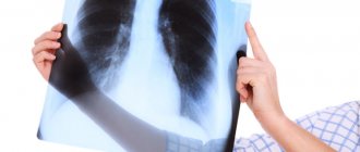
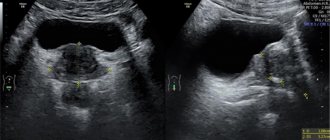
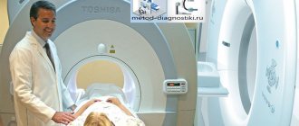
If the doctor is not satisfied with the examination results, he may additionally prescribe a number of procedures that will help make the correct diagnosis. Additional diagnostics may include: ultrasound, CT, MRI, examination of the larynx using an endoscope, etc. The doctor independently determines the type of examination.
Before undergoing the procedure, it is mandatory to consult not only with a therapist/pediatrician, but also with an ENT doctor and a surgeon.
More fresh and relevant information about health on our Telegram channel. Subscribe: https://t.me/foodandhealthru
The test is characterized by relative safety. This is due to the fact that there are no contraindications or side effects. Therefore, pharyngoscopy is allowed for children and adults. This method has no age restrictions.
The pharynx is an unpaired hollow organ that connects the cavities of the nose and mouth with the esophagus and larynx. Its walls are part of the digestive tube and respiratory tract, and consist of muscle fibers and mucous membrane. Behind this, it is in the pharynx that there is a lymphoepithelial ring, consisting of one lingual, one pharyngeal, two palatine and two tubal tonsils. In the lateral walls of the pharynx there are two funnel-shaped openings of the auditory tubes.
The examination begins with an external examination of the neck and labial mucous membranes. Next, all regional pharyngeal lymph nodes are palpated. Then the nasal, oral and laryngeal parts of the pharynx are examined in turn. The method of examining each part received its name accordingly - oroscopy, mesopharyngoscopy and epipharyngoscopy.
Oroscopy
During this part of the procedure, using a spatula, the mucous membrane of the oral vestibule, gums, tongue, hard palate, ducts of the sublingual, submandibular and parotid glands, and the floor of the mouth are examined.
During oroscopy, it is necessary to evaluate the amount of saliva, as well as the performance of the salivary glands. To do this, they may suggest thinking about something sour, such as lemon, or placing a drop of diluted acetic acid under your tongue. Normally, this manipulation can cause such strong salivation that saliva from the glands can flow like fountains.
Mesopharyngoscopy
It is during this part of the examination that the patient is asked to pronounce the sound “A”. This action helps the doctor assess the mobility of the soft palate, as well as its symmetry. Here the patient may experience gagging. Its strength depends on individual sensitivity and the temperature of the spatula, which is used to press the root of the tongue.
Doctor performs mesopharyngoscopy on a child
Next, the mucous membrane and uvula of the soft palate are examined in turn, the size is determined and the condition of the mucous membrane of the tonsils is assessed. Using two spatulas, the contents in the almond crypts are checked, which is normally scanty and may look like minor epithelial plugs.
At the end of mesopharyngoscopy, an examination of the posterior pharyngeal wall is performed. By the way, normally, on the pink, flat and smooth surface of that part of the pharyngeal wall, there are rare lymphoid granules with a diameter of 1 mm.
Epipharyngoscopy
Next, the pharyngoscopy examination is carried out using a nasopharyngeal speculum, which, like spatulas, requires pre-warming. At this stage of the examination, also called posterior rhinoscopy, the condition of the choanae connecting the nasal and oral cavities, the openings of the eustachian tubes connecting the ear and pharynx, the ends of the nasal concha and the nasopharyngeal vault are assessed.
If the tissue of this tonsil grows and becomes hypertrophied, then this formation is called adenoids, and inflammation of the third pharyngeal tonsil is called adenoiditis. Adenoids and their inflammation are rare, but can occur in adults and children 1-2 years of age.
Examination of the posterior parts of the nose and nasopharynx
If necessary, the pharyngoscopy examination procedure may end with a digital examination of the choanae, pharyngeal tonsil, fornix and lateral walls of the nasopharynx. In most cases, such palpation is performed when examining children and adolescents, or during the differential diagnosis of tumor tumors in adults.
In the vast majority of cases, a pharyngoscopy examination is immediately followed by an external examination of the larynx and indirect laryngoscopy. During this stage, the doctor will ask the patient to pronounce the sounds “I” and “E”. The main goal of this stage is to assess the condition of the lower parts and organs of the pharynx and larynx, as well as the false and true vocal cords.
Features of direct laryngoscopy
A mirror (indirect) examination cannot be performed on small children, and sometimes it is simply not enough to help the patient. In this case, the doctor performs direct laryngoscopy. This is a more complex type of examination, but it gives the doctor the opportunity to obtain more detailed and complete information. Since direct laryngoscopy is not the most pleasant procedure for the patient, it is performed under local anesthesia. The most commonly used solution is a 2% Dicaine solution.
Depending on the type of direct examination, it can be performed with a flexible fiber laryngoscope or a rigid (rigid) laryngoscope. The manipulation technique will naturally be different.
Pros and cons of laryngoscopy
- Advantages. For a doctor, it is an opportunity to assess the condition of the larynx, identify various diseases, and dysfunctions of the organ. Diagnostics allows you to select material for further study and perform a number of simple manipulations. For the patient - surgery with a minimal recovery period.
- Flaws. When moving the laryngoscope into the throat, there is a risk of injury to the vocal cords. The problem of discomfort caused by laryngoscopic examinations is eliminated by choosing an anesthetic, and bleeding by using modern laryngoscopic complexes equipped with emergency equipment.
If you are prescribed a laryngeal orthoscopy, follow your doctor's instructions exactly, notifying him of any health problems. The study is necessary to clarify the diagnosis and identify hidden throat diseases, because the lack of treatment leads to the development of chronic pathologies, which are not always easy to cope with.
Direct flexible laryngoscopy
Flexible laryngoscopy of the throat can be performed in either a sitting or lying position. Although it is somewhat more convenient for a doctor to work with a patient lying on his back. The fiber laryngoscope is inserted through the nose. The device is equipped with fiber optics and a small light source. To avoid injury to the mucous membrane, a vasoconstrictor drug is injected into the nasal passage. The examination takes about the same time as indirect laryngoscopy, that is, 5-6 minutes.
Possible complications
If you are going to undergo a procedure such as laryngoscopy of the larynx, you need to be aware of such possible complications as:
- pain;
- severe swelling or bleeding in the throat;
- allergic reaction to anesthesia;
- nosebleeds when a laryngoscope is inserted through the nose;
- nausea and vomiting;
- wounds from the teeth at the bottom of the tongue.
The laryngoscopy procedure is usually performed by an otolaryngologist.
Direct rigid laryngoscopy
Rigid laryngoscopy (what it is and how the procedure is performed will be described below) is performed in an operating room. For the patient, this type of examination is unpleasant and traumatic, but only it makes it possible to remove foreign bodies from the larynx, take a tissue sample for biopsy, remove polyps on the vocal cords, and so on.
To perform rigid direct laryngoscopy, the patient is given general anesthesia. During the manipulation, the patient is placed on his back and his head is tilted back. A rigid laryngoscope is inserted through the mouth. The special tool is introduced in 3 stages:
- the spatula is brought to the epiglottis;
- the end of the spatula, bending around the edge of the epiglottis, is passed to the entrance to the larynx;
- the root of the tongue is pressed forward a little and the instrument is moved to a vertical position.
The inspection may take approximately 30 minutes. After the manipulation, the patient is under medical supervision for several hours. Since the manipulation requires an experienced specialist, the patient should be careful when choosing the place where to perform laryngoscopy.
Reviews
Olga, 37 years old, Moscow: For about a month, the pain in the throat was accompanied by the sensation of a lump. It was painful to swallow and turn my neck. Gradually the voice became hoarse. After visiting the ENT doctor, it was suggested that I go to the hospital for examination. There the doctors suggested doing a laryngoscopy. It turned out that a tumor had appeared in the larynx. They immediately took a histology test to determine it. I'm waiting for the result to start treatment.
Vitaly, 48 years old, Samara: About 3 weeks ago we had a direct laryngoscopy under anesthesia. Of course, I didn’t feel anything during the procedure, but after it I had a sore throat, a cough and discomfort. All these unpleasant sensations passed within a few days. The consequences are not very pleasant, but as a result we found out why there had been unpleasant sensations in the larynx for a long time. After laryngoscopy, I was home a few hours later.
Marina, 33 years old, Krasnodar: About 2 months ago hoarseness of her voice appeared. I went to the ENT doctor at the clinic. He gave me a referral to a phoniatrist. This doctor immediately gave a referral for laryngoscopy, explaining that without this procedure it was impossible to determine anything. They decided to do laryngoscopy without anesthesia using mirrors. In the end, they found out that the voice was lost due to stomach reflux. The acid irritated the vocal cords and caused inflammation. Now I need to be treated by a gastroenterologist.
Patient care after rigid laryngoscopy
Upon completion of rigid laryngoscopy, the patient requires the following care:
- If for some reason the manipulation was carried out under local anesthesia, then the patient lies in the Fowler position (half-sitting). The sleeping patient should lie on his side with his head elevated to avoid aspiration.
- The nurse monitors physiological indicators every 15 minutes until they stabilize. For the next 2 hours, monitoring is carried out every 30 minutes. If longer-term monitoring is necessary, physiological parameters are determined every 2-4 hours. If the patient has tachycardia, extrasystole or other abnormalities, the doctor is notified.
- To avoid swelling, cold is applied to the larynx area after manipulation.
- A basin is placed next to the patient for spitting or vomiting. If there is a large amount of blood in the saliva, the nurse informs the doctor.
- If tracheal perforation (crepitus in the neck) is suspected, a doctor is immediately called.
- Using a phonendoscope, the tracheal area is auscultated.
How is laryngoscopy performed?
Laryngoscopy is performed in a clinic or hospital. If an endoscopic examination is planned, it is necessary to have equipment and drugs to provide emergency medical care in case of serious complications.
Preparation for manipulation
Preparation for laryngoscopy involves following these rules.
- The interval between the last meal and the procedure should be at least 8 hours.
- Smoking is prohibited on the appointed day. Smoking is a factor that increases the risk of complications. In addition, smokers have a cough with sputum, which can make the procedure difficult.
- If necessary, you may take sedatives the evening before the test. If the patient is very worried, the doctor will allow light sedatives on the day of the procedure.
- Teeth cleaning. This manipulation immediately before the examination is necessary not only to create a comfortable environment for the doctor, but also to protect the respiratory tract from bacteria living in the oral cavity.
Before starting the study, the doctor conducts a survey to identify contraindications to the procedure or anesthesia.
Patients with bronchial asthma or respiratory failure should have an inhaler with them.
Indirect technique
Hypopharyngoscopy is performed in this order.
- The laryngeal mirror is heated to an acceptable temperature and treated with an antiseptic.
- The patient opens his mouth, sticks out his tongue, and breathes through the mouth.
- The doctor wraps the tip of the tongue with a gauze napkin, takes it with his left hand and pulls it towards himself and downwards. The index finger lifts the upper lip.
- The laryngeal mirror is inserted into the oral cavity up to the soft palate and uvula. To examine the state of the larynx in two phases of physiological activity (phonation and inhalation), the patient is asked to make sounds and inhale.
- The mirror is removed from the larynx, separated from the handle and placed in saline solution.
Direct technique
Direct laryngoscopy consists of the following steps.
- The patient lies on his back, stretches his arms along his body, relaxes and breathes through his nose.
- The nurse injects a pain reliever or irrigates the mouth and throat with a local anesthetic, such as lidocaine solution.
- The doctor inserts a laryngoscope through the mouth and examines the larynx. If necessary, additional manipulations are performed - taking samples of tissue or mucus, removing polyps and nodules. The obtained and removed tissues are placed, respectively, on a glass slide or in an appropriate container.
- The laryngoscope is removed from the larynx. The conscious patient is placed in the Fowler's position, sleeping - on his side with his head slightly elevated.
Patient behavior after the procedure
After direct laryngoscopy, especially rigid laryngoscopy, the patient should not eat or drink water until the gag reflex is completely restored. This usually takes about 2 hours. First, the patient is given water at room temperature, which should be drunk in small sips.
Reviews about the procedure are mostly positive. Patients testify that after the manipulation the voice may temporarily disappear or become hoarse and a sore throat may be felt. They advise not to lose calm, since these inconveniences are temporary. When the gag reflex is restored, it will be possible to carry out softening rinses and take throat tablets.
Smoking patients should abstain from cigarettes until physiological processes stabilize and bleeding completely stops.
Choosing a clinic
Where can laryngoscopy be done? This is a rather serious issue for the patient. For example, in St. Petersburg this service is provided in 13 clinics and medical centers. In Moscow there is even more choice. You need to focus not only on the price, but also on the experience of the doctor to whom the patient will entrust his health.
Now you understand in what cases laryngoscopy may be prescribed, what it is and what types of examination modern medicine can offer. Don't panic, follow your doctor's recommendations. Some inconveniences associated with the manipulation are fully compensated by the diagnostic value of the procedure. Remember this.
Requirements for sterility during examination
In order not to become infected with HIV or viral hepatitis, it is necessary to pay attention to how the doctor prepares for the examination, as well as the condition of the instruments that will be used to conduct your particular examination. It is clear that after sterilization, medical instruments must be kept in special medical boxes, and the doctor, immediately before the examination, must wash his hands, disinfect them with an antiseptic and put on sterile gloves.
All instruments for pharyngoscopy undergo preliminary disinfection and sterilization
If necessary, and if a fiber-optic laryngoscope is available, the otolaryngologist, during a pharyngoscopy examination, can use this modern instrument to save a photo of the problem area or to take biomaterial for subsequent histological analysis.