Hysteroscopy method
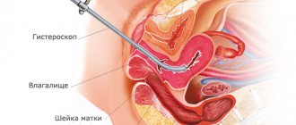
The hysteroscope is a new generation optical medical instrument for visual diagnosis of the uterus. The tool is a rectangular body with a hose system for supplying gaseous substances and liquids. The hysteroscope is equipped with a video camera and lighting, as well as a channel for introducing special medical devices - forceps, scissors, etc.
Using a hysteroscope, you can not only examine the uterine cavity, but also carry out the necessary surgical or cleansing procedures - remove polyps and neoplasms, remove a piece of tissue for biopsy. The hysteroscope provides a general and panoramic view with magnified images on the monitor. Magnifying the image allows you to examine the mucosa at the cellular level, determining the degree of change in pathological areas.
Hysteroscopy of the uterus is performed both as part of the IVF program and as a method of monitoring treatment results. A distinctive feature of hysteroscopy from abdominal operations is the possibility of performing surgical intervention under the visual supervision of a doctor without the use of a scalpel. The operation is performed in a gynecological chair under intravenous anesthesia with an individual selection of analgesics. In case of contraindications to the use of intravenous anesthesia, mask or other anesthesia is used. The examination is not performed without anesthesia.
Under general anesthesia, the uterine cavity is dilated with a gaseous/liquid substance for better visual inspection. If foreign bodies (remnants of the spiral) and polyps are detected, neoplasms or objects are immediately removed using forceps or clamps. However, if a comprehensive examination is not expected, hysteroscopy is performed not under general, but under local anesthesia.
Analogues of hysteroscopy
Among the procedures performed with similar diagnostic purposes, it should be noted:
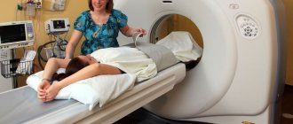
- hysterosalpingography (x-ray of the uterus with contrast) - the procedure is less informative and does not allow for surgical intervention or taking a biopsy. Does not require anesthesia, affordable;
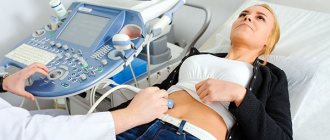
- Ultrasound - like the previous study, does not detect small tumors or foreign bodies. Tissue sampling or treatment is not possible. The procedure is carried out almost everywhere;
- diagnostic laparoscopy - manipulation requires a puncture of the abdominal cavity and strictly general anesthesia, and has a greater risk of complications. Combination with surgery is acceptable. The cost of laparoscopy is 1 or more times more expensive than hysteroscopy;
- diagnostic curettage - affordable, more traumatic and more likely to cause complications in the form of bleeding or damage to the uterine wall.
Among the analogues of operative hysteroscopy:
- surgical laparoscopy and therapeutic curettage - the disadvantages are similar to diagnostic types of interventions;
- operations with open access (through incisions) are traumatic and require recovery.
Hysteroscopy of the uterus is a modern method for diagnosing and treating gynecological diseases.
The procedure is unique (in terms of accuracy) for identifying small polyps, adhesions or foreign bodies, as well as checking the readiness of the organ for artificial insemination. Surgical interventions using a hysteroscope are low-traumatic and precise, and extremely rarely lead to complications.
Advantages of the method
More recently, for many problems with women’s health (bleeding, habitual miscarriage, chronic inflammation, etc.), diagnostic curettage (popularly “cleansing”) was prescribed. Compared to similar “blind” curettages and other invasive methods, endoscopic hysteroscopy has the following advantages:
- maximum diagnostic accuracy (up to 100%);
- low morbidity with extremely rare complications;
- painless and quick recovery after the study;
- carried out on an outpatient basis;
- the ability to take tissue for examination in a precisely planned location (targeted biopsy);
- optical control of all manipulations in the uterus;
- the possibility of treating uterine pathologies immediately after their diagnosis.
As you can see, this endoscopic method has many advantages over other methods that are less accurate in diagnosis (ultrasound) or more traumatic (diagnostic curettage).
Contraindications
The procedure is not performed if a malignant tumor is detected in the cervical canal. Despite the fact that RDV hysteroscopy is a minimally invasive surgical method, it also has a number of other contraindications:
- severe somatic diseases;
- inflammatory pathologies of the genitourinary system;
- a large tumor in the uterus or on its cervix;
- serious heart disease;
- stenosis of the cervical canal;
- 3-4 degree of vaginal cleanliness;
- diseases of the thyroid gland and internal organs;
- successful pregnancy;
- infectious pathologies in acute form (flu, pneumonia, sore throat, etc.);
- constant bleeding from the genitals.
Contraindications can be absolute or temporary. The last group includes inflammatory diseases and stenosis of the cervical canal. Then RDV with hysteroscopy is performed without dilation of the uterine cervix, and other gynecological manipulators are used during the operation.
For inflammation, 3-4 vaginal cleanliness, infectious pathologies, preliminary treatment is carried out before the procedure. Uterine bleeding is not an absolute contraindication. However, curettage and assessment of the uterine cavity will not be informative enough. Absolute contraindications include only chronic cardiovascular pathologies, serious diseases of the kidneys, liver and lungs, and normal pregnancy.
Types of hysteroscopy of the uterus
Hysteroscopy is an examination of the uterine cavity using probes equipped with a video camera and LEDs. The information obtained during the procedure helps to accurately determine the type of disorder, its stage and location. A hysteroscope equipped with special attachments helps to carry out surgical treatment and remove pathological formations in the uterine cavity. Taking these features into account, the following types of hysteroscopy are distinguished:
- diagnostic;
- surgical
Diagnostic hysteroscopy
Diagnostic or office hysteroscopy is performed to examine the inside of the uterine cavity. Based on the results of the examination, doctors make a conclusion about whether pathologies are present in the reproductive organ, determine their size, exact location and type. Using this type of hysteroscopy, specialists identify septa, adhesions, and polyps in the uterine cavity.
The information obtained can be used to further prescribe therapy. The list of pathologies identified using this procedure is wide:
- thickening of the uterine walls, the presence of septa;
- endometrial hypoplasia;
- polyps;
- adhesions;
- fibroids;
- adenomyosis;
- endometrial cancer;
- foreign bodies in the uterine cavity (remnants of the fertilized egg after an abortion, pieces of the intrauterine device).
Surgical hysteroscopy
Surgical hysteroscopy, or as it is also called, uterine resectoscopy, is prescribed for the purpose of correcting previously identified pathologies. During the procedure, additional surgical instruments are introduced through the hysteroscope to remove pathological formations. Surgical hysteroscopy of the uterus (what it is, stated above) is prescribed for women to correct the following types of gynecological pathologies:
- uterine polyps;
- myoma;
- synechia;
- dissection of the septum in the uterus;
- endometrial ablation for precancerous diseases of the uterine cavity.
What not to eat before hysteroscopy
An important part of preparing for hysteroscopy of the uterus is following a slag-free diet. It helps empty the intestines of food debris and solid contents. Such measures are necessary to ensure that intestinal loops do not compress the uterus during examinations and therapeutic procedures.
The list of what not to eat before surgery includes:
- legumes;
- all types of cabbage;
- cereals with a high content of indigestible particles;
- dark varieties of bread and bread with bran and whole grains;
- marinades, pickles, smoked meats, sausages and semi-finished products.
The list of prohibited products also includes coffee, alcohol and energy drinks. These products provoke an increase in blood pressure and also reduce blood clotting. Therefore, it is necessary to prepare very carefully for polyps, endometriosis and fibroids that are planned to be removed, avoiding their presence on the menu.
Advice! It is advisable for the patient to eat boiled, stewed, baked dishes in their own juice. Fried foods should be excluded from the menu a few days before surgery.
Preparing for planned surgery
An examination of the uterine cavity is carried out on the 4th day from the onset of menstruation. It is allowed to perform hysteroscopy on subsequent days, but no later than the tenth day after the start of menstruation. Preparation for the operation includes collecting tests, following a diet and avoiding intimate contact for three days.
What tests are needed:
- chest x-ray;
- electrocardiogram;
- general blood analysis;
- general urine analysis;
- colposcopy (under a microscope);
- vaginal smear for microflora;
- blood sampling for biochemistry and coagulation;
- cervical smear for cytology;
- Ultrasound of the pelvic organs.
Before the operation, you must follow a diet - do not eat gas-forming foods. The day before, you can eat easily digestible foods. If the procedure was scheduled for the morning, you should refuse evening meals. Can I drink liquids in the evening? You can drink as you wish, but not carbonated drinks. You should not eat or drink liquids on the day of surgery.
In the evening, clean the intestines with an enema: sometimes it is necessary to give an enema in the morning. The criterion for cleansing the intestines is light water instead of feces or pieces of undigested food. After cleansing the intestines, you should take a shower and shave your pubic and perineal hair.
During the preparatory period it is prohibited:
- rinse the vagina by douching and use suppositories without the consent of the gynecologist;
- use alkaline detergents for intimate hygiene;
- intimacy before surgery.
What should you take with you to the hospital? They take the usual set of accessories: a robe, slippers, a towel, a toothbrush, and other dental accessories at their discretion. Also take sanitary pads with you. If an outpatient minimally invasive diagnostic examination is planned without the use of general anesthesia, there is no need to take hygiene supplies. If the tests before hysteroscopy did not show a complex clinical picture, you may already be home in two to three hours.
Bottom line
Hysteroscopy is a necessary medical procedure for a detailed examination of the internal cavity of the uterus. Depending on the clinical picture of the health condition, either a diagnostic examination under local anesthesia or intracavitary surgery to remove tumors under general anesthesia may be required. There is no need to follow a strict diet before surgery - just skip dinner and breakfast. An important condition for preparation is complete emptying of the intestines and bladder.
Recommendations before diagnostic hysteroscopy
- Stop sexual intercourse two days before hysteroscopy.
- The day before the procedure, cut or shave the pubic hair, labia and perineum short.
- On the eve of the intervention, go to bed no later than 22:00. You can take a sedative: tinctures of motherwort, peony, valerian, etc., according to the instructions included with the medications.
- In the morning, on the day of the procedure, take a shower and thoroughly clean the external genitalia, and put on clean knitted underwear.
- Hunger - last meal three hours before hysteroscopy.
- Immediately before the procedure, empty your bladder.
- HAVE WITH YOU:
- Referral for hysteroscopy, examination results (ultrasound, smear for flora, RW, HIV, Hepatitis B and C).
- A clean shirt (robe) and socks, sanitary pads, slippers.
Leave jewelry (earrings, rings, etc.) at home.
Back to contents
What is office hysteroscopy?
It is practically no different from the surgical option - in both cases, it is possible to examine the woman’s genital organs and identify pathologies. But the traditional procedure is done under anesthesia and is considered a minor surgical intervention, so the patient must be observed in a hospital setting. Diagnostic office hysteroscopy can be performed in a doctor's office on an outpatient basis.
On the day of visiting a gynecologist, a woman undergoes an ultrasound, and if there is a suspicion of pathology in the uterine cavity, the doctor suggests undergoing an office hysteroscopy. It makes it possible to make an accurate diagnosis, and sometimes remove a polyp or other small formations. The uterus is easily visible using a hysteroscope - this is a special endoscopic device.
USEFUL INFORMATION: How can you lower progesterone if it is in excess?
How is the procedure performed?
A tube, the diameter of which does not exceed 3 mm, is inserted into the uterine cavity through the vagina and cervical canal. At the end there is an optical system that displays the image on the display. Due to the high degree of resolution, even small pathological formations can be seen.
The procedure lasts approximately 5-15 minutes and is virtually painless. A woman may feel slight nagging pain in the lower abdomen. Using this device, a specialist will detect adhesions in the canal and uterine cavity, polyps, and small myomatous nodes. In some cases, the doctor can remove the polyp using office hysteroscopy right in the process, as well as remove small formations and break up synechiae.
Endometrial hyperplasia, due to which a woman complains of heavy menstruation, is well diagnosed using hysteroscopy. This method also makes it possible to determine the cause of bleeding of unknown origin in girls. With hysteroscopy of the uterus, you can see oncological pathologies and begin treatment in a timely manner.
When should it be done?
If a woman is of reproductive age, the procedure is performed immediately after her period.
This is explained by the fact that the endometrium is still thin, so even small formations can be seen. By the second phase it thickens, which makes diagnosis difficult.
For postmenopausal women, the test can be performed on any day. The specialist will tell you what tests need to be taken first to rule out acute pathologies: a smear for flora, a blood test for syphilis, HIV and hepatitis.
Indications
There are a number of factors that are the basis for the procedure:
- Severe pain during menstruation;
- Uterine bleeding;
- Endometriosis;
- Uterine fibroids;
- Habitual miscarriage;
- Infertility;
- Suspicion of adhesions, endometrial pathologies.
Since general anesthesia is not performed, the woman can watch the monitor screen and monitor the progress of the study. During the procedure, she can ask questions, receive recommendations and valuable advice. If necessary, the doctor will take a piece of the endometrium for a biopsy.
Hysteroscopy is mandatory for the following diseases:
- Chronic and internal endometriosis;
- Myoma;
- Infertility;
- Any uterine pathologies.
How to prepare for the procedure?
Any intrauterine intervention should be carried out only after preparation has been made. And hysteroscopy is no exception. Before conducting it, the doctor prescribes a full clinical examination, including consultations with a therapist and other specialists, if indicated.
But one examination is not enough; preparation for office hysteroscopy is necessary with medication. It consists of sedative and antibacterial therapy. Its main goal is to protect the body as much as possible from possible consequences and ease the postoperative period.
Useful recommendations from experts:
- The day before, take a shower and epilate the pubic area;
- Before going to bed, drink 30 drops of motherwort if you are very nervous;
- The procedure is performed on an empty bladder;
- Make sure you have a clean shirt, socks and sanitary pads with you;
- Leave the decorations at home;
- A couple of hours before the operation, you need to take an antibiotic, which will be prescribed by the doctor.
After the procedure, you must regularly measure your temperature and take medications prescribed by a specialist. Their dosage and duration of use depend on the complexity of the situation and the severity of pain. For two weeks you cannot have sex, play sports or perform thermal procedures. You can only wash in the shower
It is important to monitor the nature of the discharge and for any alarming symptoms it is better to consult a doctor
We hope our tips will help you properly prepare for the procedure and go through it. Be healthy!
How to prepare for hysteroscopy
Hysteroscopy is intended to detect and eliminate pathological changes in the endometrium of the uterus, and involves penetration into the organ cavity through the cervical canal. Since it is almost impossible to avoid injury to the membranes lining the uterus, it is important to avoid infections in the wounds, as well as to prevent bleeding.
This is precisely what recommendations on how to prepare for hysteroscopy are intended for.
What a woman needs to do before diagnostic or therapeutic hysteroscopy:
- Get tested for all kinds of infections, including genital infections. This step is necessary to prevent the occurrence of infectious complications. Laboratory tests can reveal various diseases associated with an imbalance of vaginal microflora (vaginosis), STIs, and inflammatory processes, which are contraindications for manipulation.
- Check the blood for clotting to eliminate the risk of uncontrolled uterine bleeding. Perhaps drug therapy will be prescribed as a preparation.
- Consult with specialized specialists if you have chronic diseases, including pathologies of the heart, lungs, urinary system and gastrointestinal tract. If the underlying disease requires constant use of medications, it may be necessary to change their dosage or completely eliminate their use on the eve of the procedure.
- Eat according to a special plan on the eve of the intervention so that intestinal activity does not interfere with research and therapeutic procedures.
The most difficult preparation is for hysteroresectoscopy, a procedure during which tumors in the uterus will be removed. Since it requires the use of general anesthesia, which affects the entire body, you will need to undergo a detailed study with ECG and fluorography. To conduct office hysteroscopy, which differs from others in the absence of general anesthesia, a detailed detailed medical examination, as a rule, is not needed.
Recovery after hysteroscopy
Most women feel able to return to their normal activities the next day, although some women return to work the same day.
During the recovery period:
- you can eat and drink immediately as usual;
- You may experience cramping-like pain during your period and some spotting or bleeding for a few days - this is normal and nothing to worry about;
- sex after hysteroscopy should be avoided for a week, or until bleeding has stopped, to reduce the risk of infection.
Recovery tends to be very fast since there are no incisions. Most patients will require some pain medication in the immediate postoperative period, but an anti-inflammatory drug is often sufficient. Sexual intercourse should be postponed, as well as active sports for two weeks. It is advisable not to insert anything into the vagina for at least 2 weeks, including tampons. Most women can return to work within two weeks.
You should see a doctor if any of the following symptoms occur:
- heavy vaginal bleeding;
- inability to urinate;
- increase in abdominal pain.
Recovery period
After a hysteroscopic examination or surgical manipulation has been performed, complications cannot be excluded. In the postoperative period, the uterine mucosa and the natural volume of this reproductive organ, which was disrupted by artificial enlargement during hysteroscopy, should be restored. Against this background, after hysteroscopy, a woman may observe the following symptoms.
Pain syndrome. The pain is usually felt primarily above the pubis. The sensations are mild and somewhat reminiscent of pain during menstruation. In the first hours after the manipulation, the woman experiences pain, as during labor contractions, as the uterus contracts and returns to its previous size.
Vaginal discharge. Due to damage to the endometrium, in the first hours after the procedure, abundant bloody and mucous discharge may be observed. After a diagnostic procedure, discharge can be observed for 5 days, and after surgical procedures - up to 2 weeks.
A woman may experience general weakness and malaise. If a febrile condition appears, you should immediately seek medical help. How long it takes to fully recover from hysteroscopy can vary greatly from patient to patient. As a rule, this takes up to 3 weeks on average. There are those who became pregnant naturally after hysteroscopy - this happened due to the removal of a polyp or atrophied endometrium.
If the patient follows simple recommendations, the recovery period can be significantly reduced:
- To avoid causing bleeding, the patient should abstain from intimacy with a man for 14 days.
- Monitor your body temperature throughout the week so as not to miss any complications that may arise.
- Of the water procedures, only a hygienic shower is allowed. Taking baths, visiting baths, saunas, and swimming pools is contraindicated.
- Conscientiously take medications prescribed by your doctor - antibiotics, analgesics, sedatives, vitamins.
- Follow a daily routine, eat right, and exercise limitedly.
When a patient experiences severe pain, bleeding begins and the body temperature rises sharply, this is all a serious reason to urgently seek help from a doctor.
Hysteroscopy itself does not affect the ability to conceive after the procedure
Hysteroscopy before IVF
Hysteroscopy before IVF is very desirable, but not strictly necessary.
Office hysterography is especially often used for accurate diagnosis before in vitro fertilization surgery. Sometimes it is combined with a small curettage (therapeutic and diagnostic hysterography).
Diagnostic hysteroscopy before IVF allows you to choose the most optimal IVF protocol scheme and the most effective treatment method in cases of many disorders.
After all, even initially successful IVF does not always end in normal pregnancy and the birth of a full-fledged baby. And the reason for this is precisely the diseases of the female genital area that were not diagnosed in a timely manner and not cured.
Many intrauterine diseases of a woman can negate the expensive procedure of in vitro fertilization.
That is why it is best to conduct a hysteroscopic examination before repeated deaths occur during embryo transfer or termination of such a desired pregnancy. Until now, the ultrasound method has been used in this case. However, he is not able to identify all uterine pathologies in the initial stage.
The use of hysteroscopy during IVF allows:
- assess the patency of the cervical canal;
- cauterize erosion;
- diagnose undiagnosed endometrial pathologies;
- perform a cervical biopsy;
- remove polyps;
- cut the adhesions.
Hysterography is especially recommended before IVF for the following disorders:
- narrowing of the cervical canal (polyps, narrowing);
- intrauterine pathologies (hyperplasia, fibromatous nodes, endometriosis, dystrophy);
- several unsuccessful IVF attempts.
Beautiful women should not be afraid of office hysteroscopy or neglect this type of examination. In many situations, this endoscopic method is simply irreplaceable, as it can promptly identify serious gynecological pathologies, which include glandular cancer of the uterus. In other cases, the hysteroscopy method makes it possible to promptly identify and treat intrauterine pathologies, thereby facilitating the successful completion of an expensive IVF procedure.
USEFUL INFORMATION: What to do and how to treat multifollicular ovaries
Indications for hysteroscopy
Hysteroscopy of the uterine cavity is performed in the following cases:
- If a woman cannot carry a pregnancy to term and there is no other way to identify the cause.
- For uterine abnormalities.
- For control after childbirth and removal of the remnants of the fertilized egg.
- If endometriosis is suspected.
- In case of disruption of the menstrual cycle in women of childbearing age.
- If fibroid nodes are suspected.
- With endometrial pathology.
- If cancer is suspected.
- Before IVF.
- To determine the obstruction of the fallopian tubes.
- For bleeding during menopause.
- For removal of intrauterine contraceptives.
However, there are contraindications for this procedure:
- infectious diseases;
- pregnancy;
- cervical stenosis;
- inflammatory processes;
- uterine bleeding.
What happens during hysteroscopy
Hysteroscopy is usually performed in outpatient or day hospitals. This means that the patient does not have to stay in the hospital overnight. Hysteroscopy is routinely performed on days 7-9 of the cycle, and menstrual bleeding (periods) is a relative contraindication to the procedure
It may not be necessary to use an anesthetic for the procedure, although local anesthesia (where medications are used to numb the cervix) is sometimes used. General anesthesia (narcosis) may be used if the patient is scheduled for hysteroscopy for treatment during the procedure.
During hysteroscopy:
the patient lies on a chair; an instrument called a speculum may be inserted into the vagina to keep it open (the same instrument used for the cervical screening test), although this is not always necessary; The hysteroscope is placed into the uterus and fluid is gently pumped inside to make it easier for the doctor to see inside; the camera sends images to a monitor so the doctor can detect and/or treat any abnormalities.
A hysteroscopy can take up to 30 minutes, although it may only last 5-10 minutes if it is only done to diagnose a condition, or investigate symptoms.
During the procedure, patients may experience some discomfort, similar to periods of cramping, while it is performed, but it should not be painful.
What is hysteroscopy with RDV
RDV in decoding is called separate diagnostic curettage. It is done if there are suspicions of pathological processes inside the uterus or if the disease progresses. RDV is a surgical procedure, a process separate from hysteroscopy. It is performed to examine the cavity using a special device.
When combined, both procedures received a new name - RDV hysteroscopy. That is, during the examination, tumors can be removed and scrapings taken from different places. Thanks to hysteroscopy with RDV, the samples taken help determine the nature of the formations and carry out more precise removal of individual elements.
Complications
After any operation there are complications. In what case can we talk about them? You need to sound the alarm if:
- There is heavy bleeding for 5 days in a row;
- Pain in the lower abdomen does not go away for 5 days;
- the temperature is high;
- discharge with foul-smelling pus appeared;
- The blood discharge has become dark in color.
If pus appears and the temperature rises, this indicates an infection during surgery. Sharp pain and loss of consciousness - damage to the uterine wall during the operation. These pathologies require urgent treatment.
Complications are caused not by surgical procedures, but by the introduction of anesthesia or the consequence of the introduction of solutions or gases to expand the uterine cavity. For example, when gas is administered, a rapid heartbeat or embolism may occur. Air embolism is a dangerous condition in which air bubbles enter the bloodstream and impair blood circulation. This can be fatal.
After using liquid expansion solutions, blood circulation may change. The administration of a glycerin solution can provoke dizziness and encephalic manifestations.
Complications of a traumatic nature manifest themselves as heavy blood loss and abdominal pain. This may be caused by careless work of the surgeon. The consequences of surgical manipulations can be different - the appearance of intrauterine septa, adhesions, blood clots on the endometrial mucosa, burns of the intestines and bladder after laser correction.
Severe surgical complications include perforation of the uterus - damage to the walls with instruments or electrical burns from an electrode or laser beam. This depends largely on the experience of the surgeon. An inexperienced specialist may not notice the damage during the operation and continue surgical manipulations. However, such cases are an exception to the rule, since both the surgeon’s assistant and nursing staff are in the operating room.
Bleeding during surgery can be caused by poorly performed resection, which injures endometrial tissue. This complication can be prevented by prescribing a course of hormone therapy prior to surgery, which strengthens the endometrial tissue.
There are also complications associated with prolonged stay in one position on the gynecological chair. This may result in thrombosis, numbness in the lower extremities, or damage to the nerve endings in the shoulder area.
Complications after abdominal operations require intensive care, and most complications after hysteroscopy are successfully treated and are not a manifestation of a pathological condition. For example, preventive measures to prevent inflammatory processes include prescribing anti-inflammatory drugs, which ensures quick and painless restoration of damaged tissues.
Carrying out the procedure
It is of great importance on what day the hysteroscopy is done. Planned hysteroscopy is optimally done from 5 to 7 days of the cycle. At this time, the endometrium is thin and bleeds slightly. But sometimes the condition of the endometrium is assessed in the luteal phase (after ovulation), approximately 3–5 days before the end of the cycle. In mature patients, as well as in emergency situations, the time for hysteroscopy can be any.
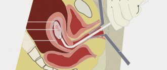
After the patient is placed on the gynecological chair, her thighs, external genitalia and vagina are treated with an antiseptic agent. A two-manual vaginal examination is performed to determine the location of the uterus and its size. The lower segment of the uterus is fixed with uterine single-tooth forceps, which pull back the body of the uterus, align the direction of the cervical canal and determine the length of the uterine cavity. And then the cervical canal is bougiened with a Hegar dilator.
The hysteroscope is treated with an antiseptic and carefully inserted into the uterine cavity, enlarged with gas or liquid. During the examination, its contents and size, shape and topography of the walls, and the condition of the area of entry into the fallopian tubes are studied. If any foreign bodies are detected, they are removed using instruments inserted through the hysteroscope channel. If necessary, a targeted biopsy is performed. The tissue sample taken is sent for histology.
According to indications, at the end of the procedure, the inner layer of the cervical canal and the uterine cavity can be removed. The anesthesiologist performs the final phase of anesthesia - brings the patient to consciousness. If there are no complications, the patient is under the supervision of specialists for another 2 hours, and then she is transferred to the general ward. Hysteroscopic surgery lasts on average 30 minutes, and if laparoscopy is performed, the manipulation can last up to 3 hours.
Patients are often interested in how long after hysteroscopy can IVF be done? Experts say that these periods fluctuate and depend on the data obtained during hysteroscopy. Some people are prescribed IVF on the 10th day after hysteroscopy, while others have to wait another six months for this moment. It all depends on the identified pathology, requiring varying degrees of surgical intervention and therapeutic measures.
With the advent of mini-hysteroscopes, which are very small in diameter, hysteroscopy and even minor surgical procedures without dilating the cervical canal have recently become increasingly common.
The medium used to expand the uterine cavity can be gas or liquid
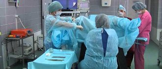
Carrying out the operation
The surgical intervention is carried out in the operating room, the patient first undergoes tests and prepares for the procedure. The operation is performed by a qualified gynecologist in the presence of an anesthesiologist, assistant and nursing staff.
The patient is given an anesthetic intravenously or mask anesthesia is used. In exceptional cases, paracervical anesthesia may be used - anesthesia of the tissues of the cervical area. After anesthesia, the doctor expands the cervical canal, inserts a hysteroscope and fills the cavity with gas or solution for better visualization.
The techniques for performing gas and liquid hysteroscopy differ. Let's look at them separately.
With gas
A portion of carbon dioxide is injected into the uterine cavity, expanding the space. The doctor monitors gas pressure and the filling level of the uterus. If the gas is supplied without violating the rules, the patient does not experience complications. If gas supply rules are violated, heart failure and even death can occur.
The gas technique is not used if there are traces of bloody discharge in the cavity - this interferes with clear visualization. Also, gas is not used during surgical interventions.
Using solutions
Hysteroscopy using solutions to enlarge the uterine cavity is the most popular procedure. Saline solution, glucose, glycine or simply distilled water are used as the basis for solutions. Can there be complications after the administration of liquid solutions?
Complications are possible with any examination technique. In this case, there is a risk of postoperative infectious tissue damage. However, hysteroscopy of the uterus using solutions is considered the most gentle compared to gas.
The advantage of therapeutic hysteroresectoscopy is the ability to preserve the integrity of the uterine body while eliminating neoplasms from it. A simple operation on the uterus does not require preliminary preparation and can be performed in a gynecologist’s office. We are talking about polyps and small processes, the elimination of which is not difficult.
During a simple operation, fragments of the fertilized egg or an ingrown intrauterine device can be removed. Foreign bodies and a thin intrauterine septum are also removed in the office.
Carrying out complex operations involves a large amount of work; they are carried out using special equipment and in appropriate conditions. Sometimes preparation for a complex operation requires a course of hormone therapy. It is also practiced to combine hysteroscopy with laparoscopy.
Type of anesthesia
The anesthesiologist chooses the anesthesia technique based on the goals of the intervention and the patient’s health condition, in particular:
- presence of chronic diseases;
- the likelihood of an allergic reaction;
- the possibility of complications associated with anesthesia or the medium supplied to expand the uterine cavity (gas or liquid).
Diagnostic procedures can be performed without anesthesia - this is permissible for certain indications (the absence of severe inflammation) and the availability of equipment (a very thin flexible fibrohysteroscope that does not require expansion of the cervical canal).
The procedure without anesthesia is not recommended for postmenopausal women, nulliparous women, or patients with hypersensitivity.
For hysteroscopic manipulations, the following types of anesthesia are used:
- paracervical anesthesia - injection of an anesthetic (Lidocaine) into the cervix. Used for diagnostic purposes only;
- mask anesthesia - chosen if there are restrictions for intravenous general. It is a type of inhalation anesthesia; ether, cyclopropane, pentran, chloroform are used as a gaseous/vapor substance entering the lungs through a mask;
- general intravenous anesthesia is optimal for long-term interventions, the presence of respiratory diseases or intolerance to mask anesthesia. In this case, a mixture of several types of drugs is administered through a venous catheter:
- analgesics (morphine, ketamine);
- sleeping pills (Relanium, Seduxen, Sibazon);
- muscle relaxants (listenone, myorelaxin);
- medications that support vascular tone and heart rate;
- endotracheal (a type of inhalation), spinal (local, injected into the space of the spinal cord) or epidural (similar to spinal, but does not relax the muscles) are types of anesthesia used when it is impossible to use general or mask anesthesia.
Regardless of the type of anesthesia, heart rate and breathing are continuously monitored during surgery.
Infertility
Is laparoscopy and hysteroscopy performed for infertility? Is there a chance to restore reproductive function using these techniques? The causes of infertility can be very diverse. For example, adhesions in the tubes prevent sperm from moving into the ovaries. With the help of hysteroscopy, adhesions can be removed, which will clear the way for sperm.
Hysteroscopy allows you to establish an accurate diagnosis, on the basis of which the gynecologist prescribes a course of therapeutic manipulations corresponding to the clinical picture. Hysteroscopy also helps to get rid of many pathologies that impede successful fertilization:
- ovarian cysts;
- adhesive obstruction;
- tumors, cysts, polyps;
- endometriosis.
USEFUL INFORMATION: Pregnancy in the absence of fallopian tubes
Conception after laparoscopy
Patients are interested in the question of the timing of possible pregnancy after surgical procedures. The chance of getting pregnant depends on the individual characteristics of the patient’s body, the severity of the existing pathology and the complexity of the surgery performed. Age, existing chronic inflammatory processes of the reproductive organs and complications after surgery also contribute.
When is it advisable to plan a pregnancy? For the body to fully recover after surgical procedures, three months or six months must pass. After a thorough examination of the patient, the gynecologist makes a conclusion about the possibility/impossibility of conceiving a child. Ovarian cyst surgery delays the time of planned fertilization by six months instead of three months.
Removal of polycystic ovary syndrome does not require a long recovery period, so after 28-30 days you can think about conceiving a child. Restoring women's health after an ectopic pregnancy requires a long period of rehabilitation - at least six months
Doctors are cautious about planning conception after an ectopic pregnancy and recommend giving the body rest for about a year.
How is childbirth after laparoscopy? This operation does not leave any scars on the body of the uterus, so the woman can give birth naturally. If doctors deem it necessary, the patient will undergo a caesarean section. One should not think that weak labor is caused by a previous operation before the birth - it depends on the characteristics of the female body.
Pregnancy after hysteroscopy
Hysteroscopy is divided into two types - diagnostic and surgical. After diagnostic procedures there are no obstacles to conception. Surgical procedures establish their own rules: the body needs a recovery period. Rehabilitation depends entirely on the severity of the pathological process and the surgery performed.
As a rule, six months is enough for the body to recover after the intervention. After surgery, intrauterine infection may develop, so premature pregnancy may result in miscarriage or premature birth.
A sluggish infectious process in the uterus can also provoke fetal hypoxia, polyhydramnios, abnormal fetal position and fetoplacental insufficiency. Therefore, it is not recommended to rush with fertilization. Before conception, the gynecologist will conduct a thorough examination using instrumental and laboratory diagnostics - ultrasound, culture of vaginal flora, blood and urine tests.
To maintain the pregnancy, synthetic hormonal drugs are used (for example, Duphaston), which promote reliable fixation of the embryo to the wall of the uterus. This drug also stimulates the endometrium, prepares the uterine cavity for gestation and eliminates contraction of muscle fibers. Duphaston is prescribed in the first trimester of pregnancy.
Necessary tests
Laboratory examination of patients before hysteroscopy is considered mandatory for all types of procedures. There are a number of tests that must be taken:
- general clinical urine and blood tests are required according to indications;
- biochemical blood tests and coagulation tests are necessary when planning therapeutic procedures within the framework of hysteroscopy, as well as to establish the overall picture of endometriosis, infertility and other gynecological diseases;
- venous blood tests for HIV, syphilis and other sexually transmitted infections;
- microscopy of discharge from the vagina and cervical canal to determine the flora of the genital tract and identify hidden infections or vaginosis.
If hysteroscopy is planned under anesthesia, the patient will have to undergo an ECG and also consult with a therapist.
Important! Almost all tests are valid for 2 weeks, and the results become known after 1-7 days, so a woman should take them approximately 10 days before the scheduled procedure.
Postoperative period
Consequences
Possible consequences after hysteroscopy:
- After the procedure for medicinal purposes, minor bleeding in the vagina is possible, because... blood vessels are injured. They usually last no more than 5 days.
- Also, after the operation, pain in the lower abdomen of mild to moderate intensity may appear (may radiate to the lumbar region). They usually last no more than the first 10 days.
- After anesthesia, the patient may also be bothered by: general muscle weakness, depressed state, depressed mood. These are the consequences of anesthesia, which may also include chills, fever, and headache.
- Possible perforation of the uterus - puncture of the wall of the organ with a surgical instrument.
Recommendations
To reduce the manifestations of discomfort after hysteroscopy of the uterus, it is recommended to follow simple recommendations for the postoperative period.
Any surgical intervention is associated with the risk of infection. Therefore, if a doctor prescribes antibacterial, anti-inflammatory, antimicrobial drugs, then you should follow the recommendations and observe the frequency, dosage and duration of use. This helps reduce the likelihood of an inflammatory process to a minimum.
If you are very concerned about painful sensations, then it is appropriate to take a drug from the NSAID group, for example, Ibuprofen, Tenoxicam, Nimesulide, Diclofenac, etc. The drugs have a combined effect on the body: they reduce inflammation, reduce temperature, and relieve pain.
If the patient has been bothered by elevated body temperature for more than 5 days, acute pain that is not relieved by painkillers, discharge mixed with pus or an unpleasant odor, heavy bleeding and other alarming symptoms, you should immediately go to the hospital.
What should not be done after hysteroscopy of the uterus?
- Use tampons. It is better to give preference to sanitary pads.
- Have sex. To exclude infection, you should limit sexual activity for the first 2 weeks after surgery.
- Take hot water treatments.
- Use vaginal suppositories.
- Perform intense physical activity.
Pregnancy
When is pregnancy possible after hysteroscopy? Depending on the purpose of the procedure and the established diagnosis, there may be several options regarding the timing of pregnancy:
- If the procedure was carried out for diagnostic purposes, then a healthy woman can become pregnant immediately after it.
- If the procedure was carried out for therapeutic purposes, then the onset of pregnancy is regulated by the characteristics of the pathological process, the volume of surgical intervention, as well as the recommended recovery time (usually 3-6 months).
The time when you can plan a pregnancy without fear for your health and the health of the child is individual in each case and can only be determined by a doctor, having previously assessed all the risks for the woman and child.
Complications and consequences after surgery
The consequences of hysteroscopy completely depend on the physiological characteristics of the patient’s body, but complications, as a rule, do not occur for more than 5 days. During this period, flatulence is observed in the gastrointestinal tract, which is caused by the ingress of gas that affects the internal organs, as well as the release of ichor coupled with cramps reminiscent of menstrual pain.
Bloody discharge
After diagnostic hysteroscopy, discharge from the uterus is insignificant. If a medical abortion was performed, spotting will be observed at first, and yellow or bloody discharge will be observed in the next 3-5 days. After removal of a fibromatous node or endometrial polyp, bleeding is also insignificant if there are no complications, otherwise uterine bleeding may be profuse.
In this case, doctors prescribe repeated surgery, hemostatic drugs, or drugs that contract the uterus. If after hysteroscopy of the uterus the patient experiences bloody-purulent discharge, which is accompanied by an increase in temperature, this means that the woman has developed inflammation after the procedure, requiring immediate treatment.
Nagging pain
Rehabilitation after hysteroscopy of the uterus lasts for the patient a maximum of 10 days, during which she feels aching pain. They are localized in the lumbosacral region or lower abdomen and are of moderate or weak intensity. If pain after surgery is very bothersome, doctors prescribe nonsteroidal medications that relieve acute pain. If the pain in the lower abdomen does not go away within 10 days, then you need to consult a doctor - this is an inflammatory process.
Characteristics of hysteroscopy
Endometrial hysteroscopy is a method for examining the uterine cavity, thanks to which it is possible to identify various gynecological diseases. During the procedure, a special device called a hysteroscope is used. It is a tube equipped with a video camera. Thanks to this, the doctor can examine the organ enlarged and in good lighting.
During the procedure, it is possible to identify developmental anomalies, assess the condition of the uterine tissue, detect various neoplasms, determine their size and location. The method turns out to be more informative than ultrasound.
Hysteroscopy of the uterus is a minimally invasive method in which the abdominal wall is not cut. Instruments are inserted into the organ cavity through the vagina. Thanks to this, the procedure is completed without injury or cracks. Only in rare cases is accidental damage possible.
One of the main advantages of the method is the ability to remove pathological areas directly during the study. Due to this, the speed of recovery increases significantly.
Recovery
During the recovery period after a hysteroscopic examination, women experience discomfort in the abdominal area for several hours. Painkillers or No-spa help relieve pain.
Within ten days after this procedure, spotting is also observed, gradually decreasing in volume. During the recovery period, antibiotics are often prescribed.
The arrival of menstruation should occur 30–40 days after the operation. In some cases, there is a delay. Regulations become more meager or intense. This largely depends on what kind of manipulations were performed.
To avoid deterioration of the condition during the postoperative period, a woman must adhere to certain rules. It is extremely important to avoid thermal procedures, taking hot baths, applying a hot heating pad to the stomach, and overheating in the sun. Such actions can lead to an increase in the intensity of bleeding.
It is not recommended to use tampons, douching, or use vaginal suppositories and ointments during this period. Hygiene procedures should be carried out with special care.
A routine gynecological examination should be completed in a timely manner, regardless of whether the operation was performed during menopause or during reproductive age. Even minor discomfort in the abdominal area is a reason to visit a gynecologist.
Sexual activity should be excluded for a whole month. Pregnancy after hysteroscopy becomes possible within a couple of weeks, but doctors strongly recommend planning conception at least three months later.
Risks of hysteroscopy
Hysteroscopy is generally very safe, but like any procedure there is a small risk of complications. The risk is higher in women who undergo treatment during hysteroscopy.
Some of the main risks associated with hysteroscopy are as follows:
- Accidental injury to the uterus - this is very rare, but may require treatment with antibiotics in hospital or, in rare cases, other surgery to repair it.
- Accidental injury to the cervix is a rare complication and the injury can usually be easily repaired.
- Excessive bleeding during or after surgery – this may occur if the treatment was carried out under general anesthesia; very rarely, the uterus may need to be removed (hysterectomy)
- Uterine infection – can cause smelly vaginal discharge, fever and heavy bleeding; it is usually treated with a short course of antibiotics.
- Feeling weak – affects 1 in every 200 women who have a hysteroscopy, done without anesthesia or with local anesthesia only.
Bleeding or infection can occur after any surgery. Sometimes the surgeon cannot complete the procedure safely due to excessive bleeding, fluid absorption, or the size of the fibroid. Complications common with hysteroscopy include uterine perforation and disproportionate fluid retention. The liquid is used to stretch the uterine cavity during hysteroscopy. Sometimes this fluid can be absorbed into the general circulation (lungs and brain). If excessive fluid absorption occurs, the procedure should be discontinued.
Emboli and death are rare but potential complications of any surgery.
Laparoscopy or hysteroscopy?
Despite the difference in the purpose of these procedures, sometimes they pursue the same goals. Then the question arises - what to prefer? Only a gynecologist can answer this question, based on an analysis of the patient’s health status.
For example, during hysteroscopic diagnostics, a tissue sample can be removed for research or the remains of the fertilized egg can be removed - these manipulations are impossible with laparoscopy. Laparoscopy copes with the elimination of ectopic pregnancy, which hysteroscopy of the uterus is not capable of.
If, nevertheless, the choice has become relevant, it should be taken into account that after laparoscopy there will be small scars from punctures in the abdominal cavity. If it is necessary to perform surgery inside the uterus, it is better to prefer hysteroscopy. The only contraindication to hysteroscopy is blood clotting pathology.
The recovery period after hysteroscopy is many times faster than after laparoscopy. You need to monitor your stitches, follow a diet and adhere to other restrictions. Recovery after hysteroscopy does not require all this.
However, there are cases of combined laparoscopy with hysteroscopy. The combination is prescribed if necessary to diagnose the uterus from the inside and outside. This saves examination time and provides a complete clinical picture when determining the causes of infertility. It is also easier for the patient to tolerate both diagnostics performed together than sequentially.
Possible risks
If the study was carried out with high quality, the woman strictly followed all the rules for preparing for it, then in most cases there are no complications. Nevertheless, certain risks do exist in the postoperative period. Among the main gynecological disorders are the following:
- organ damage from instruments;
- bleeding caused by a violation of the integrity of large vessels of the mucous membrane;
- penetration of infection into the reproductive organ and the onset of the inflammatory process.
Severe pain in the lower abdomen, hyperthermia, an increase in the volume of discharge, the acquisition of an unpleasant odor, and an admixture of pus in the blood that comes out of the genital tract may indicate the development of complications.
If, during the treatment of uterine pathologies, perforation of the organ occurs, then the pain syndrome in the abdominal area becomes excessively pronounced. In this case, internal bleeding begins and such unpleasant symptoms as nausea, vomiting, dizziness and low blood pressure appear.
The development of hematometra (a pathological condition in which blood accumulates in the organ and its outflow is disrupted) is also possible. As a result, pronounced pain occurs. In order to remove clots and prevent the onset of the inflammatory process, curettage is performed.
If unpleasant symptoms appear during the postoperative period, you should immediately contact a medical facility. It is extremely important to eliminate complications in a timely manner and take all necessary measures.
Alternatives to Hysteroscopy
Hysteroscopy will only be performed if the benefits are considered to outweigh the risks.
The uterus can also be examined using:
- pelvic ultrasound – where a small probe is inserted into the vagina and uses sound waves to create an image of the inside of the uterus;
- endometrial biopsy – where a narrow tube is passed through the cervix into the uterus, with suction used to remove a sample of uterine tissue.
These alternatives can be performed alongside a hysteroscope, but do not provide as much information and cannot be used to treat problems in the same way as hysteroscopy.
Carrying out diagnostics
Let us consider in detail how the diagnostic procedure is carried out. First, the gynecologist dilates the cervix with a medical instrument and injects a special solution or gas into the cavity to increase the examination space. Then an examination instrument equipped with a micro video camera is used to examine the organ. The image is displayed on the monitor and can be saved to disk.
If necessary, tissue is collected for laboratory testing. If the diagnosis is carried out without anesthesia, the patient may experience discomfort. Sometimes diagnosis is performed under local anesthesia.
How long does a hysteroscopic examination take? No more than forty minutes.
Patient preparation
Before performing hysteroscopy, a woman undergoes special training. She must be ready for the operation both physically and psychologically. Due to the fact that anesthesia is used during hysteroscopy, there are a number of special requirements. Before administering anesthesia, the doctor must be sure that anesthetics will not cause complications.
In addition, there are certain restrictions before the procedure, which the gynecologist informs in advance. The duration of the operation itself does not exceed half an hour. As a rule, the woman goes home on the day of the procedure.
Psychological attitude
It is extremely important that the patient is psychologically prepared for the operation. Fear of pain, the thought that hysteroscopy of the uterus is performed without anesthesia - all this can become a serious problem.
The doctor should explain that this is a painless procedure that uses anesthesia. There are no unpleasant sensations during the procedure.
In addition, hysteroscopy surgery is considered one of the safest examinations. The risk of developing complications after it is minimized. The woman needs to be told about this in advance.
Basic Research
Preparation for hysteroscopy involves the following studies:
- general blood analysis;
- bacterial culture;
- determination of blood clotting rate (coagulogram);
- Analysis of urine;
- blood chemistry;
- determination of blood sugar levels;
- chest x-ray;
- Ultrasound of the pelvic organs and peritoneum;
- cardiogram;
- collection of smears;
- bimanual examination.
The purpose of these studies is to identify diseases for which the use of a hysteroscope is contraindicated. If they are detected, a course of treatment is initially carried out aimed at eliminating existing pathologies. A woman is considered ready for the procedure when test results show the absence of diseases that would prevent it from being performed.
Medication preparation
During preparation for surgery, certain medications are necessary. Taking antibiotics is necessary in order to reduce the risk of developing infectious processes after the study. In addition, seven days before the procedure, antifungal agents and douching using medications such as Octenisept or Miramistin are also prescribed.
If the problem is hormonal, then the endometrium is prepared by medication before the hysteroscopic examination. For this purpose, hormonal drugs are prescribed.
Sanitary and hygienic measures
Before hysteroscopy of the uterus is performed, during which it will be possible to examine the condition of the mucous membrane and perform cleaning, the gynecologist prescribes certain sanitary measures. They require careful sanitation of the genital tract. For this purpose, suppositories are used that have antifungal, antiprotozoal and combined effects.
Immediately before the procedure, the woman needs to thoroughly wash herself and shave her intimate area.
Benefits of hysteroscopy
At the moment, this is the most progressive method for removing uterine cavity tumors. The surgeon can monitor the progress of the resection on the monitor and control his actions. This makes it possible to completely remove the polyp without leaving a single fragment. Fragmentary particles cause the growth of a new polyp at the site of the removed one, so visual control allows you to monitor the quality of the resection performed.
Another advantage of resectoscopy is the simultaneous cauterization of the polyp site after its removal. This ensures the preservation of the injured area from the development of the inflammatory process, especially in the presence of multiple polypous formations. Also, resection and simultaneous cauterization of the pathological area preserves the adjacent unaffected endometrial tissue from infection or injury.
Important! After hysteroresectoscopy, no adhesions are formed.
Resectoscopy is many times more effective than polypectomy, in particular in terms of eliminating pedunculated polypous formations. The minimally invasive nature of the procedure eliminates cutting into the abdominal cavity and organs adjacent to the uterus. This ensures a quick recovery period and minimal trauma during surgery.
The recovery period after resectoscopy is short and does not require additional therapy, as after abdominal operations. All that is required is restorative and strengthening manipulations.
Flaws
These include, first of all, possible complications:
- infection during surgery;
- injury to organs adjacent to the uterus;
- poor-quality removal of pathological tissue;
- heavy bleeding.
However, complications are largely the body's reaction to surgical procedures. This cannot be avoided with any gentle method of surgical correction.
Despite these complications, hysteroresectoscopy is considered the most progressive method of cleansing the uterine cavity from tumors.
When is the procedure needed?
There are certain indications for hysteroscopy. Various studies, examinations, ultrasounds and tests do not always reveal problems in the body. Some pathologies may be asymptomatic; most diagnostic methods turn out to be uninformative. The safest and most reliable way to identify and treat fibroids, polyps, endometriosis and endometrial hyperplasia is considered to be a hysteroscopic examination.
No matter how large the list of tests that a doctor prescribes for a woman, the most accurate information can be obtained using a hysteroscope.
Hysteroscopic surgery is necessary if pathological growth of the endometrium, the formation of fibroids, and polypous growths are observed. Eliminating endometriosis by other means turns out to be very problematic.
As a rule, a hysteroscopy with a biopsy is performed. After the operation, the excised tissue is sent to the histology laboratory. Thanks to this, it is possible to identify cells that have degenerated into malignant ones, and then accurately determine the tactics of further therapy.
Indications
A hysteroscopic examination of the uterine cavity is indicated in the presence of the following problems:
- endometrial hyperplasia;
- endometriosis and adenomyosis;
- abnormal development of the uterus and fallopian tubes;
- uterine fibroids;
- intrauterine synechiae;
- infertility;
- failure of the menstrual cycle;
- determination of the location of the spiral;
- several unsuccessful IVF attempts;
- suspected cancer;
- control examination of the field of hormonal therapy and surgical intervention.
Contraindications
The method has a number of undeniable advantages, but there are many contraindications to its use. The procedure is not performed if the woman has a progressive pregnancy. This is due to the fact that any intervention in the area of the cervix and reproductive organ can lead to spontaneous miscarriage. There is also a risk of fetal damage.
Hysteroscopy in nulliparous women is contraindicated if there is a history of inflammatory or infectious diseases of the genitourinary system. If such manipulations are performed, there is a high risk of developing infectious complications.
The procedure is not performed for cervical stenosis. Oncology is considered an absolute contraindication. If cancer is detected, surgery is not performed.
Also, the study is not used in cases of severe kidney pathologies, heart and lung failure, problems with blood clotting, and after a heart attack.
The method is not used if you are intolerant to medications used for anesthesia.
When can hysteroscopy be performed?
Hysteroscopy can be used for:
- Research into symptoms or problems - such as heavy periods (periods), abnormal vaginal bleeding, postmenopausal bleeding, pelvic pain, recurrent miscarriages, or difficulty getting pregnant.
- Diagnosis of conditions – such as fibroids and polyps (non-cancerous growths in the uterus).
- Carrying out curettage.
- Treatments for conditions and problems such as removal of fibroids, polyps, displaced intrauterine devices (IUDs), and intrauterine adhesions (scar tissue that causes missed periods and decreased fertility).
A procedure called dilation and curettage was commonly used to examine the uterus and remove abnormal tumors, but hysteroscopy is now being performed.
Indications and contraindications
Hysteroscopy - what is this procedure and what are the indications for it? This is a modern, minimally invasive diagnostic procedure that is used to remove various tumors, foreign bodies, and also identify many gynecological diseases. The examination is carried out using a modern device - a hysteroscope. It is equipped with a unique optical system that provides the doctor with the opportunity to fully examine the uterine cavity, its cervix, as well as assess the condition of the endometrium.
The main indications for hysteroscopy of the uterus:
- Suspicion of the development of endometriosis or uterine fibroids.
- Monitoring the effects of hormonal or surgical therapy.
- Pathologies of the menstrual cycle - heavy or prolonged periods, the appearance of bleeding between menstruation.
- The appearance of bleeding during menopause.
- Curettage of the uterine cavity to remove fetal remains during a frozen pregnancy, various postpartum complications.
- Endometrial hyperplasia.
- The presence of foreign bodies in the uterine cavity.
- Infertility or spontaneous abortion.
- Diagnosis and treatment of congenital anomalies in the structure of the uterus.
- Surgical removal of cervical canal polyps.
Hysteroscopy of the uterus has several types and can be used for both diagnostic and therapeutic purposes. The procedure makes it possible to identify pathological conditions in the early stages of development - and this will help to begin treatment procedures in a timely manner and avoid serious complications.
Before performing hysteroscopy, it is imperative to familiarize yourself with the existing contraindications, which include:
- All trimesters of pregnancy.
- Inflammatory process in the female genitourinary system.
- During exacerbation of infectious diseases.
- Cervical stenosis, open uterine bleeding and menstruation.
- Diagnosed cancer of the uterus.
- Blood clotting disorder.
Hysteroscopy of the uterus is performed by a specialist in an outpatient clinic or in an operating room.