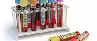
Women's health Urine tests Genitourinary system Men's health
09/02/2020 Chernenko A. L. 39 Views urine analysis, genitourinary system
In this article, we will analyze the situation when red blood cells are elevated: what does this mean, why do they rise, what is the norm of red blood cells in a urine test. And what to do if they increase. Red blood cells in the urine, increased levels of leukocytes, casts and protein are included in the concept of “urinary syndrome” - a laboratory syndrome that characterizes the presence of qualitative and quantitative changes in urine tests.
Normally, protein, casts, leukocytes and red blood cells are either not detected in the urine or are detected in minimal quantities.
Increased red blood cells in the urine can be detected with any pathology in the genitourinary system. Therefore, when red blood cells are detected in a urine test, additional examination is always carried out. A diagnosis cannot be made based on just one urine test.
- 1 Red blood cells in urine - what are they? 1.1 What do increased red blood cells in urine mean?
- 1.2 What are red blood cells in urine analysis according to Nechiporenko?
- 1.3 What do red blood cells in urine indicate: microhematuria and gross hematuria
Red blood cells in urine - what are they?
Red blood cells in urine tests are the detection of red blood cells (red blood cells containing hemoglobin) by microscopy of urine.
Depending on the reason for red blood cells getting into the urine, they can be changed or unchanged.
Unchanged red blood cells in the urine are detected during bleeding in the genitourinary tract below the glomerular filter in the kidneys (postglomerular hematuria).
If altered cells are identified, we can conclude that the source of bleeding is located above the glomerular filter. In this regard, when passing through the basement membrane of the capillaries of the renal glomeruli, red blood cells are damaged and “leached.” Such red blood cells in the urine are called “red blood cell shadows” and indicate glomerular hematuria. Leached red blood cells in the urine are small, have a deformed shape, do not contain hemoglobin and are surrounded by a faint, colorless ring.
What do increased red blood cells in urine mean?
Increased red blood cells in the urine (hematuria) is an increase in the level of blood cells in the urine, more than 1-2 red blood cells per field of view.
Detection of 1-2 red blood cells in the urine per field of view (single red blood cells in the urine) is the norm. Such indicators are not considered pathology.
However, in the presence of symptoms of kidney and urinary tract diseases, as well as proteinuria (protein in the urine), leukocyturia (leukocytes in the urine) and cylindruria (casts in the urine) - even 2 red blood cells in the urine (per 1 field of view) require mandatory additional examination and repeated urine analysis, as they may indicate incipient microhematuria.
In young children, the detection of up to 5 red blood cells in the urine can be considered normal. This is in the absence of symptoms of damage to the genitourinary tract, as well as an increase in protein, casts and leukocytes.
What are red blood cells in a urine test according to Nechiporenko?
Urinalysis according to Nechiporenko is carried out to identify inflammatory and infectious processes in the genitourinary system. When conducting a urine test according to Nechiporenko, the number of leukocytes, casts and red blood cells in the urine per unit volume is determined. Most often, the calculation is carried out per 1 milliliter.
Normally, the presence of: erythrocytes in urine up to 1000 per milliliter, leukocytes up to 2000 per milliliter, and cylinders up to 20 per 1 milliliter of urine is allowed.
Accordingly, increased leukocytes and erythrocytes in the urine mean that more than 1000 erythrocytes and 2000 leukocytes were detected in 1 milliliter of urine.
What do red blood cells in urine indicate: microhematuria and macrohematuria
Elevated red blood cells in urine are divided into:
- microhematuria (increased levels of red blood cells in the urine are detected only during microscopic examination);
- macrohematuria (there are a lot of red blood cells in the urine, hematuria is noticeable due to changes in the color of the urine).
For example, if up to 10 red blood cells appear in the urine, its color will not change. Such a number of red blood cells will be noticeable only during a microscopic examination, therefore such an increase in the level of red blood cells in the urine will be called microhematuria.
With gross hematuria, the color of the urine becomes reddish or takes on the color of meat slop. Gross hematuria always indicates severe diseases of the genitourinary system (glomerulonephritis, the presence of malignant neoplasms in the kidneys or bladder).
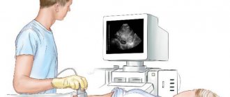
Description of the study
In traditional general urine analysis with sediment microscopy, the procedure is carried out after centrifugation (separation into liquid and solid parts). The methods differ in different laboratories, since there is no standardization in the CIS countries. This also leads to variation in results and norms.
Methodology
Currently, most hospitals use special automation equipment for examination - urinary stations, which include the following:
- Analysis of test strips (chemical indicators).
- Study of formed elements of urine according to the principle of identification.
- Channel for assessing the presence of bacteria.
The physicochemical properties of urine are also studied using the “dry chemistry” method (reflection photometer). When detecting latent leukocyturia, an analysis for esterase activity using the same test strips is relevant, since damaged cells are not detected by microscopy.
Rules preparation
To avoid mistakes on your part, proper preparation is required. Urine must be collected according to the following algorithm:
- Two days before you are going to take the material, avoid drinking alcohol, smoking, sexual intercourse and taking medications (as directed by your doctor).
- For general urine analysis with sediment microscopy, the middle portion is most often used.
- After a night's sleep (on an empty stomach), take a shower, especially paying attention to the toilet of the external genitalia.
- The first few milliliters are flushed down the toilet. Without stopping the process, substitute a container, collecting 100-150 ml of urine. The rest is also lowered.
- To transport the material, it is best to use a disposable container (sold at any pharmacy) or a dry, clean container with a tight-fitting lid.
Advice! Urine must be brought to the laboratory within 1-1.5 hours, otherwise the result will be unreliable. At the same time, every woman should know that urine is donated before the onset of menstrual bleeding or after 2 days.
If there are increased red blood cells in the urine, what does this mean?
Depending on the sources of bleeding, the causes of increased red blood cells in the urine are divided into: renal (glomerular) and extrarenal (non-glomerular, post-glomerular).
Glomerular causes of increased red blood cells in the urine include:
- acute glomerulonephritis (characteristic is the appearance of gross hematuria and urine the color of meat slops, the development of microhematuria is less common);
- chronic glomerulonephritis. During the remission stage, the appearance of single red blood cells in the urine is typical, but with an exacerbation of the disease, a sharp increase in hematuria is characteristic;
- kidney infarction. There is the appearance of a large number of red blood cells in the urine, as well as pronounced pain in the lower back);
- renal tuberculosis (constant microhematuria is noted);
- malignant renal neoplasms (the development of recurrent painless hematuria is noted);
- hemorrhagic diathesis. This group includes autoimmune thrombocytopenia, hemophilia, thrombocytopenic purpura, etc.;
- overdose of anticoagulants and antiplatelet agents;
- polycystic kidney disease (most often accompanied by asymptomatic microhematuria);
- severe infectious diseases accompanied by kidney damage. An increase in red blood cells in the urine can be observed with leptospirosis, scarlet fever, typhoid and typhus, malaria);
- toxic vasculitis of the renal vessels;
- traumatic kidney damage (most often there is a sharply increased number of red blood cells in the urine);
- “stagnant kidney” (there is an increase in red blood cells and protein in the urine)
What is this research used for?
This research is carried out for the following purposes:
- To perform a comprehensive examination of the human body.
- For differential diagnosis of pathologies of the kidneys and urinary tract.
- To evaluate the effectiveness of therapy for diseases of the urinary system.
- To diagnose metabolic pathologies, and, in addition, disturbances in water and electrolyte balance.
- For diagnosing pathologies of the digestive system.
- To diagnose infectious and inflammatory pathologies in a patient.
- To assess and monitor the patient’s clinical well-being during medical or surgical treatment.
Protein and red blood cells in urine?
Proteinuria (increased protein in the urine) in combination with an increase in the level of red blood cells in the urine can be observed with:
- “stagnant kidney”;
- glomerulonephritis;
- severe pyelonephritis (increased leukocytes and red blood cells in the urine may be observed);
- heavy physical activity (short-term minor proteinuria and microhematuria may be observed with the appearance of 2-3 red blood cells in the urine in the field of view);
- cystitis (in both acute and chronic inflammation of the bladder, increased protein and red blood cells in the urine may be observed).
Purpose of analysis
Urine is a type of biological fluid that is secreted by the kidneys. Along with urine, many metabolic products leave the human body, and therefore, by its characteristics, one can indirectly judge the blood composition, and, in addition, the condition of the urinary canals and kidneys. Urine includes substances in the form of urea, uric acid, ketone bodies, amino acids, creatinine, glucose, protein, chlorides, sulfates and phosphates. Analysis of the microbiological and chemical composition of urine is important in diagnosis.
The fact is that any deviation from the norm indicates abnormal metabolic processes in the patient’s body. When is a general urine test prescribed? People need such research for any disease of the endocrine and genitourinary system. It is also advisable to carry it out in case of abnormalities in the functioning of the cardiac, vascular and immune systems and in case of suspected diabetes. In addition, a general urine test is prescribed to patients who have suffered a streptococcal infection. In addition, it is carried out for preventive purposes and directly to monitor the dynamics of pathology.
A transcript of urine sediment microscopy is presented below.
Extrarenal causes
The causes of extrarenal increase in red blood cells in urine analysis include:
- stones in the urinary system (red blood cells appear in the urine due to stone damage to the urinary tract). In this case, hematuria is accompanied by severe pain);
- acute cystitis (characterized by the appearance of blood at the end of urination);
- acute urethritis (blood may be detected in the first drops of urine);
- malignant lesion of the bladder (a large number of red blood cells appear in the urine);
- damage to the urinary tract.
Interpretation of the research result
Interpretation of urine sediment microscopy should only be carried out by a qualified specialist.
Neutrophils in the analysis occur in the presence of urinary tract infection and other conditions. Lymphocytes may be associated with chronic inflammation, viral disease, and renal transplant rejection.
Hematuria is an important sign of urinary tract and kidney disease. Hematuria may also reflect the body's general tendency to bleed. Epithelial cells trapped in this biomaterial can help determine the location of the disease of the urinary canals. Squamous epithelial cells are located on the side of the external genitalia and are therefore a sign of improper urine collection. An exception is considered to be pregnant women, who have increased detachment of squamous epithelium.
What else does deciphering microscopy of urine sediment in adults suggest?
Particles of transient epithelium can cover the urinary canals in several layers, starting from the renal pelvis up to the bladder in women and to the initial region of the urethra in men. Epithelial cells often arise in the presence of urinary tract infections and against the background of one or another non-infectious urological disease.
Renal epithelial cells can appear predominantly in the urine of patients suffering from tubular acute necrosis and interstitial nephritis, and, in addition, during rejection reactions of renal transplants. They can also appear with glomerulonephritis.
Cylinders are formed, as a rule, in the convoluted distal and collecting tubules. In the cylinders you can find plasma protein along with lipids, various cells, microorganisms, pigments (we are talking about hemoglobin, myoglobin, bilirubin) and crystals. Single hyaline casts occur in the concentrated first urine of a healthy person. Microbes may be present in urine sediment. Sometimes Candida or Trichomonas fungi can be found in the urine.
In most situations, crystals found in urine are not clinically significant, as this may be due to the consumption of foods that contain oxalates with urates. This can also be explained by the sample being in a cold place or it depends on the acidity of the urine. What matters are those crystals that arise against the background of recurrent kidney stones or in patients suffering from insufficiency of this organ.
What does a general urine test show: deciphering the results
Decoding the results of such an analysis helps to understand the relevant indicators before visiting a doctor. However, under no circumstances should you engage in self-medication and self-diagnosis based on the information received. In order to correctly interpret a general urine test and sediment microscopy, as well as to determine a diagnosis, you need to contact a specialist. Urine is analyzed according to several criteria, among which are organoleptic properties along with physicochemical indicators, biochemical characteristics and microscopic examination.
When and why do you need a urine test?
For research, a morning portion of urine is given before meals. After collection, the liquid must be delivered to the laboratory no later than 2 hours, until chemical reactions change its composition. Indications:
- pathologies of the kidneys, excretory tract, diseases of the urethra and bladder, when it is necessary to make a diagnosis and select treatment;
- the need for differential diagnosis;
- suspicion of infectious, inflammatory diseases;
- monitoring the functioning of body systems as treatment progresses, checking the effectiveness of prescriptions;
- monitoring the patient's condition to prevent complications;
- pre- and postoperative period, assessment of the patient’s condition before blood transfusion and after the procedure;
- preventive examination (once a year).
A morning sample of urine is taken for testing before meals.
Varieties
Doctors classify the release of red blood cells in urine in a certain way. This helps in assessing the severity of the patient's condition and making a diagnosis. Depending on the number of red cells in the urine, the following are distinguished:
- microhematuria - moderate excess of the norm, the color of urine is not changed;
- macrohematuria - a significant content of red blood cells, which is noticeable to the naked eye (color from pale pink to dark brown, the presence of blood clots).
Depending on where the cells enter the urine, they distinguish between glomerular hematuria (blood passes at the level of the glomeruli, which is often accompanied by an increase in protein) and non-glomerular (penetration occurs at other stages of urine formation, outside the filtration system).
Table - Classification of hematuria by place of origin
| Name | Mechanism | Possible reasons |
| Extrarenal | Not associated with renal dysfunction | Pathologies of the heart, blood vessels, hemolysis |
| Renal | Organic or functional disorders of the kidneys | Nephroptosis, glomerulonephritis, pyelonephritis, etc. |
| Postrenal | Red blood cells come out “below” the kidneys | Pathologies of the urinary tract (ureters, urethra, bladder), genital organs |
Using a three-glass sample, the time at which red cells enter the urine is determined. This may indicate the localization of the pathological process:
- initial (initial) - red blood cells are found in the first portion of the biomaterial, which indicates damage to the urethra;
- final (terminal) - cells are found in the last urine excreted, which often accompanies pathologies of the bladder or prostate in men;
- total - the indicator is approximately equal in all portions (happens for any violations).
To obtain accurate test results, it is important to correctly take a three-glass sample. To do this, you need to prepare 3 sterile containers into which to separately collect urine at the beginning, middle and end of emptying the bladder. All containers are labeled and sent to the laboratory. Before collecting biomaterial, it is important to perform hygiene; during this, you should not touch the container to the skin. All samples are delivered for analysis within 1.5 hours.
Urine analysis with sediment microscopy
Urine, or urine, is a product of the kidneys, the final component of metabolic processes. Urine contains water, as well as hormones, electrolytes dissolved in it, dead cells of the mucous membrane of the urinary tract, salts, mucus, leukocytes, etc. A general urine test (UCA) provides a set of information about the physical and chemical parameters of urine and the presence of various metabolites in it.
A general urine test allows you to evaluate the activity of the kidneys, bladder and other organs of the system; these are its most important, but not exhaustive purposes. The study will also help to identify disturbances in the activity of internal organs not related to the urinary system.
Microscopy of urine sediment is one of the routine diagnostic methods, which is used both for screening various diseases and for monitoring the course of diseases and the results of therapy.
The data that will be obtained after completing the study is as follows:
- General analysis (urine examination using dry chemistry) - urine specific gravity, hue, transparency, acid-base index, protein, sugar, nitrites, hemoglobin, ketone bodies, bilirubin, urobilinogen.
- Sediment microscopy (qualitative and quantitative assessment of a number of insoluble components) - leukocytes, casts, erythrocytes, epithelial cells, salts, bacteria.
What else should you know about sediment microscopy?
At this stage of a general urine analysis with sediment microscopy, unorganized and organized sediment and cylindruria are examined. In total, microscopy of urine sediment makes it possible to determine approximately a dozen components of the liquid being examined. Conducting a study of organized sediment gives an idea of the presence of four components in the urine:
Correct interpretation of urine analysis with sediment microscopy is very important.
The study of the unorganized version of sediment is aimed at studying salts and ions in urine. In total, they are found in numbers up to ten. But often urates are found in the urine (which can be a manifestation of leukemia, gout, hepatitis or diathesis). Phosphates (this indicates cystitis) and oxalates (this indicates diabetes or pyelonephritis) can also be found. In addition, ammonium urate is isolated in the unorganized sediment along with uric acid and tripelphosphates, but they do not have a clear specification.
The term cylindruria refers to the study of the protein cast that forms in the urinary canals. Cylinders are classified according to their area of origin and appearance:
Casts of the epithelial, waxy, pigmented, and leukocyte varieties may also occur, but they are usually less common. Now let’s find out how experts decipher the condition of urine in children.
Also, a separate interpretation of urine examination with sediment microscopy in young patients is carried out.
Preparing to collect urine for analysis
When issuing a referral for analysis, the doctor explains how to collect biological material and how to prepare. The accuracy of the results obtained will depend on how correctly the patient does everything. The quality and composition of urine is affected by the food eaten, drinks, psycho-emotional state, physical activity, use of medications, dietary supplements, and vitamins. To prevent the listed factors from distorting the results of OAM, doctors give recommendations:
- 24 hours before urine collection, foods that can change its color are removed from the diet. These are beets, carrots, sweets, smoked meats;
- exclude the consumption of alcoholic beverages, coffee, carbonated water;
- stop taking diuretics, vitamins, dietary supplements;
- If possible, do not take medications. When this is not possible, you need to tell the laboratory assistant or doctor about it. This way, specialists will know how to correctly perceive the results of the analysis;
- do not go to the sauna, refuse to go to the gym.
When urine contains foreign matter, the result will be incorrect
You need to reschedule the test if you have a fever, menstruation, an attack of hypertension, or an infectious process. The listed factors can distort the results. When cystoscopy was performed, OAM was postponed for a week.
When urine contains foreign matter, the result will be incorrect, and the accuracy of the diagnosis depends on this. Therefore, it is important to follow the collection rules, namely:
- collect the morning portion of urine;
- Before taking the liquid, you need to wash your genitals, preferably without hygiene products, with warm water;
- It is not recommended to take a test during menstruation, but if there is an urgent need, insert a tampon, wash the genitals, and be sure to warn the doctor that the urine may contain blood particles;
- For collection you need a sterile container. It is advisable to use a medicine container with a screw cap;
- they don’t start urinating, they urinate in the toilet to let bacteria through. Next, place the container and take about 150 ml;
- the collected urine must be delivered to laboratory technicians within 1.5-2 hours. When the urine has been sitting in a warm place for 2 hours, there is no point in sending it for testing;
- Fluid is collected from the child using a urine bag from a pharmacy. This is a plastic bag with a convenient fastening. After collection, the liquid is transfused and taken for analysis. Older children scald the pot to collect urine, but such manipulations lead to an increase in white blood cells in the results.