Myelocytes: structural features and types
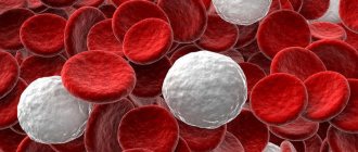
A myelocyte is an immature representative of leukocytes. It consists of a large round kernel. Its cytoplasm is saturated with granular inclusions or granules.
Under natural conditions, myelocytes having reached maturity should degenerate into segmented granulocytes.
Leukocytes
Cytological analysis shows that during the period of maturation, myelocytes are painted in a rich red-violet color. Protoplasm acquires a blue tint, and during ripening it has a pink color.
Varieties
Experts distinguish the following types of myelocytes:
- Neutrophilic. Mature representatives have pink protoplasm, younger ones have pink-violet protoplasm. It contains both fine grain and larger granules.
- Eosinophilic. They have weakly basophilic protoplasm. It contains a large number of large grains. The color of eosinophilic cells is pinkish-red.
- Basophilic. The protoplasm of these cells is oxyphilic, and the grain is violet.
Stages of maturation
To become full-fledged granulocytes, myelocytes undergo the following stages of maturation:
- myeloblast,
- Promyelocyte,
- Metamyelocyte.
Reference! Promyelocytes are larger than myeloblasts. They have “primary granules” in the cytoplasm and condensed chromatin.
Why can they be detected in an adult?
The normal composition of blood never changes just like that. The development of such a disorder always requires a specific reason. Certain unfavorable conditions that provoke the formation and reproduction of myelocytes in the protoplasm of bone marrow tissue.
Expert opinion
Kovaleva Elena Anatolyevna
Doctor-Laboratory Assistant. 14 years of experience in clinical diagnostic services.
Ask a question to an expert
To cope with the problem and bring blood counts back to normal, you need to eliminate the provoking factor.
There is no other way to get rid of such a disorder, since myelocytes are a consequence, and not the disease itself. The reasons for the rapid proliferation of myelocytes are most often the following factors:
- infections;
- purulent processes;
- low-grade inflammation;
- intoxication of the body;
- the presence of a malignant tumor or cancer in the decay stage;
- viral diseases;
- food poisoning;
- any pathologies in acute and advanced stages;
- radiation or chemical exposure;
- hematogenous diseases;
- a side effect from taking any medications.
Myelocytes and the pathological process
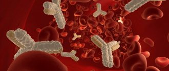
When pathogenic flora invades the body, mature segmented granulocytes are the first to come to defense.
If there are not enough of them or they cannot cope with the harmful agent, band granulocytes are sent to their aid. Normally, their quantity in the blood is limited.
The stronger the disease, the more young immature cells are neocytized (accumulated). First, the level of band myelokaryocytes increases; as the condition worsens, metamyelocytes appear in the blood (one of the stages of myelocyte development).
If things are really bad, very young cells – myelocytes – join the fight. This means that the body has no strength left to protect itself.
How to determine myelocyte level
A myelogram will show the exact level of myelocytes
Determination of the level of myelocytes, as well as other components of the bone marrow, is carried out by taking a bone marrow puncture. The resulting myelogram will show the exact cellular composition of the bone marrow.
Bone marrow samples are taken from:
- sternum,
- iliac crest,
- from the calcaneus (this is how puncture is performed in small children).
After receiving the punctate, the myelogram data must be compared with the data of the general clinical blood test.
Why do myelocytes appear in the blood?
The reasons for the appearance of myelocytes in the blood are different.
The most common of them:
- Infectious diseases of an acute nature, which occur with the presence of inflammatory and purulent foci. This is observed with pneumonia, sore throat, tuberculosis, and sepsis.
- Poisoning with heavy metal salts (lead) or alcoholic beverages.
- Cell death during a pathological process. It occurs in patients with stroke, gangrene, and extensive burns.
- Benign or malignant tumors.
- Pathological changes in the blood system. Example: leukemia.
Other etiological factors for the occurrence of myelocytes are:
- Acute bleeding
- Poisoning with toxic substances,
- Radiation therapy and chemotherapy carried out to treat cancer,
- Radioactive radiation,
- Insufficient intake of vitamin B in the body or its absence,
- Intestinal infections
- Diseases caused by viruses (influenza, rubella)
- Increased physical activity
- Comatose and shock states,
- Using certain medications in large quantities (anti-depression pills, painkillers),
- Acid-base balance disorders.
Reasons for the increase
There can be quite a few factors for the growth of indicators. Here are just a few of the possible culprits.
Acute infectious processes
It doesn’t matter what type, bacterial, viral or fungal. Accompanied by corresponding symptoms. Sometimes they occur without manifestations. Why does the concentration of precursors, immature cells, increase in this case?
The reason is simple - the body strives to cope with the disorder as quickly as possible. Localize the area, separate it from others and fight the infection.
For these purposes, many neutrophils and other structures are synthesized. But with such active work, the tissues malfunction; some of the immature cytological units enter the bloodstream.
Treatment. Is therapy for this particular process necessary? No, as you recover, hematopoiesis will return to normal. Everything will return to normal.
It is important to correct the underlying pathological process—the infectious disease itself. Antibiotics and antivirals are used for this purpose.
If fungal agents are to blame, then fungicides. Also non-steroidal anti-inflammatory drugs (glucocorticoids suppress the immune system), antipyretics to reduce fever. For example, Nurofen, Ibuprofen. It is important to consult a doctor to prescribe a course of therapy.
During pregnancy
Myelocytes in the blood are normal.
Granulocytes are increased, and a large number of their immature forms are released into the bloodstream. In peripheral blood their content reaches 3%.
However, the detection of myelocytes in pregnant women may also indicate pathological processes. Their appearance is associated with the body's response, for example, to inflammation.
In general, the appearance of myelocytes in the blood of expectant mothers does not negatively affect either their health or the health of the fetus.
Bone marrow assessment
Obviously, the word “normal” can only be applied to the bone marrow, since myelocytes a priori cannot be present in the blood. And they are elevated there only for certain reasons, and not just like that. Therefore, further we will talk about the place of myelocytes in the bone marrow.
Currently, a bone marrow biopsy and its examination (cytological analysis) is a mandatory procedure if the development of hematological pathology is suspected. Morphological characteristics of the bone marrow after testing are compared with peripheral blood parameters.
It should be noted that when examining the bone marrow (myelogram), doctors consider both generations of myelocytes together, without dividing them into daughter and maternal, since such a division has absolutely no meaning for either normality or pathology.
The norm of myelocytes in the bone marrow ranges from 7 to 12.2%. The table below will help tell you about the norms of other participants in hematopoiesis, which originated from a white sprout.
Table: normal cellular composition of bone marrow (white lineage of hematopoiesis)
| Bone marrow elements | Limits of normal values,% | Average values,% |
| Reticular cells (reticular stroma cells) | 0,1 – 1,6 | 0,9 |
| Blasts | 0,1 – 1,1 | 0,6 |
| Myeloblasts | 0,2 – 1,7 | 1,0 |
| Neutrophils: - promyelocytes - myelocytes - metamyelocytes - band - segmented | 1,0 – 4,1 7,0 – 12,2 8,0 – 15,0 12,8 – 23,7 13,1 – 24,1 | 2,5 9,6 11,5 18,2 18,6 |
| All neutrophil elements | 52,7 – 68, 9 | 60,8 |
| Neutrophil Maturation Index | 0,5 – 0,9 | — |
| Eosinophils (all generations) | 0,5 – 5,8 | 3,2 |
| Basophils | 0 – 0,5 | 0,2 |
| Lymphocytes | 4,3 – 13,7 | 9,0 |
| Monocytes | 0,7 – 3,1 | 1,9 |
| Plasma cells | 0,1 – 1,8 | 0,9 |
Basophilic and eosinophilic myeloblasts in healthy bone marrow, as a rule, are not detected (they are difficult to recognize), but they become quite noticeable with a high eosinophilic reaction or chronic myeloid leukemia. Approximately the same thing happens with promyelocytes - the young cells that strive to become neutrophils show themselves the most.
As for myelocytes (eosinophilic, basophilic and neutrophilic), the situation here changes somewhat if there are no complaints about the main hematopoietic organ. The eosinophilic myelocyte, although the nucleus is similar to the neutrophil, is distinguished by a dense granularity that fills the entire cytoplasm; the basophilic myelocyte is also easily recognized; it is the first to acquire a specific granularity that sparsely covers the cytoplasm. When a pathological process arises in the bone marrow, representatives of 3 generations of myelocytes are difficult to distinguish from each other and all resemble neutrophils.
At the metamyelocyte stage, the cells have already “decided” in their “profession”, so it is not difficult for a specialist who knows their characteristics and main features to understand “who is who”. Meanwhile, a description of the nucleus, cytoplasm and other characteristics is unlikely to interest the reader; it is difficult to understand all this, especially if there is no microscope nearby and the cell cannot be seen with one’s own eyes. Therefore, you should not waste time; it would be more useful to talk about those situations that can really worry a person, for example, about the appearance of myelocytes in a child or their presence during pregnancy in a woman.
Rules for donating blood
To get a correct blood test and determine or refute the presence of myelocytes, patients must adhere to several rules before donating.
- You can't eat. In the evening before going to the laboratory, it is also recommended to avoid a heavy dinner. At least 8 hours must pass before donating blood and the last meal.
- For a week, you must refrain from drinking alcohol and limit your intake of salty, fried and fatty foods.
- Before collecting biological material, you should not smoke for an hour.
- It is prohibited to donate blood after intense physical activity or after undergoing an X-ray examination.
The designation of myelocytes in a blood test is Mie. They are counted using a leukogram.
If immature forms of granulocytes are present in the blood, doctors talk about the so-called “shift of the leukocyte formula to the left,” in other words, about myelocytosis.
Diagnostics
It is impossible to detect myelocytes in a blood test from a healthy person. There are only a few exceptions. A small number of young leukocytes in pregnant women in the third trimester of pregnancy (no more than 3%) should not cause serious concern.
Myelocytes in a blood test under a microscope
Experts believe that this is an expected reaction of the body to the transfer of maternal data to the fetus. To exclude the possibility of infections, it is recommended to conduct a second blood test, and then repeat it every week until delivery.
The presence of white immature cells (up to 0.5%) in the blood of a newborn in the first 4 days after birth is also considered a relative norm. But already on the fifth day this may indicate a pathological process. In older children, young forms of white blood cells report increased immune function, which requires careful examination.
If myelocytes appear in the plasma of an adult, it means that an inflammatory process is occurring in the patient’s body, which the immune cells can no longer cope with. First of all, it is necessary to identify the possible cause of the release of immature cytological structures into the plasma.
A general blood test is the beginning of a long research journey. The patient is referred to an appointment with a hematologist, under whose supervision he must undergo a number of special examinations.
Initially, the doctor conducts a survey, finds out the general condition of the patient, clarifies complaints, identifies symptoms and puts forward hypotheses about the alleged pathological process.
The patient is then advised to undergo extensive blood testing to screen for possible infection. The next stage is biochemical tests showing the presence or absence of anemia.
Regardless of the results, the patient is referred for examination of the digestive tract to exclude disturbances in the gastrointestinal tract. If oncology is suspected, they are referred for MRI and CT. Moreover, in cases where the localization of a probable tumor process is unknown, the entire body is scanned.
In addition, a visual examination of the skin is carried out to identify inflammation, necrotic areas, purulent and other damage. After a thorough examination and diagnosis, the patient is prescribed treatment, the effectiveness of which will determine the restoration of the hematopoietic process.
Myelocytes detected in the blood: what to do
When myelocytes are detected in the blood, the doctor prescribes a series of special studies to establish the cause of its occurrence, and, accordingly, the disease. No PVP required.
Only after a diagnosis has been established can treatment begin.
Doctors recommend:
- Stop taking medications that cause myelocyte synthesis. This helps bring the indicators back to normal.
- Stick to a diet for some time (in case of vitamin B deficiency).
- Use special vitamins and medications (for a direct effect on myelocytes).
What to do?
We have already learned what myelocytes are in the blood, and we have examined the reasons for their appearance. Now let’s figure out what a person should do, what to do.
As a rule, the presence of myelocytes in the blood indicates that some acute disease is present in the body. This indicator also affects the functioning of the human immune system.
If a blood test shows the presence of myelocytes, the doctor will look for the cause of their appearance. For example, if it turns out that they arose due to taking medications, the patient will be asked to change the medications to others or give them up altogether.
When the reason lies in a lack of vitamin B, a special diet will be developed for the person. Special medications were also prescribed.
If a serious disease is present in the body, the doctor will order an examination, make a diagnosis and develop a treatment regimen for the patient. A cure for the disease will bring the indicators back to normal. You should know that when a person recovers, his blood test will still contain myelocytes for some time. They will leave after two weeks after the patient has recovered.
Reasons for having a child
Children need regular blood tests . The child’s body is susceptible to infectious and viral diseases, and the immune system cannot always cope with pathogenic bacteria that attack healthy tissues and organs. This may cause myelocytes to be detected in the next laboratory test result.
The child is referred to a pediatrician, who finds out the exact diagnosis and prescribes treatment. However, you should not relax, since the reason for the presence of myelocytes may be much more serious. We are talking about leukemia or leukemia (blood cancer). This pathology is most often detected in children, so parents should be especially vigilant. If the analysis shows a large number of myelocytes, the child should be taken to a pediatrician, and then for a comprehensive diagnosis of the body.