How and under what conditions is it produced?
CA 15-3 is a highly specific antigen, so it is used as a tumor marker. The antigen is found in 95% of cases of malignant breast lesions. In rare cases, the appearance of a specific protein in the blood is associated with other types of cancer, for example, when a tumor grows in the intestinal area.
CA 15-3 tumor marker
It is synthesized by the membranes of cancer cells located in the mammary gland. In addition to blood, the antigen can be detected in the secretion of the mammary gland.
Which specialist should I contact?
If you suspect breast cancer, you need to contact an oncologist, mammologist, or surgeon. Timely diagnosis using modern methods will allow the doctor to prescribe effective treatment. As a rule, contacting a specialist at an early stage of the disease is a guarantee of complete recovery.
Important! Breast cancer is a deadly disease, so it cannot be ignored!
Refusal of treatment can lead to the growth of a malignant tumor into other parts of the body, and metastases develop. First of all, metastases affect the lymph nodes located under the collarbone, under the scapula, and armpits, since they are closest to the mammary glands. Pathogenic cells enter the liver through the blood, affecting the pleura, bones, and even the brain. By affecting the bones, metastases provoke severe pain. Bone cancer is known to be the most painful form of the disease.
If cancer has reached the metastasis stage, it is practically incurable!
Indicator table is normal
| Tumor marker level | |
| normal amount | up to 20 units/ml |
| threshold limit | 30 units/ml |
| high level | over 30 units/ml |
| very tall | above 50 units/ml |
CA 15-3 is a tumor marker; the norm for women is not the same and depends on the laboratory in which the study was carried out. It is necessary to compare the existing result with the standards accepted in this medical institution.
Reference values
Important! Standards vary depending on the reagents and equipment used in each particular laboratory.
Therefore, when interpreting the results, it is necessary to use the standards adopted in the laboratory where the analysis was carried out. You also need to pay attention to the units of measurement. Below are the most commonly used ranges:
- less than 31.3 U/ml (Invitro)
- 0 - 25 U/ml. (Helix)
- Less than 30 U/ml (European standard)
The average norm for CA 15-3 is 0-27 U/ml. Alternative unit of measurement is IU/ml, threshold value is 0-22. The indicated range indicates the absence of a malignant process or its tendency to decrease with successful therapy.
A value from 22 to 30 U/ml is borderline, so additional studies are used to interpret them, for example, cytology or CEA analysis. The higher the level of CA 15-3 in the blood, the greater the likelihood that the process of metastasis has begun, and the tumor itself occupies a significant proportion in the body. Moreover, with a high level of tumor marker, metastases are more often found in liver tissue and bones.
Exceeding the norm by more than 40 U/ml is critical and suggests breast cancer.
Reasons for the increase
In addition to breast cancer, there are a number of pathologies in which it is possible to detect a specific protein in the blood.
These conditions include:
- inflammation of the bile ducts
- cirrhosis, viral hepatitis
Cirrhosis of the liver - some types of changes in the lungs
- kidney pathologies,
- rheumatic changes in the body.
Occasionally, the appearance of the antigen is observed in pregnant women. This phenomenon is observed during hormonal changes in the body, immune stress associated with fetal growth and the body’s reaction to the presence of foreign cells in the body.
CA 15-3 tumor marker is normal for women; it is restored after the birth of a child to the required 20 units/ml. Such a reaction is individual and depends on the state of the body of the expectant mother.
The development of breast cancer is most often promoted by increased activity of the glands that produce the hormone estrogen. Its high concentration leads in some cases to the formation of a tumor. During pregnancy and breastfeeding, estrogen levels drop to minimal levels, so the likelihood of breast carcinoma growing is minimal.
Much more often, an increase in the level of tumor markers in the blood occurs against the background of frequent abortions.
In what cases do they donate blood for tumor markers?
A blood test for CA 15-3 is strongly recommended for those who experience symptoms characteristic of benign or malignant tumors located near the mammary glands. These include:
- compactions that feel like stones (small stones);
- retraction, inflammation, displacement or deformation of the nipples;
- the presence of discharge from the nipple canal;
- breast swelling;
- itching in the chest area;
- small nodular formations;
- increased body temperature;
- bust asymmetry with pathological reduction of one of the mammary glands;
- irritation and peeling of the skin on the chest;
- sudden appearance of deep folds and wrinkles in the décolleté area;
- enlargement of lymph nodes, their fusion into certain conglomerates;
- fragmentary yellowing, blueness or redness of the dermis;
- sores near the areola.
A blood test for CA 15-3 is also taken if nagging, aching pain in the back is detected along with abnormalities affecting the chest directly
The disease is often accompanied by vomiting, chronic nausea, acute weight loss and poor appetite. A blood test is necessarily carried out when the mobility of one mammary gland is extremely limited, which swells, turns purple and then hardens. Sometimes the arm near which the tumor focus is located swells due to impaired metabolism in it; if it is raised, the woman may feel unpleasant pain inside the armpit.
Determination of a tumor marker in the blood is required for women who have a predisposition to the oncological process and have crossed the 40-year mark. A blood test is often prescribed during treatment for diagnosed breast carcinoma to monitor the effectiveness of the developed therapy.
Indications for the study
The purpose of a blood test is to detect a malignant tumor of the mammary glands at a stage when there are no visible signs of ongoing changes.
A blood test to identify this type of tumor marker is prescribed in cases where:
- it is necessary to differentiate breast cancer from mastopathy;
- there is a threat of metastasis with the risk of formation of a secondary malignant tumor;
- it is necessary to determine how effective the course of treatment is;
- trying to determine how aggressive the tumor is. The higher the concentration of antibodies, the greater the number of cells the tumor consists of.
A mandatory examination is prescribed after the patient has recovered, to exclude relapses of the disease. Secondary appearance of the tumor is possible several years after remission.
In the presence of proven mastopathy, testing for tumor markers is not carried out. If breast cancer is suspected, the secretions released from the nipples are examined in parallel with the blood.
The decision to send a patient for testing is made by an oncologist, gynecologist or mammologist. The material is collected in the laboratories of an oncology clinic or in specialized clinics. In ordinary district clinics there is no equipment that allows blood serum testing to be carried out at the proper level.
Algorithm for obtaining results using the dynamic method
The given data on the amount of antigen and its decoding were obtained on the basis of reference values (taking into account the relationship to the norm at the time of taking the analysis). This approach does not always show the true picture of the disease. To establish an accurate diagnosis or direction of tumor reduction, it is customary to conduct a dynamic study of the level of CA 15-3. This method is applicable in patients who are systematically observed by an oncologist, that is, in cases where a specific oncological disease is involved or when the antigen level is determined at a borderline value, between a threshold and a high level, and benign tumors in the mammary gland are clinically diagnosed. The most popular sampling scheme is the following:
- first year – 1 time per month;
- second year – once every two months;
- third year - once every three months.
Scheme for donating blood from a vein for testing for tumor markers
The resulting dynamics can be displayed graphically and, based on its analysis, control is carried out over the course of the disease, relapses, remission or the formation of malignant cells as a result of the degeneration of a benign tumor.
Monitoring therapy based on the determination of CA 15-3 is carried out with almost every patient diagnosed with breast cancer. The high information content of this manipulation is achieved if data on the amount of antigen is available before an accurate diagnosis is made - breast carcinoma.
The tumor marker CA 15-3 performs a whole list of important tasks as part of the treatment regimen for breast cancer. The main issues resolved by controlling the amount of antigen:
- making a decision on the advisability of conservative treatment of the disease;
- based on the growth rate of the antigen, the root cause of the disease is established;
- the formation of metastases is established several months before their clinical diagnosis;
- the effectiveness of conservative treatment with various drugs and methods is determined (in inoperable cases);
- timely detection of relapse of a malignant tumor;
- differentiation of benign and malignant tumors.
How to determine
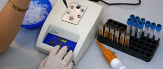
Chemiluminescence immunoassay is used to detect tumor markers in serum. This method is similar in its method to enzyme-linked immunosorbent assay. The essence of immunochemiluminescence research lies in the use of specific magnetic particles that have a high degree of sensitivity.
During the study of serum, immune reactions are detected combining a specific antigen with a specific antibody. Such reactions are unique, which makes it possible to prove with high accuracy the presence of pathological changes.
Plasma testing is carried out using an automatic chemiluminescence immunoassay analyzer.
The likelihood of receiving false data is low.
The cause of the error may be:
- incorrect actions of the laboratory assistant when drawing blood;
- failure of the patient to comply with the conditions of preparation for the examination.
For a more accurate diagnosis, an additional blood test for carcinoembryonic antigen is performed. If the test is negative and there are visible signs of breast cancer, the examination is repeated.
In the initial stages of pathology, the concentration of antigens may be low and specific proteins will not be detected by traditional laboratory tests. In this case, a repeat analysis is carried out after a short period of time.
Preparation for delivery
The fear of getting cancer, which contradicts logic, is called cancerophobia, but one cannot trust the distortions of the mind. The number of people suffering from such psychological difficulties is constantly increasing. A common symptom that interferes with a peaceful life is false pain in the areas of suspected foci of the disease. It is better for people with unhealthy anxiety to consult a psychologist or psychiatrist, and not to create barriers to the early start of therapy for those who have a serious suspicion of cancer.
The patient begins to prepare for the analysis 2-3 days before the test. The patient stops eating pickled, spicy, salty, fatty and smoked foods, drinks only still water. Doctors recommend not smoking on the day of the test; in extreme cases, only 30 minutes before the actual test. The procedure takes place early in the morning; food intake is stopped 12 hours before.
If a person takes medications, then the appointment on the day of delivery is agreed with the treating doctor. It is also better to report about chronic diseases: if they are associated with immunity problems, they will affect the result. In 24 hours, it is unacceptable to: sunbathe, swim, visit a solarium, warm the chest, or unnecessarily palpate the mammary gland on your own. Violation of prohibitions leads to incorrect research.
Blood is drawn in a completely sterile environment. The collection occurs without severe pain, but for people with increased sensitivity, the event will be inconvenient. The action itself follows a standard scenario: a tourniquet is applied, the skin is treated with a substance with a disinfecting effect, and pierced with a needle. After the procedure the patient is free. The results, according to the standard, are ready within 24 hours.
Preparing and conducting analysis
Collecting material for research is a completely safe procedure. Its task is to obtain the most accurate result.
To do this, a number of conditions must be met:
- Blood sampling is carried out in the morning strictly on an empty stomach.
- The last meal should take place twelve hours before the examination.
- At the last meal, dishes containing large amounts of fat and spicy seasonings are completely excluded from the diet.
- A few days before the visit to the laboratory, the consumption of alcohol, alcohol-containing drinks, and energy drinks with high levels of caffeine is prohibited. These substances can affect the composition and condition of the blood.
- Smoking is prohibited for several hours prior to your visit to the laboratory.
- On the eve of the examination, avoid stress and limit the level of physical activity.
- For several days before the examination, it is recommended not to take medications, especially those that suppress the immune system, as well as drugs belonging to the group of corticosteroids and tranquilizers. An exception is made for essential medicines. Their name and dose must be indicated on the directions.
- If necessary, drinking still water before analysis is allowed.
- Receiving material
- The material is collected in a treatment room under highly sterile conditions.
- Blood is taken from the antecubital vein only by a medical professional.
- Minor discomfort may appear if the patient’s pain threshold is elevated.
How to take the test and properly prepare for it?
The examination today is carried out in a medical institution, modern medical centers. To do this, blood is drawn from a vein. On the day of the examination, you must refrain from eating; only non-carbonated water is allowed as a drink. It is also important to avoid smoking and drinking alcohol, as this can affect the results. In addition, before the study, it is important to refrain from undergoing various physiotherapeutic procedures and manual examination of the mammary glands.
Decoding the results
CA 15-3 tumor marker, which is normal in women, does not yet indicate the absence of the disease.
The results obtained are assessed by an oncologist, gynecologist or mammologist:
- The doctor ascertains the absence of a tumor if there are no tumor markers in the blood or their insignificant detection is not more than 27 IU/ml.
- A negative test result is possible if the patient recovers.
- There are no antibodies in the serum if tumor growth has just begun.
A one-time increase in titer does not indicate the presence of the disease. Changes occurring in the body must be confirmed by other diagnostic methods. A repeat examination must be carried out four weeks after receiving the first result. If there is no dynamics, then they look for other causes not related to breast cancer.
Repeated increase in titer. At levels above 27 IU/ml.
Assuming availability:
- stomach cancer
- liver cancer
- pancreas cancer
- cancer of the ovaries, endometrium, uterus, mainly in the later stages of tumor development.
When diagnosing benign processes:
- with localization in the mammary glands;
- in liver cirrhosis, the number of tumor markers does not exceed the level of 50 units/ml;
- physiological increase in CA 15-3 in the presence of pregnancy in the third trimester. The tumor marker concentration approaches 50 U/ml;
- autoimmune diseases.
CA 15-3 tumor marker, the norm for women in the normal state is 27 units/ml, during gestation against the background of hormonal changes in the blood, sometimes reaches over 35–39 units/ml. The level of tumor marker in serum can be affected by the presence of sarcoidosis and tuberculosis in the body.
Why is blood taken for CA 15-3?
Although CA 15-3 is not considered the only indicator of the presence of cancer, an elevated level of protein in a blood sample indicates the need for additional examinations.
Remember! Breast tumor markers CA 15-3 are examined not only for the presence of cancer, but also to evaluate the effectiveness of treatment, for example, for breast carcinoma.
Of course, a final diagnosis cannot be made from this sample alone, due to the fact that high levels of this protein can also be observed in other diseases: autoimmune disorders or tuberculosis.
When to see a doctor
In the initial stages of breast cancer:
- Unusual formations are felt in the mammary gland. Very often the tumor can be small and not paid attention to.
- A characteristic feature of this structure is its painlessness. Most often, a growing cancer tumor in the initial stages does not cause concern.
- Areas with skin retraction are identified.
- In later stages of the disease, pain appears in the area where the tumor grows.
- A change in the nipple is detected, which thickens and swells.
- The nipple is retracted into the breast. In this case, you can look for a tumor in this area.
- Discharge from the nipple appears that is not associated with pregnancy or breastfeeding. Normally, the presence of minor discharge associated with changes in hormonal levels is acceptable. At the same time, the amount of discharge is insignificant and there are no blood impurities in them. In the presence of pathological changes, the amount of discharge increases sharply.
- There is an increase in the lymph nodes in the axillary region, they form a bunch of grapes.
- In case of advanced pathology, a blood test for tumor markers is carried out in the presence of the following symptoms:
- A tumor that is easily detected by palpation of the chest.
- Changing the location of the seal with a retracted nipple.
- Thickening of the skin around the nipple.
The signs of cancer are the same for all women; the symptoms that occur as the tumor grows may differ.
There are many forms of breast cancer, but they all have one common symptom - the appearance of enlarged lymph nodes in the armpit. If such a symptom is detected, contacting a doctor is mandatory. As a rule, when studying blood, even in normal conditions, an increase in the concentration of a tumor marker is detected.
At the discretion of the attending physician and in the presence of alarming symptoms, persons who are at risk for developing breast cancer and other pathologies are sent for a blood test:
- Patients whose families have already been diagnosed with breast cancer
- Women over forty years of age
- If the patient has been taking hormonal contraceptives for a long time
- Persons with atypical hyperplasia
- Patients with a persistent increase in estrogen levels in the blood
- Pregnant for the first time after thirty years
- Women suffering from long-term infertility
- Patients with a history of breast and ovarian cancer
- Women with late menopause
- Persons by occupation who come into contact with radioactive substances.
- How to get it back to normal
- If there are elevated tumor marker values in the blood, measures are taken to identify the reasons that led to the changes.
- When a diagnosis is made, a course of treatment is prescribed, which can be conservative or require surgical intervention. Upon completion of therapy, the level of antibodies drops to minimal values or specific proteins disappear completely.
Conservative treatment includes:
- Radiotherapy. Refers to the category of maintenance treatment. It is carried out before or after removal of the tumor. During the procedure, degenerated cells are destroyed.
- Intrabeam. Intraoperative irradiation. Radiation therapy is carried out during surgery, affecting the area where the tumor was located. In this case, the risk of damage to healthy tissue is significantly reduced and relapses of the disease are eliminated. The body's recovery period is reduced by six weeks.
- Chemotherapy. Which refers to medicinal methods. The patient is prescribed drugs of a toxic group that destroy mutated cells. They resort to a similar technique if surgical intervention is impossible. The result of the action of chemotherapy is the destruction of proteins responsible for the growth and division of cancer cells. In some cases, the DNA and RNA chains of diseased cells are destroyed.
- Hormone therapy is rarely used, only in situations where the tumor is susceptible to the influence of drugs belonging to this group.
- Targeted therapy is considered the mildest form of treatment for breast cancer, since the effect is exclusively on cancer cells. For this purpose, special drugs are prescribed.
Among the medications, patients are prescribed:
- Maximim in injections. The solution is administered intramuscularly daily for two weeks.
- Penicillin is prescribed when a diagnosis of fibrocystic mastopathy is established. In this case, an antibiotic is necessary to eliminate the symptom of inflammation and tissue swelling. The daily dose is two tablets.
- Tamoxifen, drug related to hormonal. Prescribed to restore hormonal levels. Which contributes to the disappearance of the tumor.
- Femoden. It also affects hormone levels. Under its influence, the tumor disintegrates.
Even after receiving a negative result, monitoring of the patient continues. She is prescribed preventive examinations and laboratory tests are carried out. Most often, such observation is carried out for life, since the likelihood of the disease returning remains.
Decoding
Analyzing the readings of the CA 15-3 tumor marker will help the oncologist select the necessary conservative treatment and monitor the effectiveness of its use (in cases where surgery is contraindicated). The test results are usually ready no less than 3 hours after donating blood for analysis and no later than 2 days. To more accurately decipher the analysis, it must be carried out according to a certain scheme.
After a positive test and treatment, the patient should donate blood for analysis every month for a year, in the next year this should be done once every couple of months, and in the 3rd year they donate blood once a quarter. The doctor conducting the treatment records all the readings in a special chart and monitors the dynamics of the disease. A growing graph indicates that the prescribed treatment is ineffective and then another therapy is selected.
Note! In the process of deciphering the analysis, it is possible to determine the cause of the disease and the effectiveness of treatment.
If tumor marker values border on normal values, then additional tests are prescribed, for example, biopsy and mammography.
You should also keep in mind that a single jump in the indicator is not the final analysis, since the results could be influenced by external factors (for example, jumps in hormone levels occur during breastfeeding), and this means that you need to take a blood test again.
Possible complications
If abnormalities are detected in the blood, self-medication is prohibited.
A high volume of tumor markers in the blood is observed in advanced forms of cancer and developing mammary glands.
- This means that breast tissue is being destroyed. A growing tumor compresses and destroys healthy tissue that forms the mammary gland and milk ducts, which leads to the development of severe inflammation, even necrotic changes.
- There is a high probability of metastases occurring in organs and systems located far from the mammary glands. Cancer cells travel through the blood throughout the body and serve as the basis for the emergence of new malignant tumors. In this case, doctors give an extremely unfavorable prognosis.
First of all, metastases germinate:
- Into the lung tissue, which is actively supplied with blood and nutrients. In this case, signs of suffocation will be observed, often occurring against the background of ongoing pneumonia.
- Into liver tissue. When metastases form in the liver, symptoms of hepatitis appear.
- Into the brain. When metastases grow in the brain, vision, hearing, and sleep deteriorate, convulsions appear mainly at night, consciousness is impaired and the thinking process changes.
When metastases form, the probability of death is always high. CA 15-3 is a tumor marker, the norm in women without pathology is quite low, in this case it will be many times higher than normal values. Most of the methods used in the treatment of cancer at this stage of cancer are ineffective. Treatment of such patients is carried out in specialized hospices.
Indications for analysis
The CA 15-3 cancer antigen test is considered auxiliary among specialists and is prescribed for the following purposes:
- confirmation or refutation of a malignant process in the mammary glands;
- differential diagnosis of benign and malignant tumors, mastopathy, other inflammatory or infectious processes of the mammary glands and internal organs;
- dynamic assessment of the effectiveness of surgical or conservative treatment of confirmed breast cancer;
- assessment of the likelihood of metastasis and relapse;
- making a prognosis for the course of the disease, remission, monitoring the rehabilitation of patients;
- obtaining data regarding the shape and volume of the tumor, its location and extent of spread (the greater the specific gravity of CA 15-3 in the plasma, the greater the volume of malignant cells produced by mammary tissue).
The need to prescribe this test as part of a comprehensive examination of the mammary glands may be indicated by the following clinical signs:
- the appearance of lumps, lumps or other formations in the breast tissue, which is determined visually or during palpation;
- pain in the chest, feeling of fullness or heaviness;
- pathological discharge from the nipples (including in males);
- change in the shape of the nipple (retraction, hardening, redness), the appearance of cracks or wounds on it that may bleed;
- changes in the structure of tissues and skin (redness, generalized swelling, roughness, “lemon peel” effect);
- immobility of the mammary gland (the tumor has grown into the sternum, which leads to deformation of muscle tissue and glands);
- inflammation of regional lymph nodes, their enlargement.
You can read more about breast self-examination techniques for women in our separate article.
The study is also prescribed to patients at risk for oncology:
- the presence of direct relatives who have a history of being diagnosed with cancer of internal organs;
- age over 40-50 years;
- long-term hormonal support, oral contraceptives, hormone replacement therapy;
- chronically elevated levels of estrogen in the blood;
- chronic menstrual irregularities, secondary amenorrhea;
- earlier onset of menstruation;
- late menopause;
- history of ovarian cancer.
Interpretation of the test results for cancer antigen CA 15-3 is carried out by an oncologist, mammologist, surgeon or functional diagnostician.
Additional Research
An analysis of 1301 simultaneous determinations of the tumor markers CA153 and CEA was reported in 405 patients with breast cancer. CA153 was superior to carcinoembryonic antigen in sensitivity and specificity (ROC presentation).
At reference levels of 30 U/mL for CA 15-3 and 5 ng/mL for CEA, sensitivities of 75.7% versus 61.8% and estimated specificities of 93.8% versus 83.4% were found.
In 73 patients who experienced their first relapse after primary tumor marker-guided therapy, an increase in CA 15-3 above the control value preceded clinical detection in 43.8% of cases.
The local recurrence rate was 10-18%, so the benefit was negligible. On the other hand, hematogenous metastases were predicted in 33-69% of cases.
Although tumor marker diagnostics are most useful for monitoring larger tumors, research findings also suggest their use in the early detection of breast cancer.
Marker system
Laboratory tests use a breast cancer tumor marker to detect the tumor or its recurrence, determine the extent of the disease and monitor treatment. All of these readings mean that tumor cells are being released into the patient's bloodstream in measurable amounts. Their levels can be calculated from a blood sample.
As already indicated, a sharp jump from normal to a high value exceeding established norms does not necessarily represent the process of cancer development and only indicates the onset of acute inflammation of the mammary glands. However, systematic analysis of the appearance of the antigen and changes in its amount in blood samples make the diagnosis more accurate.
The system means regular studies depending on the treatment period:
- One-time analysis before starting treatment.
- Monitoring after radical treatment for the first 3 years quarterly.
- After this period, every six months.
- If high titers of specific markers are observed, monitoring should be carried out monthly or even twice a month.
- When carrying out chemotherapy, an analysis is carried out before each course, as well as when changing treatment methods.
- During radiation therapy, tests are ordered midway through treatment and after completion.
It should be remembered that normal levels of antigens do not exclude the presence of cancer, and their moderate increase does not always indicate a tumor. Correct conclusions about the concentration of breast tumor markers allow us to obtain blood tests done sequentially at certain intervals under the same laboratory conditions.
Measuring tumor marker levels helps doctors plan appropriate therapy. Some of them use the results of marker studies as early indicators of acceleration of the tumor process in the mammary gland, others to make decisions about changing treatment, etc.