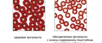
Hemoglobin
Hemoglobin (Hb, Hgb) in a blood test is the main component of red blood cells, which transports oxygen from the lungs to organs and tissues. For analysis, a cyanide complex or cyanide-free reagents are used (as a replacement for toxic cyanide). It is measured in moles or grams per liter or deciliter. Its definition has not only diagnostic, but also prognostic significance, since pathological conditions leading to a decrease in hemoglobin content lead to oxygen starvation of tissues.
The normal hemoglobin content in the blood is:
- men - 135-160 (130-170) g/l (grams per liter);
- women - 120-140 (120-150) g/l;
- children - 120-140 g/l.
Anemia (low hemoglobin) - a decrease in the total amount of hemoglobin in the blood.
Hyperchromemia (high hemoglobin) is an increase in the concentration of hemoglobin in the blood.
Thrombocytosis in children
Thrombocytosis is an increase in the number of platelets in the blood to values several times higher than normal. Most often, thrombocytosis is a consequence of a viral or bacterial infection. In particularly severe cases, an increase in blood cells may indicate the development of cancer.
How to determine: symptoms
The symptoms of thrombocytosis are similar to the clinical manifestations of thrombocytopenia, although there are still some differences. If a child has at least one of the listed signs, you should immediately consult a doctor and take a blood test.
Symptoms of thrombocytosis in childhood:
- frequent and severe nosebleeds;
- blue skin;
- the appearance of subcutaneous hemorrhages;
- itching and burning in certain areas of the skin;
- tingling sensation on the skin;
- cold hands and feet at a comfortable ambient temperature;
- dyspnea;
- increased heart rate.
The child may also complain of frequent headaches. In some cases, pressure surges and blood clots may form.
Causes
To correctly determine the cause of the increase in platelets, it is necessary to diagnose what type of thrombocytosis is developing in the child. The treatment regimen and tactics for managing a small patient will depend on this.
| Type of thrombocytosis | Causes |
| Clonal | Bone marrow stem cell defects |
| Primary | Pathologies in the functioning of bone marrow cells, the formation of several sites of hematopoiesis |
| Secondary |
|
Recommendations for thrombocytosis
- Consult your doctor about the use of products that contain acetylsalicylic acid (for thinning the blood).
- During the day, offer your child compotes and fruit drinks made from blueberries, lingonberries and cranberries as often as possible.
- Monitor the iodine content in the body, and if there is a deficiency, ensure sufficient consumption of fish and seafood.
- If the child does not have problems with the gastrointestinal tract, it is recommended to add fresh garlic and ginger (in minimal quantities) to food.
- In the morning, you can give your baby cocoa without sugar (a prerequisite is that the drink must be consumed on an empty stomach).
- Olive and flaxseed oils, as well as foods rich in magnesium and B vitamins, should be mandatory products in a child’s diet for thrombocytosis.
- Avoid dehydration (the water norm for a child is 1 liter per day).
- Increase the amount of fruits and vegetables (tomato juice and lemons are especially useful).
Leukocytes
White blood cells (L, WBC) are blood cells produced in the bone marrow and lymph nodes. There are 5 types of leukocytes: granulocytes (neutrophils, eosinophils, basophils), monocytes and lymphocytes. The main function of leukocytes is to protect the body from foreign antigens (including microorganisms, tumor cells; the effect also manifests itself in the direction of transplant cells).
Normal leukocyte content in the blood: (4-9) x 109
Leukocytosis is an increased level of leukocytes in the blood.
Leukopenia is a decrease in the number of leukocytes in the blood.
What is thrombocytopenia?
Thrombocytopenia is an absolute decrease in the platelet count below 150 thousand cells in 1 microliter. The upper limit of normal is 400 thousand/µl. Thrombocytopenia, like other deviations from the norm in the cellular composition of the blood, can be absolute and relative. With relative thrombocytopenia in the blood volume, a decrease in their number is detected for the reason that the content of other cells is increased “to the detriment” of platelets. Or, for example, thrombocytopenia can occur after massive intravenous fluid administration, when the blood is “diluted” and the platelet count is reduced due to hemodilution (dilution) of the blood. And absolute thrombocytopenia is detected against the background of normal parameters of volume and cellular composition.
As a rule, pronounced clinical signs occur if the platelet count is still three times lower than normal - their number is less than 50 thousand / μl. Symptoms of thrombocytopenia consist of various hemostasis disorders, such as:
- intradermal and subcutaneous hemorrhages from the most minor injuries;
- bleeding gums;
- heavy and prolonged periods (menorrhagia);
- nosebleeds.
In addition to these relatively harmless symptoms, conditions that actually threaten life may occur, for example, massive gastric bleeding due to a stomach ulcer (in the case of erosion of a large vessel). With chronic alcoholism and frequent urge to vomit, the development of Mallory-Weiss syndrome is possible, in which bleeding occurs from multiple small submucosal ruptures in the wall of the cardia of the stomach. Bleeding from varicose veins of the esophagus in severe thrombocytopenia is often the cause of hemorrhagic shock and can be fatal, especially in old age and against the background of severe anemia.
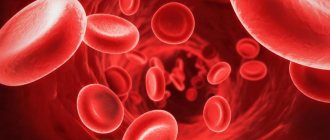
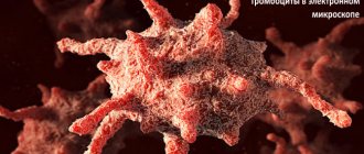
Platelets
Platelets (PLT) (blood plates, Bizzocero plaque) (from ancient Greek θρόμβος - lump, clot; κύτος - receptacle, here: cell) - small flat colorless bodies of irregular shape, circulating in large quantities in the blood
Norm: 150-400 G/l
The main function of platelets is to participate in the process of human blood clotting. The number of platelets varies depending on the time of day and throughout the year. A natural decrease in platelet levels is observed during menstruation and pregnancy, and an increase after physical activity.
Physiological causes of thrombocytopenia
Based on clinical data and test results, the doctor must recognize the causes of thrombocytopenia in the blood and begin treatment. You can immediately note the physiological reasons for which low platelets occur:
- Pregnancy. During pregnancy, the cause of thrombocytopenia is an increase in circulating blood volume (due to the fetus and fetoplacental blood flow), as well as a shortening of the lifespan of platelets.
- Heavy and painful periods, which often occur in women with an unsteady menstrual cycle.
What are the dangers of high and low platelet levels in the blood?
Platelets (Bizzocero plaques) are blood formed elements, the main function of which is to stop bleeding and restore the vascular wall under the influence of growth factors secreted by them.
Their precursors are megakaryocyte cells, which are formed in the bone marrow. Blood plates are not cells, but their fragments. A platelet is a nuclear-free result of fragmentation of a fragment from a megakaryocyte cell.
Life expectancy is 8–12 days.
When the endothelium (the lining of a vessel) is damaged, Bizzocero plaques adhere (stick) to its surface.
Then, with the help of special signaling molecules, these blood platelets attract their fellows, the process of their accumulation is called aggregation. After which the thrombus thickens (retraction), plugs form.
Platelets contain serotonin in their granules, which, when released, constricts blood vessels, preventing bleeding.
https://www.youtube.com/watch?v=_t2qBdtNnjc
The substance thrombopoietin, synthesized by muscles, liver and kidneys, stimulates the formation of platelets. The body has a well-established negative feedback mechanism: when the number of platelets increases, the production of thrombopoietin decreases, and when there is a lack of blood platelets, the synthesis increases.
Platelets in the spleen are destroyed.
Causes of thrombocytopenia
Thrombocytopenia is a decrease in the content of blood platelets in the blood below normal. Causes of thrombopenia:
- Decreased platelet formation (their synthesis can be reduced by taking certain medications, infectious diseases, etc.).
- Increased consumption of blood platelets during thrombus formation in intravascular coagulation syndrome (DIC). DIC syndrome can result from infectious and autoimmune processes.
- The appearance of antibodies against platelets, for example, in thrombocytopenic purpura. This disease is characterized by a red rash on the body.
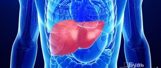
- Liver diseases. With increased pressure in the portal vein system, platelets accumulate in greater numbers in the spleen. Therefore, there are few of them left in the peripheral blood. During inflammatory processes in the liver, IgG antibodies are sometimes formed, which leads to autoimmune lysis of platelets. With alcoholic hepatitis, hemolysis develops (destruction of blood cells).
- Hypersplenism is an increase in the function of the spleen, destruction of the blood platelets in it. Hypersplenism occurs with portal hypertension, as well as with thesaurismosis (storage diseases), when various substances accumulate in the spleen, disrupting its function.
- The use of antiplatelet agents and anti-inflammatory drugs (Aspirin, Ketorol, aminosalicylates), direct anticoagulants. The drugs can reduce blood clotting by reducing the number of blood platelets.
- Hemolytic poisons. Poisoning with acetic acid or heavy metals causes lysis of blood platelets and other formed elements. In this case, indirect bilirubin will be increased in the biochemical analysis.
- Radiation damage to the bone marrow.
- Cytostatics inhibit hematopoiesis.
- Leukemia and increased spleen function with them.
- Prosthetic valves in the heart. Hemolysis occurs.
- Hemolytic-uremic syndrome due to dysentery bacillus infection.
- Vasculitis.
- Deficiency of vitamins B12 and folic acid reduces the number of platelets and red blood cells in a general blood test (CBC).
- Hereditary thrombocytopenia.
Immune thrombocytopenias are divided into 4 groups:
- Isoimmune. Occur due to a conflict of blood group antigens between the donor and recipient.
- Transimmune. They occur when antibodies to the fetus penetrate through the hematoplacental barrier from a mother who has autoantibodies to platelets.
- Heteroimmune. In some viral diseases, viral antigens accumulate on the surface of the platelet membrane and cause its immune lysis.
- Autoimmune thrombocytopenia. Autoantibodies bombard self-antigens.
Why is thrombocytopenia dangerous?
A lack of Bizzocero plaques in the peripheral blood can reduce blood clotting and lead to bleeding.
Causes of thrombocytosis
Thrombocytosis is an increased content of Bizzocero plaques in the peripheral blood (analysis shows their number is more than 400,000 per cubic mm).
Causes of thrombocytosis:
- Polycythemia (malignant tumor process in the blood).
- Splenectomy, or congenital absence of the spleen. The spleen deposits 30% of all Bizzocero plaques in itself. When it is removed, all these elements end up in the bloodstream.
- Damage to blood vessels during surgery and trauma.
- Inflammatory processes in the body. Proinflammatory cytokines increase thrombopoietin production. Platelets in the blood during general analysis are increased.
- Iron deficiency in acute blood loss, radiation therapy.
- Infectious thrombocytosis in meningococcemia, fungal, parasitic, viral infections, tuberculosis.
- Myeloproliferative syndrome.
- Pregnancy. In women, the platelet count increases due to hormones.
- The use of glucocorticoids, adrenergic agonists, and oral contraceptives in women.
- Physical exercise.
- Inflammatory bowel diseases.
- Osteomyelitis, bone fracture in children.
- Tumor diseases of the lungs and pancreas.
- Cirrhosis of the liver.
- Amyloidosis.
- Lymphoma.
- Endocarditis.
- Hereditary thrombocytosis.
Why is thrombocytosis dangerous?
An excessive number of Bizzocero plaques worsens blood rheology and increases the likelihood of developing thromboembolism and intravascular coagulation syndrome.
Average platelet count as the norm for children in the table
The platelet content in a child’s blood depends on many factors, for example, health status, nutritional habits, age, etc. Age is the main criterion when determining compliance with standards.
The normal platelet count depending on the age of the child (the table shows values for healthy children!)
| Age | Blood cell content (per liter of blood) |
| Newborn (1-3 days after birth) | 100-420*109 |
| 7-10 days | 150-400*109 |
| 1 month | 160-400*109 |
| 6 months | 180-400*109 |
| 12 months | 160-380*109 |
| From 1 to 4 years (inclusive) | 160-400*109 |
| From 5 to 7 years (inclusive) | 180-450*109 |
| Over 7 years old | 180-460*109 |
How to determine the level of platelets in the blood?
To determine the volume of blood cells, a general blood test is used, which is done even to newborn children 1-3 days after birth.
For the purpose of early detection of pathologies, healthy children are recommended to donate blood at least once a year.
If there is a hereditary predisposition to the development of thrombocytopenia or thrombocytosis, the pediatrician may prescribe a complete blood count more often, for example, 2-4 times a year. Bone marrow pathologists, as well as some diseases, also require more frequent monitoring.
Do I need special preparation before taking the test?
In order for the analysis to be as accurate as possible, the baby should be properly prepared for blood donation. To do this, you must follow the following rules:
- the fasting period should be at least 8 (for children under 7 years of age) or 12 hours (for children over 7 years of age);
- collection of material should be carried out in the morning (60-120 minutes after waking up);
- Drinking and eating food before the analysis is not allowed (you can give the baby some clean drinking water);
- 1-2 days before donating blood for analysis, it is necessary to limit the child’s physical activity and activity (if the child goes to school, you should skip physical education class);
- Under no circumstances should you rub your fingers before entering the nurse's office, as this may affect test results (for example, white blood cell levels, etc.).
Participation in folding
A special feature of the platelet is its ability to activate - a rapid and, as a rule, irreversible transition to a new state. The activation stimulus can be almost any environmental disturbance, even simple mechanical stress. However, the main physiological activators of platelets are collagen (the main protein of the extracellular matrix), thrombin (the main protein of the plasma coagulation system), ADP (adenosine diphosphate, appearing from destroyed vascular cells or secreted by the platelets themselves) and thromboxane A2 (a secondary activator synthesized and released by platelets; its additional function is to stimulate vasoconstriction).
Activated platelets become able to attach to the site of damage (adhesion) and to each other (aggregation), forming a plug that covers the damage. In addition, they participate in plasma coagulation in two main ways - exposure of the procoagulant membrane and secretion of α-granules.
Sequence of primary biochemical and morphological changes upon activation
The initial stages of platelet activation under the influence of external factors are associated not only with the appearance of biochemical markers, but also with morphological changes in the form of the platelet. As has been shown by flow cytometry and electron microscopy, the most sensitive sign of activation (when platelets are exposed to ADP) are morphological changes[6]. The appearance of biochemical and morphological changes, arranged according to the degree of decrease in sensitivity, is as follows: change in platelet shape, conformational changes in glycoprotein IIb/IIIa, expression of P-selectin, expression of phosphatidylserine.
Exposure of the procoagulant membrane
Under normal conditions, the platelet membrane does not support clotting reactions. Negatively charged phospholipids, primarily phosphatidylserine, are concentrated in the inner layer of the membrane, while phosphatidylcholine in the outer layer binds coagulation factors much less well. Although some coagulation factors can bind to non-activated platelets, this does not lead to the formation of active enzymatic complexes. Activation of the platelet presumably leads to the activation of the scramblase enzyme, which begins to quickly, specifically, bidirectionally and ATP-independently transfer negatively charged phospholipids from one layer to another. As a result, thermodynamic equilibrium is established, in which the concentration of phosphatidylserine in both layers is equalized. In addition, upon activation, many transmembrane proteins in the outer layer of the membrane undergo alignment and/or conformational changes, and they acquire the ability to specifically bind coagulation factors, accelerating reactions involving them.
Platelet activation has several degrees, and procoagulant surface expression is one of the highest. Only thrombin or collagen can produce such a strong response. A weaker activator, especially ADP, can contribute to the work of stronger activators. However, they are not able to independently cause the appearance of phosphatidylserine; their effects are limited to changes in platelet shape, aggregation and partial secretion.
Secretion of α-granules
Platelets contain several types of granules, the contents of which are secreted during activation. Essential for coagulation are α-granules containing high molecular weight proteins such as factor V and fibrinogen.
Tests to evaluate the vascular-platelet component of hemostasis
- Bleeding time;
- The number of platelets in the blood;
- Induced platelet aggregation.
Qualitative platelet defects, which underlie a large number of hemorrhagic diathesis, are divided into the following groups:
- disaggregation thrombocytopathies caused by the absence or blockade of platelet membrane receptors (Glanzmann’s thrombasthenia, etc.);
- diseases of the absence of dense and α-granules;
- granule release disorders;
- disturbances in the formation of cyclic prostaglandins and thromboxane A2;
- deficiency, anomalies and disorders of the multidimensionality of von Willebrand factor;
- disorders of nucleotide metabolism and calcium transport.
Bizzocero plaque
Bizzocero plaque
(G. Bizzozero, 1846-1901, Italian doctor) - see
Platelet
.
Articles on the topic Bizzocero plaque
- Bizzozero plaque Bizzozero plaque (G. Bizzozero, 1846-1901, Italian doctor) - see Platelet.
- Platelet Platelet (thrombocytus; thrombo- + hist. cytus cell; synonym: Bizzocero plaque, blood plate) is an oval or round shaped blood element, which is a cytoplasmic fragment of a bone marrow megakaryocyte; at.
- Bizzocero - Neumann crystalsAtherosclerotic plaqueAmniotic plaques
News about Bizzocero plaque
- Chlamydia pneumoniae is found in atherosclerotic plaques in unstable angina pectoris Neither serological markers nor clinical characteristics are reliable predictors of the presence of Chlamydia pneumoniae in coronary atherosclerotic plaques, according to the results of a new study. “Even if a cause-and-effect relationship is proven in the near future
- Amyloid plaques removed from the brains of living animals with Alzheimer's disease A remarkable discovery has been made by scientists at the Mayo Clinic in Jacksonville, Florida, that could lead to a new treatment for Alzheimer's disease that actually removes amyloid plaques, considered a hallmark of the disease, from the brains of patients.
- Atherosclerotic plaque morphology influences the outcome of anterior myocardial infarction
Functions
Platelets perform two main functions:
- Formation of a platelet aggregate, a primary plug closing the site of vessel damage;
- Providing its surface to accelerate key plasma coagulation reactions.
Relatively recently, it was found that platelets also play a critical role in the healing and regeneration of damaged tissues, releasing growth factors into damaged tissues that stimulate cell division and growth. Growth factors. They are polypeptide molecules of various structures and purposes. The most important growth factors include platelet-derived growth factor (PDGF), transforming growth factor (TGF-β), vascular endothelial growth factor (VEGF), epithelial growth factor (EGF), fibroblast growth factor (FGF), insulin-like growth factor (IGF) [ 5] .
The physiological plasma concentration of platelets is 180-360*10^9 platelets per liter.
A decrease in the number of platelets in the blood can lead to bleeding. An increase in their number leads to the formation of blood clots (thrombosis), which can block blood vessels and lead to pathological conditions such as stroke, myocardial infarction, pulmonary embolism, or blockage of blood vessels in other organs of the body.
A deficiency or disease of platelets is called thrombocytopathy, which can be either a decrease in the number of platelets (thrombocytopenia), a violation of the functional activity of platelets (thrombasthenia), or an increase in the number of platelets (thrombocytosis). There are diseases that reduce platelet counts, such as heparin-induced thrombocytopenia or thrombotic purpura, which usually cause thrombosis instead of bleeding.
Due to the inaccuracy of descriptions, the lack of photographic equipment and the confusing terminology of the early periods of the development of microscopy, the exact time of the first observation of platelets is unknown. Most often, their discovery is attributed to Donna (1842, Paris), but there is evidence that they were observed by the creator of the microscope himself, Antonie van Leeuwenhoek (1677, the Netherlands). The term “blood platelets,” which is still preferred in English-language literature (blood platelets), was introduced by Bizzocero (1881, Turin), who also played a leading role in identifying the connection of platelets with homeostasis and thrombosis. This subsequently led to the emergence of the term “platelet” (Dekhuizen, 1901), which became the main one in the Russian language. In the English-language literature, the term is used exclusively for nucleated platelets in non-mammalians (thrombocytes). In addition, in Russian literature the term “Bizzocero plaque” may be used for platelets.
Decoding the PCT indicator in a blood test
Have you been struggling with HYPERTENSION for many years without success?
Head of the Institute: “You will be amazed at how easy it is to cure hypertension by taking it every day.
The PCT value in a blood test allows you to determine the quantitative content of platelets. The purpose of counting blood cells is to establish thrombocytopenia, which can be hypoproliferative and hyperdestructive. Platelet indices are a simple diagnostic method that has become available through automatic cell counting.
PCT indicator in blood test
Thrombocrit refers to the percentage of platelet mass in the blood volume. Deviation from the norm is a prerequisite for further research. It is important to compare the obtained value with the number of platelets responsible for the coagulation indicator in a general blood test.
Our readers successfully use ReCardio to treat hypertension. Seeing how popular this product is, we decided to bring it to your attention. Read more here...
The level decreases against the background of B12 deficiency, folic acid, as well as in hematopoietic disorders (Fanconi and aplastic anemia), viral infections, leukemia and granulomatosis after taking corticosteroids and cytostatics. An increase is observed in immune thrombocytopenic purpura, but more often in diseases of the cardiovascular system. Inflammatory processes and infections, tuberculosis, tumors, blood loss, ulcers and necrosis, anemia and diabetes mellitus, and surgical interventions can change the indicator upward. Platelets are important for wound healing because they help:
- stop blood loss;
- stimulate tissue regeneration.
Platelet indices are useful markers for the early diagnosis of thromboembolic diseases. Thrombocrit is considered together with two other indicators - the average volume and range of platelet distribution. Their values make it possible to identify the causes of thrombocytopenia, as well as to determine immune thrombocytopenic purpura.
The range of platelet distribution increases due to their activation with an increase in cell volume and the formation of pseudopodia. An increase in the average value is associated with the following conditions:
- inflammatory processes in the intestines;
- immune thrombocytopenic purpura;
- myeloproliferative diseases;
- preeclampsia and recovery from hypoplasia.
A decrease in the level of blood platelets occurs against the background of bleeding.
Analysis transcript
Thrombocrit is normally between 0.2 and 0.36%, but is not considered alone.
It is necessary to focus on the platelet indices presented in the table:
Platelet crit (PCT)0.2–0.36%.Mean platelet volume (MPV)7–10 femtolitersPlatelet distribution range (PDW)56.6%A blood test with PCT decoding is useful to confirm certain diseases:
- suspicion of sepsis and determination of its danger;
- severe bacterial infections and their complications;
- unknown fevers;
- suspicion of meningitis, peritonitis and pneumonia.
The indicator helps to evaluate the effectiveness of therapy in shock, sepsis, immunosuppression and neutropenia, after organ transplantation and spleen removal, while on a ventilator.
Factors influencing the indicator
Thrombocrit, by analogy with hematocrit, determines the mass of settled cells. Platelets in the external environment undergo changes: they form pseudopodia, increase in size, stick together or undergo aggregation, and therefore they are difficult to count. After the development of automatic blood analyzers, this became much easier, which is why thrombocrit appeared in the list of research results. The number of platelets is counted only as directed by a doctor, since the process requires the use of special conditions and reagents.
A false decrease in thrombocrit may be due to the following factors:
- Partial clotting of a blood sample with platelet activation and aggregation during injection.
- Platelet aggregation when using ethylenediaminetetraacetic acid (EDTA) as a stabilizer.
- Platelet satellitetism, or their adherence to neutrophils when using various anticoagulants for analysis, which is associated with the production of IgG or IgM antibodies.
- Detection of giant cells that cannot be identified by automatic counters.
A false increase in level occurs against the background of the following conditions:
- Fragmentation of red blood cells, when small cells or their fragments were counted as platelets.
- Cryoglobulinemia (detection of immunoglobulins in serum) is associated with pseudothrombocytosis and pseudoleukocytosis.
Low and high values are confirmed with a repeat blood test.
Differences in thrombocrit levels for children and adults are not clinically determined. All platelet indices generally change proportionally with age, and PCT is determined by multiplying mean platelet volume (MPV) by cell count.
It is necessary to pay attention to fluctuations in the number of platelets, which can be physiological and pathological. The indicator is influenced by the following factors:
- In the spring and during depression, as well as at night, the level of platelets and thrombocrit decreases by 10% compared to normal.
- During menstrual bleeding and pregnancy, the norm in women decreases by 50%.
- Thrombocrit doubles after exercise.
Reasons for deviation from the norm
The thrombocrit indicator determines the risk of bleeding and thrombosis - conditions that threaten a person’s life. The blood cell ratio reveals hematopoietic disorders or hidden pathologies that cause the body to produce more platelets.
An increased level of thrombocrit is characteristic of the following pathologies:
- hyperthyroidism and diabetes mellitus;
- atherosclerosis, heart attacks and strokes;
- iron deficiency anemia (a common cause in children);
- viral and inflammatory processes;
- myeloproliferative diseases.
Smoking also increases your PCT score.
A sharp decrease in thrombocrit means a severe hematological condition - thrombocytopenia, which is associated with two pathologies:
- damage to the megakaryocyte lineage, leading to reduced production of blood cells;
- accelerated destruction and utilization of platelets.
The cause of low thrombocrit is blood diseases, chronic pathologies of internal organs and conditions that affect cellular functions. Low thrombocrit indicates or confirms the following ailments:
- megaloblastic and aplastic anemia;
- viral infections;
- liver cirrhosis and renal failure;
- intoxication;
- tumors of lymphoid and hematopoietic tissues;
- autoimmune lesions of connective tissues.