The main way to detect diseases of the larynx is laryngoscopy (examination of the throat, upper trachea, oropharynx and nasopharynx using special instruments). When additional examination is necessary, the doctor may prescribe a computed tomography scan. One of the types of CT of the larynx is MSCT.
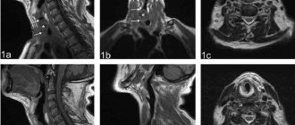
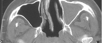
Computed tomogram of the larynx
What does a CT scan of the larynx show?
A CT scan of the larynx and neck will show:
- developmental anomalies;
- pathologies of blood vessels;
- consequences of external and internal injuries - fractures of cartilage, hyoid bone, larynx tear, muscle damage, blood accumulation (hematoma) around neck formations;
- lymphadenitis (inflamed lymph nodes), abscesses, adenophlegmons, you can see the extent of the spread of the purulent process into the surrounding tissues;
- tumors, metastases - CT images of the larynx will show heterogeneous volumetric formations of soft tissues and deformation of adjacent bone structures caused by their growth;
- the presence of foreign bodies in the examined area.
For the purpose of differential diagnosis to exclude the possibility of a tumor, examination is carried out in patients with:
- hyperplasia of the palatine tonsils (adenoids);
- laryngocele;
- chronic atrophic laryngitis;
- pharyngoesophageal diverticula;
- cysts.
Laryngeal tumor (marked with an asterisk) in the area of the left vocal cord (indicated by an arrow) on a computed tomogram
Thyroglossal duct cyst on CT scan
Indications
A CT scan of the throat and larynx is prescribed strictly according to indications during the diagnosis process. At the initial visit to the doctor, painful organs are examined and palpated, and the patient’s complaints are clarified. A CT scan of the throat is prescribed after safer methods required to make a preliminary diagnosis - laboratory tests, ultrasound, x-rays.
Complaints and symptoms that may cause a CT scan of the throat and larynx to be prescribed:
- swelling of the neck area, enlarged lymph nodes of unknown etiology;
- traumatic neck injuries;
- visible increase in size and change in shape of the thyroid gland;
- sore throat, feeling of a lump when swallowing;
- difficulty breathing, hoarseness, asthma attacks;
- signs of vascular pathology in the neck - headaches, dizziness, loss of consciousness, memory loss, darkening of the eyes.
CT scan of the throat and larynx provides complete information about the condition of the soft tissues and organs of the neck after injuries, the degree and prevalence of malignant neoplasms, the localization and boundaries of inflammatory processes. The study allows you to successfully carry out differential diagnosis and establish an accurate diagnosis. Based on the data obtained, the doctor chooses a treatment tactic - conservative therapy or surgical intervention.
CT scan of the larynx with contrast
The best way to diagnose tumors is a contrast-enhanced CT scan of the larynx, which involves the use of iodine-containing substances to improve the clarity of the images. The drug accumulates in tissues with increased blood circulation and clearly highlights them on tomograms.
The procedure with contrast shows edema, abscesses, primary tumors of the larynx, vocal cords, endocrine glands, lymph nodes at all stages of development, as well as metastatic tumors from the nasopharynx and distant organs.
The examination is used before operations to assess the effectiveness of surgery, radiation and chemotherapy in the treatment of cancer.
Laryngeal cancer on a CT scan. In the area of the right pyriform sinus, the arrow indicates a tumor that differs from neighboring tissues in its denser structure.
CT scan of soft tissues of the larynx and neck - what kind of technique
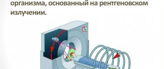
CT of the larynx is understood as a radiographic method for assessing the morphological changes that occur in a specified area of the body. The method is based on the property of X-rays to penetrate soft tissue, linger in dense structures and allow them to be visualized on medical photographs. Computed tomography of the larynx is the most accurate instrumental diagnostic method, giving an idea of absolutely any type of pathology:
- Dystrophic
- Inflammatory
- Degenerative
- Tumor
- Destructive
During the procedure, the tomograph takes many layer-by-layer images with a section size from 1 mm to 0.5 cm, depending on the type of device. CT images will be presented in the form of a three-dimensional image, and the scanning is carried out in several projections. This approach makes tomography data very accurate, and it will be almost impossible to miss even the slightest changes in the condition of the tissues. Even more detailed information is provided by MSCT of the larynx, where the number of sections is greater.
A CT scan of the larynx will show not only the upper respiratory tract. The tomography observation area includes:
- Thyroid
- Epithelial body
- Regional lymph nodes
- Blood vessels
- Upper esophagus
CT scan of the pharynx and larynx - advantages of the method
Unlike laryngoscopy, computer diagnostics of the larynx is a non-contact study that does not cause discomfort in patients with narrowing of the airways. CT and MSCT make it possible to study in detail the structure of organs and examine the smallest pathologies that are not noticeable on X-rays and ultrasound. Three-dimensional reconstruction allows you to get a clear picture of the location of structures in space, improves the recognition of details important for diagnosis, and helps plan surgical operations.
CT scan of the larynx, color 3D reconstruction
Alternative Methods
If the patient has contraindications, the specialist prescribes an alternative hardware examination method, such as MRI. Diagnosis is made using a similar method, but instead of X-rays, a phenomenon called nuclear magnetic resonance is used. It is completely safe for the body, therefore it is used for diagnosis even in young children. The only contraindication for MRI is considered to be metal elements in the body, which can heat up or become displaced during operation of the device.
As an alternative to CT, MSCT is used; this study is also carried out using radiation. The device creates multiple slices of the body area being examined, but the radiation damage is lower when compared with a standard CT procedure.
There is linear tomography of the larynx, in which the device reads data using a beam of X-rays, moving along the depth specified by the radiologist. This allows you to maximize the detail of the desired area, while the remaining tissues that do not provide informational benefit for the study remain blurred. Most often prescribed for the study of hollow organs - lungs, larynx, bronchi.
Preparation for examination of the larynx
There is no need to prepare for a CT scan without contrast. You should come to the clinic according to your appointment, taking with you your passport, a referral (you can do without it), and the results of previous diagnostics. If contrast is expected, it is necessary to donate blood for creatinine. Exceeding the norm is an absolute contraindication to the use of amplifiers.
Before a CT scan with contrast, a blood test for creatinine is required.
It is advisable to have a light snack before the contrast examination to reduce nausea and other side effects. Patients with infants need to take care of nutrition for their babies, since due to the accumulation of the contrast agent in the milk after the procedure, you will have to skip two feedings.
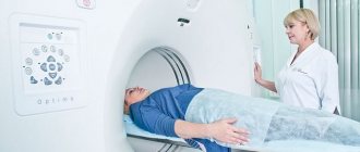
How is CT diagnostics performed?
No special preparation is required when performing CT without contrast. You should arrive at the appointed time, having with you the previous examination results or data from other instrumental techniques. If you plan to use a contrast agent, you should not eat 4-6 hours before the examination to avoid pathological reactions from the gastrointestinal tract (they sometimes happen, especially in patients susceptible to them).
Before the examination, you should remove all metal jewelry, accessories, put away your phone, and all removable medical devices. Diagnosis is carried out in the supine position. The person is placed on a table (couch), which is then pushed under the arc of the tomograph. You are not allowed to move during the CT scan. The device, or rather its attachment, begins to move smoothly around the head, taking a series of pictures.
The procedure takes about 10-15 minutes. If the study involves the use of contrast, preparation, administration of the drug and the CT scan itself may take 30-40 minutes.
The price of a CT scan of the neck, throat, and larynx will be 3,000-7,000 rubles, depending on the status of the clinic, the type and amount of contrast agent. An MSCT examination can cost about 5,000 rubles.
How is a CT scan of the larynx done?
Before the CT scan of the larynx, you should remove jewelry, glasses, and metal dentures: dense objects appear on the images and interfere with the perception of the structures being studied.
The patient lies down on the mobile table of the device. The operator turns on the tomograph, and the conveyor smoothly moves into the ring of the scanning system. The doctor controls the examination process from the next room - through glass, on a computer screen and through a speakerphone.
In contrast tomography, a control sample is first obtained, then an enhancer is injected. After distributing the drug to the tissues, a series of images of the organs is taken at different phases of blood filling. Real-time data is recorded on the monitor. The examination takes 15 minutes, and half an hour if contrast media are used.
Laryngeal CT procedure
Contrast examination of the neck and head (CT angiography) - how it is done
After intravenous enhancement with Omnipaque and Ultravist, the cervical vessels are contrasted. The contrast passes from the ulnar vein to the vessels of the upper shoulder girdle in a few seconds.
CT scan of the soft tissue lymph nodes of the neck visualizes the jugular vein, vertebral artery, internal and external carotid arteries. An informative image is obtained after three-dimensional modeling (3D mode).
The software application creates a spatial display that clearly shows aneurysms (dissections of the vascular wall), blood clots inside the lumen, external compression of the artery or vein, inflammatory and destructive changes.
Main indications for contrast CT of the neck:
- Diagnosis of venous and arterial anomalies;
- Studying the characteristics of injuries to the neck and larynx after injuries for vascular compression;
- Fatty deposits inside the artery wall, blood clots;
- Identification of the causes of progressive headaches, loss of concentration, visual acuity, hearing;
- Inflammatory changes in the vascular wall, assessment of healing after surgery, radiation exposure of the tumor.
The advantage of CT scan of the neck is visualization of the spinal cord canal. With neoplasms and displacement of the cervical vertebrae, infringement occurs, leading to disability and stroke. Concomitant examination of the larynx and other anatomical formations of the cervical region helps determine the number of tumors, metastases, and the degree of involvement of surrounding organs.
CT scan of the larynx with functional tests
A functional test is an observation of the functioning of the speech organs in dynamics, which allows one to obtain more information about their condition. A CT scan of the neck area may include additional scanning when the patient pronounces the “i” sound. At this point, the images better display the laryngeal ventricles and determine the symmetry of the vocal folds.
CT with functional tests is recommended in the following cases:
- injuries;
- prolonged sore throat;
- vocal cord dysfunction;
- breathing problems;
- swelling of the larynx;
- difficulty swallowing;
- compression of the esophagus;
- enlarged lymph nodes in the neck.
The CT scan of the larynx with functional tests is performed in a standard manner and does not require specific preparation.
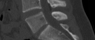
CT scan of the larynx: preepiglottic space (1), plate of thyroid cartilage (2), anterior commissure (3); vocal fold.
Decoding the research results
A medical specialist compares the scan images with standard indicators. The doctor identifies a discrepancy between the norms. Next, the patient is given the scan results in his hands. After receiving the conclusion, he must transfer it to the attending physician, who ordered a tomography. The doctor will assess the extent of the disease, its characteristics and prescribe the necessary drug therapy.
Based on the images, doctors identify the causes of pain and swelling, difficulty breathing, and impaired blood flow. Using a tomographic image, the doctor assesses the condition of blood vessels and soft tissues. In case of vascular pathology, the direction and speed of blood flow, any malfunctions and disturbances present are identified.
CT scan of trachea and larynx
Multislice computed tomography of the neck reveals the consequences of injuries, inflammatory and tumor processes in the larynx, and partially in the trachea. Using MSCT, you can accurately determine the location, nature and relationship of the pathological process with neighboring organs.
The examination is indispensable for:
- assessing the degree and extent of stenosis of the pharynx and airways;
- clarification of the condition of the walls of the larynx and the upper part of the trachea (thickening, compaction);
- identifying changes in paratracheal tissue.
Larynx and trachea on CT scan
Larynx
Larynx
,
larynx, belongs to the lower respiratory tract and is a voice-forming organ.
Topography
Holotopy: The larynx is located in the middle part of the front of the neck, it protrudes under the skin, forming a protrusion of the larynx
,
prominentia laryngea , more pronounced in men (Adam's apple).
Skeletotopy: in adults, the larynx is located at the level of the IV-VI cervical vertebrae.
Syntopy: at the top the larynx is suspended from the hyoid bone, at the bottom it continues into the trachea. The thyroid gland lies in front and to the sides of it. The main neurovascular bundle of the neck (carotid arteries, internal jugular vein and vagus nerve) runs laterally. In front, the larynx is not completely covered by the sublingual muscles with the pretracheal plate of the cervical fascia. The laryngeal part of the pharynx is located at the back. Here is the entrance to the larynx
,
aditus laryngis ; it is limited by the epiglottis and two folds of the mucous membrane that extend from the epiglottis downward and posteriorly. At the posterior end of these folds protrude a corniculate tubercle
,
tuberculum corniculatum , and a wedge-shaped tubercle
,
tuberculum cuneiforme , which correspond to the cartilages of the same name located in the thickness of the fold.
Structure of the larynx
The skeleton of the larynx is formed by unpaired and paired cartilages.
Thyroid cartilage
,
cartilago thyroidea , unpaired, hyaline. It consists of two plates that converge at an angle to each other. For men this angle is acute. At the junction of the plates there is a notch
,
incisura thyroidea . From the posterior edge of each plate, the upper horns
,
cornu superius , are long and narrow, and the lower horns, cornu inferius , are short and wide. The inferior horns connect to the cricoid cartilage. On the outer surface of the thyroid cartilage, an oblique line
,
linea obliqua , is visible - the place of attachment of the sternothyroid and thyrohyoid muscles.
Cricoid cartilage
,
cartilago cricoidea , unpaired, hyaline, lies at the base of the larynx.
Its front part forms an arc, the back part – a plate. On the sides of the plate there is a paired articular surface for articulation with the thyroid cartilage, and in its upper part there is a paired surface for articulation with the arytenoid cartilages. Arytenoid cartilage
,
cartilago arytenoidea , paired, hyaline, pyramid-shaped.
It has a top and a base. At the base there is an articular surface for articulation with the cricoid cartilage. Two processes extend from the base: 1) vocal process
,
processus vocalis , – the place of attachment of the vocal cords, built of elastic cartilage;
2) muscle process
,
processus muscularis , is the place of attachment of the muscles of the larynx, built of hyaline cartilage.
Epiglottis
,
epiglottis , unpaired, elastic. At the bottom it tapers, forming a stalk
,
petiolus .
Wedge-shaped and cornicular cartilages
,
cartilagines cuneiformes et corniculatae , paired, elastic, located above the apex of the arytenoid cartilages.
The cartilages of the larynx are connected to each other and to neighboring formations through ligaments, membranes and joints.
Between the larynx and the hyoid bone is the thyrohyoid membrane.
,
membrana thyrohyoidea , in which the median and paired lateral thyrohyoid ligaments are distinguished.
The latter arise from the superior horns of the thyroid cartilage. The epiglottis fixes two ligaments: 1) sublingual-epiglottic, ligamentum hyoepiglotticum;
2) thyroid-epiglottic, ligamentum thyroepiglotticum .
The thyroid cartilage is connected to the arch of the cricoid cartilage via the cricothyroid ligament
,
ligamentum cricothyroideum . The cricoid cartilage is connected to the trachea by the cricotracheal ligament
,
ligamentum cricotracheale . Under the mucous membrane there is a fibroelastic membrane of the larynx
,
membrana fibroelastica laryngis ; in the upper part of the larynx it forms a quadrangular membrane
,
membrana quadrangularis , and in the lower part - an elastic cone
,
conus elasticus . The lower edge of the quadrangular membrane forms the paired vestibular ligament
,
ligamentum vestibulare , and the upper edge of the elastic cone forms the paired vocal ligament
,
ligamentum vocale , which is stretched between the angle of the thyroid cartilage and the vocal process of the arytenoid cartilage.
The joints of the larynx are paired, combined:
1. cricothyroid joint
,
articulatio cricothyroidea , formed by the articulation of the articular surfaces of the cricoid cartilage with the lower horns of the thyroid cartilage.
Has one transverse axis of rotation. When the thyroid cartilage moves forward, the vocal folds lengthen and tighten, and when it moves backward, they relax. 2. Cricoarytenoid joint
,
articulatio cricoarytenoidea , formed by the articulation of the articular surfaces of the cricoid cartilage with the articular surfaces of the arytenoid cartilages.
Has a vertical axis of rotation. When the arytenoid processes rotate inward, the vocal cords come closer together (the glottis narrows), and when they rotate outward, they move away from each other (the glottis widens). The muscles of the larynx are striated, voluntary; move the cartilages of the larynx relative to each other, change the size of the glottis and the tension of the vocal cords (folds). There are external and internal muscles of the larynx.
According to function, the muscles of the larynx are divided into three groups (Fig. 1).
| Fig. 1. Muscles of the larynx:1 – cricoid cartilage, (cartilago cricoidea); 2 – vocal process of the arytenoid cartilage, (processus vocalis); 3 – muscular process of the arytenoid cartilage (processus muscularis); 4 – fibro-elastic membrane, (cone elasticus); 5 – thyroid cartilage, (cartilago thyroidea); 6 – vocal ligament, (ligamentum vocale); 7 – vocal muscle, (musculus vocalis); 8 – thyroarytinoid muscle, (m. thyroarytenoideus); 9 – cricothyroid muscle, (m. cricothyroideus); 10 – arytenoid muscles, (m. arytenoideus); 11 – cricoarytenoid lateral muscle, (m. cricoarythenoideus lateralis); 12 – cricoarytenoid posterior muscle, (m. cricoarythenoideus posterior). (according to F. Netter, 2003) |
1. Muscles that narrow the glottis (constrictors):
a) lateral cricoarytenoid muscle
,
musculus crycoarytenoideus lateralis.
Start
: upper edge of the cricoid cartilage arch.
Attachment
: muscular process of the arytenoid cartilage.
Function
: rotates the arytenoid cartilage around a vertical axis; in this case, the vocal process moves medially and the vocal cords come closer together.
b) thyroarytenoid muscle , musculus thyroarytenoideus .
Start
: inner surface of the lamina of the thyroid cartilage.
Attachment
: anterolateral surface of the arytenoid cartilage.
Function
: similar to the previous muscle.
c) transverse arytenoid muscle
,
musculus arytenoideus transversus.
d) oblique arytenoid muscle
musculus arytenoideus obliquus
. Start and Attachment
: posterior surfaces of the arytenoid cartilages.
Function
: Both muscles bring the arytenoid cartilages closer to the midplane, promoting closure of the glottis.
2. Muscles that expand the glottis (dilators):
a) posterior cricoarytenoid , musculus cricoarytenoideus posterior .
Start:
posterior surface of the cricoid cartilage plate.
Attachment:
muscular process of the arytenoid cartilage.
Function:
rotates the arytenoid cartilage around a vertical axis, turning the vocal processes laterally, while the glottis expands.
3. Muscles that change the tension of the vocal cords:
a) cricothyroid muscle
,
musculus cricothyroideus.
Start
: arch of cricoid cartilage.
Attachment
: the lower edge of the thyroid cartilage and its lower horn.
Function:
tilts the thyroid edge forward, increasing the distance between it and the vocal process, while the vocal cords lengthen and stretch;
b) vocal muscle , musculus vocalis .
Start:
inner surface of the thyroid cartilage.
Attachment:
vocal process.
Function:
the muscle contains longitudinal, vertical, and oblique fibers. Longitudinal fibers shorten the vocal cord, vertical fibers strain it, and oblique fibers strain individual parts of the vocal cord.
Laryngeal cavity
,
cavitas laryngis , resembles an hourglass and is divided into three sections: the vestibule of the larynx, the ventricles, and the subglottic cavity.
Vestibule of the larynx
,
vestibulum laryngis , extends from the entrance to the larynx to the vestibular folds, which include the vestibular ligaments.
Ventricles of the larynx
,
ventriculi laryngis , located in the form of depressions from the vestibule to the vocal folds; between them is the narrowest place of the larynx, up to 1 cm high. Vocal folds, plicae vocales , contain in their posterior part the vocal processes of the arytenoid cartilages, and in the anterior part - an elastic vocal fold and vocal muscle. Both vocal folds limit the glottis
,
rima glottidis s. vocalis . It distinguishes between the posterior - intercartilaginous part
,
pars intercartilaginea , and the anterior - intermembranous part
,
pars intermembranacea .
The glottis,
rima glottidis ,
is the narrowest part of the laryngeal cavity, bounded by two vocal folds.
Subglottic cavity
,
cavitas infraglottica , extends from the vocal folds to the beginning of the trachea.
The mucous membrane of the larynx is lined with stratified ciliated epithelium. The exception is the vocal folds, which are covered with stratified squamous epithelium. The function of the larynx as a respiratory and vocal organ.
The muscles attached to the hyoid bone (supra- and hyoid) raise, lower, or fix the larynx. When swallowing, the larynx is raised by the action of the suprahyoid muscles, the root of the tongue moves posteriorly and presses on the epiglottis so that it covers the entrance to the larynx. This is facilitated by the contraction of the thyroepiglottic and aryepiglottic muscles.
During quiet breathing and whispering, the intermembranous part of the glottis is closed, and the intercartilaginous part is open in the form of a triangle by the action of the lateral cricoarytenoid muscle (Fig. 2). During deep breathing, both parts of the glottis are opened in a diamond shape by the action of the posterior cricoarytenoid muscle. At the beginning of vocal production, the glottis closes and the vocal cords become tense. The flow of exhaled air causes vibrations in the vocal folds, resulting in sound waves. The strength of sound is determined by the strength of the air flow, which depends on the lumen of the glottis, and the timbre of the voice is determined by the frequency of vibration of the vocal folds. The installation of the vocal folds is carried out by the cricothyroid muscle and the muscles attached to the muscular process, and more precisely, it is modeled by the vocal muscle.
| Rice. 2. Action of the muscles of the larynx. (after F. Netter, 2003) |
The resonators of sound produced by the vocal apparatus are the pharynx, oral and nasal cavities, and paranasal sinuses. The pitch of the voice depends on the individual structural features of the sound resonators. Due to the position of the larynx in a person, the sounding air flow is directed to the speech organs - palate, tongue, teeth and lips. When coughing, a closed glottis opens with expiratory impulses.
Age characteristics.
In newborns, the larynx is located at the level of the II-IV cervical vertebrae. The epiglottis touches the uvula. The larynx is short and wide, its cavity is funnel-shaped, and there is no laryngeal prominence. The vocal folds are short, the ventricles of the larynx are shallow. Rapid growth of the larynx occurs in children 3 years old, at 5-7 years old, and especially during puberty. At 12-13 years old, the length of the vocal folds in girls increases by 1/3, and in boys at 13-15 years old, by 2/3. This causes a mutation (fracture) of the voice in boys. In men, growth of the vocal folds continues until the age of 30. Gender differences in voice are due to the greater length of the vocal folds and glottis in men. In old age, the cartilage of the larynx becomes calcified, the vocal cords become less elastic, which leads to a change in voice.
Anomalies of the larynx
1. Atresia, stenosis.
2. Formation of septa in the laryngeal cavity.
3. Aplasia of the epiglottis. In this case, the entrance to the larynx is not closed.
4. Laryngeal-esophageal fistulas. They are formed when the laryngeal primordium is incompletely separated from the digestive tube.
Trachea
Trachea
,
trachea , (windpipe), is an unpaired tubular organ that serves to conduct air.
Topography
Holotopia: cervical part,
pars cervicalis , located in the lower part of the anterior cervical region; the thoracic part
,
pars thoracica , lies in the anterior part of the upper mediastinum.
Skeletotopy: in adults, it begins at the level of the VI cervical vertebra and ends at the level of the V thoracic vertebra (2-3 rib), where it forms a bifurcation, bifurcatio tracheae , that is, it is divided into two main bronchi.
Syntopy: the thyroid gland is adjacent to the cervical part in front and on the sides, and the hypoglossal muscles are also located. There is a gap between the edges of the muscles in the midline, where the trachea is covered only by the pretracheal plate of the cervical fascia. Between this plate and the trachea there is a pretracheal cellular space that communicates with the mediastinum. The thoracic part of the trachea borders in front with the aortic arch, brachiocephalic trunk, left brachiocephalic vein, left common carotid artery, thymus gland, laterally with the mediastinal pleura, behind with the esophagus throughout the trachea.
Structure of the trachea
The tracheal skeleton consists of 16-20 hyaline half-rings
,
cartilagines tracheales . They are connected to each other by fibrous annular ligaments
,
ligg. anularia . At the top, the trachea is connected to the cricoid cartilage of the larynx by the cricotracheal ligament. The cartilages of the trachea form the anterior and lateral walls, the posterior wall of the trachea is membranous, paries membranaceus , contains connective tissue, circular and longitudinal bundles of smooth muscles.
The tracheal cavity is lined with a mucous membrane with stratified ciliated epithelium; it contains branched mucous glands and lymphatic follicles. Externally, the trachea is covered with an adventitial membrane. Age characteristics
. In newborns, the trachea begins at the level of the IV cervical vertebra, and its bifurcation projects to the III thoracic vertebra. Tracheal cartilages and glands are poorly developed. The growth of the trachea occurs most intensively in the first 6 months after birth and during puberty. The final position of the trachea is established after 7 years. In old age, atrophy of the mucous membrane, glands, lymphoid tissue, and calcification of cartilage are observed.
Tracheal anomalies
1. Atresia and stenosis.
2. Deformation and splitting of cartilage.
3. Tracheo-esophageal cartilages.
Examination of the larynx - MRI or CT
When referred for examination, patients are interested in how informative a CT scan of the larynx is, what the analysis will show, and whether it would be better to do an MRI. Let's compare these diagnostic methods:
| Criterion | CT | MRI |
| Operating principle | The work of a computed tomograph is based on the properties of body tissues to absorb or reflect x-rays. | The device records the distribution of hydrogen atoms in the composition of water dipoles in tissues located in a magnetic field |
| Visualization quality | Bone and cartilage tissue are better displayed (the latter serves as the frame of the larynx). | The photographs show the soft tissues of the larynx and neck more clearly. |
| pros |
|
|
| Minuses |
|
|
CT and MRI are complementary types of diagnostics. In some situations, both studies may be required to complete the picture.
Cancer of the right vocal fold on a tomogram. CT on the left, MRI on the right. The same patient.
Ligaments and muscles
The muscular system consists of two large groups:
- External, which set the entire larynx in motion.
- Internal, which causes cartilage to move relative to each other.
Internal muscles are also involved in the processes of breathing, swallowing and sound production.
External muscles, in turn, are divided into 2 groups:
- Sternothyroid and thyrohyoid. These are paired formations, which at one end are attached to the largest thyroid cartilage, and at the other to the bones of the skeleton.
- Sternohyoid, geniohyoid, digastric, omohyoid and stylohyoid. These structures are attached to the hyoid bone and skeletal bones.
The functions of the internal muscles of the larynx are:
- In changing the position of the epiglottis during swallowing.
- They change the tension of the vocal folds, as well as the width of the glottis during speaking or singing.
The two pairs of muscles that perform opposite actions are called the aryepiglottic and thyroepiglottic. The first structure, contracting, pulls the epiglottic cartilage back to close the entrance to the respiratory tract. When the thyroepiglottic muscle contracts, the entrance to the larynx opens.
Vocal muscles of the larynx
Vocal folds of the larynx
This is a large group of muscles, which consists of structures that narrow, dilate, tense and relax the glottis:
- When contracted, the cricoarytenoid muscle lateralis narrows the glottis.
- When inhaling, the posterior cricoarytenoid muscle expands the lumen of the larynx. This is the most important structure in vocal production. With paralysis, the lumen closes, the person is unable to utter a sound. Respiratory functions may also be impaired, leading to asphyxia. Most often this happens during a stroke.
- The transverse arytenoid muscle, contracting, narrows the posterior part of the glottis by a third.
- The oblique arytenoid muscle enhances the action of the transverse muscle. They are located behind it, crossing at right angles.
- The thyroarytenoid muscle moves the arytenoid cartilage.
- When the cricothyroid muscle contracts, it causes the thyroid cartilage to tilt, which narrows the glottis.
- The vocal paired triceps muscle, the contraction of which thickens or lengthens the vocal folds.
The ligamentous apparatus connects the cartilages and parts of the larynx:
- Between the pharynx and the cricopharyngeal cartilage plate is the cricopharyngeal ligament.
- Between the cricoid cartilage and the upper ring of the trachea is the cricotracheal ligament.
- Cricoarytenoid and cricothyroid joints.
- The cricothyroid ligament is between the cricoid and thyroid cartilages.
- Vestibular and vocal cords.
- Hypoepiglottic, thyroepiglottic, thyrohyoid ligaments.
- Thyrohyoid membrane.
In case of weakening of the ligamentous apparatus of the larynx or disease, the arrangement of the structures relative to each other changes, so the timbre of the voice may also change.
Indications and contraindications
CT scan of the larynx and neck is recommended in the following cases:
- anomalies (congenital defects) of organs in the area of interest;
- injuries;
- foreign objects in the larynx;
- attacks of suffocation;
- feeling of a lump in the throat;
- problems with breathing and swallowing;
- changes in lymph nodes;
- swelling of the larynx;
- voice change;
- inflammatory diseases of the nasopharynx and throat;
- calcification (calcification) of the cartilage of the larynx;
- pathologies of the thyroid gland;
- diseases of the esophagus;
- suspicion of any neoplasms.
Contraindications to computed tomography:
- pregnancy at all stages;
- early age (due to the sensitivity of the child’s body to radiation, the examination should not be done before 5 years of age);
- body weight more than 150 kg, chest (abdominal) circumference more than 150 cm.
The use of contrast agents is prohibited when:
- allergies to iodine;
- hyperthyroidism;
- severe renal failure;
- diabetes mellitus if the patient is taking metformin.
Taking an anamnesis for computed tomography
How to prepare for a CT scan of the larynx
No special preparatory measures are needed to perform a CT scan of the larynx without a contrast agent. It is necessary not to overload the stomach at dinner the day before the test. You will first need to undergo a number of additional tests.
If contrast is used, do not consume food or water 4 hours before the start of the session. This will eliminate side effects from the digestive organs when performing a computed tomography scan of the larynx.
CT scan of the throat is a safe diagnostic test that requires minimal radiation exposure. The emission of X-rays is somewhat different from the usual X-ray. With CT there are no radiation or ionization lesions; the method is used to examine any acute and chronic pathologies of the anatomical region.
CT safety
Computed tomography can indeed cause harm to the health of a living organism, but it is insignificant; no cases of pronounced disorders that resulted from a CT examination of the larynx were found. This research method is based on x-ray radiation. This phenomenon is recognized as relatively safe for human health if he is not constantly near the device without special protection.
Fear of undergoing the procedure is unfounded, so if necessary, you should not refuse this functional study. When performing a CT scan of the larynx, the price is moderate; the study costs about 4,000 rubles. Modern devices are created with a modified design, so the dose of direct radiation is even lower. It is allowed to carry out this study more than once, without any disturbances in the functioning of internal organs.